Types of body coverings
Ingument or animal skins are designed to perform various functions to protect the skin of mammals and animals. These include protection against loss of water and nutrients, thermoregulation. In addition, the skin of animals serves them for breathing and excreting substances. Invertebrates are known for the fact that their skin does not develop significantly and is a layer of ectoderm with additional substances. The evolutionary process of their dermis developed from ciliated epithelium to squamous epithelium. This can be observed in most varieties of worms. In this case, the squamous epithelium serves as an elementary organ of movement.
Arthropods have a superficial type of epithelium, which evolved during the process into a chitinous cuticle and shell in all crustaceans. In the process of development in mollusks, it developed into a shell containing lime. In animals of some groups, multicellular glands are locally formed in the epidermis, which help form cobwebs, poison, saliva, etc.
Chordates went through stages of evolution in the direction from single-layered to multilayered epidermis and skin itself. In chordates, the skin contains a layer of epidermis, often called the cuticle and the skin itself.
Vertebrates went a longer way and acquired a cover of two layers: the superficial epithelial layer and the skin itself, which attaches to the subcutaneous tissue, including the muscles and skeleton. Amphibians use their skin as a breathing apparatus, among other things. Due to the water film, multilayer glands gradually developed on the skin, producing mucus with the function of moisturizing the outer integument. Also, some species have actively developed poisonous glands that perform a protective function.
The development of the skin in mammals led to the formation of derivatives: caratine, hooves, wool and hair, horns and nails. They perform important vital defense functions. And the skin sweat glands allow you to maintain the level of thermoregulation through sweat channels.
Functions of the body
The outer cover of the body has a number of functions that are essential for the life of humans and animals:
• respiratory function. A small amount of air passes through the pores of the skin per day, which in turn leads to the removal of steam and carbon dioxide. This property of the integument often complements the function of the lungs. Children's skin conducts much more oxygen than adult skin. Solid substances and water almost do not pass through healthy skin, which cannot be said about some chemicals and medications. These include esters, iodine, chloroform, salicylic acid and more;
• receptor function. The terminal nerve apparatus on the skin, as well as the nerve endings, interact directly with the cerebral cortex. The skin is capable of transmitting signals of irritation from mechanical influences to the central nervous system. The signals it sends turn into various sensations: warmth, touch, pain, cold and much more. The reference points in the external environment are hearing, smell and vision, also interconnected with skin receptors;
• thermoregulatory function. The functioning of this skin property is fully controlled by the body’s nervous system. Reduced sweat production and dilation of blood vessels help increase heat transfer. Through the pores of the skin, heat is transferred from the body to the external environment. Temperature regulation is important not only during the activity of the body, but also during the rest period, when a small amount of heat is generated. Its volume increases during physical activity, however, according to research results, it has been established that heat transfer to the body is a constant figure and changes in exceptional cases. Thermoregulatory function is divided into physical and chemical. Physical is intended to transfer heat to the external environment, and chemical is aimed at creating it;
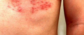
• protective function. The skin is a factor that protects the tissues and organs of the body from environmental influences. The existence of the stratum corneum and fatty tissue preserves the internal organs during mechanical impact on them in the form of shock and friction, chemical irritants and physical influences. The acidic reaction of fatty tissue prevents the penetration of harmful microorganisms and helps reduce the impact of external chemicals. The sweat glands of the skin protect the internal environment of the body from bacteria. The sebaceous glands have the same property. Sterility is ensured by the upper horn cells. Solar radiation from the earth in moderate quantities does not cause visible harm to the body due to the melanin pigment contained in the skin;
• secretory function. Vitamins, hormones, organic substances, mineral products, water, enzymes and much more are removed from the body due to the work of the sebaceous and sweat glands located in the layers of the skin. It is these processes that determine the metabolic processes of the whole organism. During fever, high temperature or physical exertion, the glands are capable of removing up to 700 ml of water and up to 4 liters of sweat. 20 g of sebum are removed by the sebaceous glands through their channels during normal functioning of the body.
Protective function of the skin
| Skin function | |
| Receptor | Tactile: sensation of touch; Temperature: perception of cold and hot; Protective:
|
| excretory | During the day, 0.5 liters of water, salt, and lactic acid are released through the skin |
| Thermoregulatory | More than 80% of heat is lost through the surface of the skin |
| Participation in blood circulation | At the same time, the skin contains up to 1 liter of blood |
| Participates in mineral metabolism | Produces vitamin D and melanin |
The skin is equipped with a large number of different sensory nerves and corresponding nerve endings (receptors), due to which it functions as a complex sensory organ. Skin receptors perceive various irritations from the external environment and signal them to the cerebral cortex. That is why we are able to feel touch and feel objects, feel heat and cold, experience pressure and pain. Skin receptors make it possible to obtain ideas about the objects around us, their size, shape, the nature of their surface, temperature.
The structure of the body
The skin of humans and vertebrates consists of 3 layers: epidermis, dermis and subcutaneous fat.
• Epidermis is an epithelial tissue in which there are no blood vessels, and nutrition occurs due to lymph in the intercellular gaps. The epidermis consists of 5 layers:
- the deepest main layer bordering the dermis. It consists of only one row, which includes cylindrical cells containing melanin. The amount of melanin determines the skin tone. The amount of this substance varies not only among people of different races and places of residence. It can vary significantly in the same person on different parts of the body. The substance helps protect cells and their nuclei from the negative effects of solar radiation. In this area, the process of formation of the epidermis occurs;
– above the main layer there is an awl-shaped layer. It is formed by cubic cells with a light core, arranged in 6 rows. They are connected to each other by fibers - bridges, through which lymph flows, bringing nutrients and carrying away waste products of the body;
– 1-2 rows of oblong-shaped cells form the granular layer. The cells contain a special protoplasm with a protein involved in the initial stage of the formation of keratinization of cells;
– flat cells without a nucleus, arranged in 2 rows, form a shiny layer. Eleidin, stored in cells, plays a large role in cell keratinization;
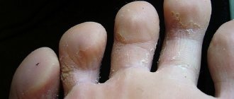
– the stratum corneum, consisting of 5 to 15 layers of cells, depending on the area of the body, is formed by flat cells without nuclei. The layer contains keratin and keratinized substance, which is rejected by plates closer to the surface of the skin or flakes off.
• Dermis. The second name is the skin itself. It is created from two layers: mesh and net. The epidermis is deepened into the dermis, a strong connection between which is provided by the papillae. It consists of cells, connective fibers and intercellular substance. Connective fibers provide elasticity, strength and quality of the skin. The dermis also participates in processes in which allergic and inflammatory processes can be monitored, preventing pathogenic microbes from entering the body.
• Subcutaneous adipose tissue consists of bundles of connective fibers in the form of a lattice. Each loop of such a lattice is equipped with fat cells. This fabric effectively protects the body from cold and mechanical influences, secures hair and fur, and has sweat glands, nerves and blood vessels. The layers of skin are also equipped with sweat and sebaceous glands. Sweat glands consist of a body and an excretory duct. There are more than 3 million of them on the human body, but they are not evenly distributed. Some areas of the body are completely deprived of them, and the bulk of them are on the face, scalp, palms and back. Sebaceous glands with a lobular structure surround hair follicles, needles, and feathers. They are abundantly supplied with nerve endings and blood vessels. Additional skin formations include hair, fur, nails and claws.
Structure and functions of the skin
Epidermis - the outer layer is formed by flat epithelium and has a thickness of 0.07 to 2.5 mm and depends on the nature and strength of environmental influences. The epidermis is thickest on the palms and soles, thinnest on the eyelids. The surface layer of the epidermis consists of keratinized dead cells (it also forms hair and nails), which look like scales and fit tightly to each other.
The epidermis is based on one row of prismatic cells that constantly divide and form new cells; these cells move to the outer layers, changing their shape, becoming flat, keratinizing, dying and exfoliating. The epidermis contains pigment cells (melanin) that give the skin its specific color. Pigmentation helps absorb short-wave rays and thereby protects internal organs from damage.
Skin color depends on several factors. The pigment melanin gives the skin a brown tint, the stratum corneum of the epidermis is gray-yellow, and the granular layer is whitish. The pink or reddish color of the skin is due to the fact that blood vessels are visible through the stratum corneum. Tissue fluids, refracting light rays, give the skin a matte white hue. The combination of all these shades forms the normal skin color, which changes with age and with certain diseases. There is no pigment in the skin of newborns. Even among blacks, the skin of a newborn child is light, reddish and receives its color only some time after birth. Pigmentation of human skin develops during the first months and years of life and depends not only on the number of pigment cells, but on their functional ability, i.e. on the degree of pigment formation in them. In people with fair skin at a young age, the pigment is distributed evenly. The blood supply to the skin at this age is well developed. The skin is rich in tissue fluid, giving it a whitish-matte tint.
In older people, the stratum corneum and granulosa of the epidermis thicken, and blood circulation worsens. White-yellow or gray-yellow color begins to predominate over the predominantly pink color. In addition, the tissues in aging skin become dehydrated and begin to transmit light rays deeper, which determines the yellowish color of the skin. Under the influence of endocrine factors, the nervous system and external phenomena (sunlight, air temperature, wind, mechanical stimuli), the amount of melanin pigment also increases, giving the skin a yellowish-brown tint. Moreover, the distribution of pigment in the skin becomes disrupted with age: age spots appear. Thus, as a result of all these changes, aging skin acquires a pale yellow or gray-earth color, often with pigment spots of various sizes in certain areas of the face, neck, and hands.
The dermis, or inner layer (the skin itself), is much thicker than the epidermis. It consists of dense connective tissue formed by cells and closely intertwined fibers. The fibers give the skin elasticity: it stretches and moves easily.
This layer of skin is abundantly supplied with blood vessels and nerve endings. There are numerous formations here: hair follicles, sebaceous and sweat glands, receptors. The hair follicles are connected to blood vessels, nerves and muscles. Contraction of these muscles occurs reflexively when cooling and straightens the hair, which helps improve the thermal insulation properties of the skin. In this case, a person develops “goose bumps”. Very sparse and short hair covers almost the entire human skin (except for the palms and feet). They have lost their protective significance and are vestigial. Together with nails, hair belongs to the horny formations of the skin.
The sebaceous glands, located at the roots of the hair, secrete oil that lubricates the hair and skin and protects it from drying out. Sweat glands produce and secrete sweat, which is similar in composition to urine, but less concentrated. They have a tubular structure and are a tube twisted into a ball, braided with blood capillaries.
Hypodermis - subcutaneous fatty tissue - is the deepest layer of the skin, consisting of loose connective tissue, which contains fibers and many fat cells. Fat reserves are stored here. In addition, subcutaneous tissue serves to protect the organs covered by it from shock and cooling.
Covers of the human body
The skin of the human body has an average surface area of 1.7 square meters. It protects organs from temperature changes, mechanical stress and infections. The overall health of a person depends on its functioning. It consists of 3 main layers:
– epidermis, the first outer layer;
– the dermis, in the structure of which there are vital sebaceous and sweat glands;
– subcutaneous base or connective tissue of loose structure.
The volume and condition of each layer depend on various factors: gender, age, genetics, lifestyle, physical activity and diet. Derivatives of the skin are the mammary, sebaceous and sweat glands. They are directly responsible for removing salt and inorganic substances, protecting and moisturizing the skin.
A special part of the human skin is occupied by nails and hair. Nails form a strong plate that creates protection on the fingers and toes. They grow evenly throughout life, without slowing down. The uniformity of cover and color can indicate the state of human health. Hair protects the most vulnerable parts of the human body. Their bulbs are immersed in the dermis. During life, the rate of hair growth may change, and the number of hair follicles may not increase. Their numbers are established in the womb and cannot increase.
Human outer coverings
Skin is the outer covering of the human body. Its area is 1.5-1.6 m2. It consists of three layers: the outer - epidermis, the middle - dermis and the inner - subcutaneous fatty tissue (hypodermis). The skin performs a protective function: it protects the body from many harmful influences (mechanical, chemical, physical), and also creates an obstacle to most microbes. It produces special substances that promote its self-disinfection.
The skin is rich in muscle and elastic fibers that have the ability to stretch, giving it elasticity and resist pressure. Thanks to these fibers, the skin can return to its original position after stretching. The extensibility of the skin in different directions is not the same. Upon careful examination, you will notice that the entire surface of the skin is dotted with a large number of tiny folds, grooves and lines, forming a complex pattern of triangular and rhombic fields. In addition, there are subtle indentations on the skin. These are the so-called pores. The skin pattern of lines, grooves and pores in young people is less noticeable. With age it becomes more distinct. The skin grooves on the palms and soles stand out especially sharply, forming a complex individual pattern in their areas.
Animal body coverings
The covering of an animal's body is an external formation that is a barrier between the body and the environment. The characteristics of animal covers are determined by their species and life factors.
The protective functions of the integument are associated with the main secretions: wax, fat, salt and carbohydrates. Thus, a film of fat and wax on the surface of the body of insects protects their body from dehydration, and the skeleton of crustaceans consists of secreted calcium. Animal integument cells can be dead or alive, with the ability of secretion and pigmentation.
The functioning of the skin of multicellular animals is directly related to blood vessels, glands, muscles and pigment cells. In addition to the main functions of the integument of the body of animals, they also have the following factors:
– attack function in the form of stinging cells on the skin, claws, hooves, horns, needles and others;
– the function of movement and support, which gives the body shape, supports internal organs in the right place, and forms barriers between the fingers and limbs;
– secretory function, manifested in the ability to secrete substances for territorial designation and protection from parasites and enemies;
– a function of irritability, in which the integument has receptors that react to light, the composition of water, touch and much more;
- a storage function of substances that helps mammals store water and fat.
The skin of many animals has a sign of adaptation that promotes survival and reproduction. Various environmental influences on the skin of animals determine the type of device:
– coloring and camouflage due to pigment substances, helping to quickly become invisible depending on environmental conditions;
– mimicry, which allows unprotected animals to take on the colors of more protected species;
– toxicity as a protective property;
– electrical organs of fish that allow the generation of current for the purposes of protection and warning.
Skin thermoregulation
The skin takes part in heat regulation. It protects the body from overheating and cooling, helps maintain a constant body temperature. About 80% of the heat generated in the body is released through the skin. The skin is the main organ of thermoregulation. Through the skin, the body releases heat to the environment through heat conduction, heat radiation, and a significant part through the evaporation of sweat. If, for example, you hold a wet hand in the air, a feeling of cold appears because the evaporation of water cools the hand.
Heat transfer through the evaporation of sweat occurs in different ways: with the evaporation of sweat, the formation of which depends on the amount of blood passing through the skin vessels; as a result of radiation of heat into the surrounding air; due to the transfer of heat to objects in direct contact with the body, such as clothing; due to the mixing of air currents, in which the warm air surrounding the surface of the body is replaced by cooler air.
Heat transfer through the evaporation of sweat occurs continuously in the form of perspiration, imperceptible to us. During the day, the skin secretes twice as much water through sweat as the lungs do during the same time. In hot weather, the body loses more heat because the blood vessels dilate, the blood flow in them accelerates, and the amount of blood flowing near the surface of our body increases, where it cools. In cold weather, the skin vessels narrow, the skin turns pale, and sweat production decreases sharply, resulting in reduced heat transfer.