The article was prepared by a specialist for informational purposes only. We urge you not to self-medicate. When the first symptoms appear, consult a doctor.
Red moles are benign neoplasms that grow from blood vessels. Red moles are also called hemangiomas.
According to statistics, red moles most often appear in childhood. They are observed somewhat less frequently in the adult population.
Until now, scientists have not decided whether hemangioma should be considered a vascular tumor or a pathology of a congenital nature. However, recent data indicate that red moles are the result of proliferation of the vascular endothelium, which allows hemangiomas to be considered vascular tumors.
Prevalence and localization
According to available statistics, hemangiomas are detected immediately after the birth of a child. This happens in 87% of cases. Girls are at risk; they account for about 70% of all diagnosed red moles. By the way, it is hemangiomas that make up up to 48% of all soft tissue formations that are detected in childhood.
A red mole can be found in any part of the body, but 80% of all such defects are localized in the upper part of the body. It is also possible to detect hemangiomas, although extremely rarely, on the bones, in the brain, lungs, and liver.
- 95% of all hemangiomas are simple formations;
- 3% – cavernous formations;
- 2% – mixed type formations.
Diagnosis of moles during pregnancy
What to do if you notice the appearance of new age spots on the skin or an old mole has enlarged during pregnancy?
It is necessary to make an appointment for an in-person consultation with a dermatologist.
The doctor will conduct a visual examination of the pigment formation, assess its condition and appearance.
Dermoscopy is mandatory to determine the possible pathogenicity of the nevus.
Dermatoscopy is a non-invasive method for the differential diagnosis of pigmented skin lesions and early diagnosis of malignant melanoma.
This technique is also known as dermoscopy, reflected light microscopy, and epiluminescence microscopy.
Dermatoscopy is an integral part of the dermatological examination of the skin.
Today, the dermatoscope is considered the “dermatologist’s stethoscope.”
In addition to the diagnosis of pigmented and non-pigmented skin tumors, the method will be able to establish the diagnosis of infectious, parasitic and inflammatory lesions of the dermis.
In the field of dermato-oncology, dermatoscopy contributes to preoperative planning for the removal of pigmented skin lesions.
Helps you choose the best treatment option and subsequent care.
The main task of a dermatological examination is to find out the following points:
- whether the lesion is melanocytic or non-melanocytic;
- whether the formation is benign, suspicious or malignant.
In modern dermatology, digital dermatoscopy is more often used.
Where diagnostics displays the results of the procedure on a computer monitor.
This allows you to create reports with patient data and images of his pigmented lesions.
This makes it easier to make a diagnosis and makes it possible to consult with other specialists.
Another advantage of this method is that the images can be printed and given to the patient.
Alternatively, the digital images can be transferred to a CD and sent to the appropriate physician.
Causes of red moles
The reasons for the development of hemangiomas have not yet been precisely established. It is unknown why they appear most often in the face area. Scientists attribute this to the fact that it is here that there is an abundant vascular network.
Childhood
Researchers agree that red moles are a consequence of pathology in the formation of vascular tissue, which occurs due to disturbances in the process of fetal development.
Vascular tissue passes through all parts of the fetal body along a chain of pericytic cells at the time of organ formation. When there is a lack of oxygen to any organ or tissue of the fetus, pericytic cells quickly react to hypoxia and try to eliminate it. To do this, using the production of special proteins, they attract a large number of pericyte cells to the problem area. Cells begin to form new areas of blood supply, eliminating existing hypoxia. Sometimes, due to imperfections in the mechanism, the reproduction of specific proteins does not stop even when oxygen starvation has already been eliminated. As a result, the vascular tissue continues to grow, forming a hemangioma.
Red moles are also called vascular hyperplasia, which indicates that they are a consequence of a disruption in the growth process of vascular tissue. Answering the question of how this happens is quite problematic, since it is necessary to carefully monitor how the intrauterine development of fetal tissue occurs. And the only data available are those obtained from the study of stillborn babies or aborted fetuses.
Adult patients
- The appearance of red moles in adulthood is explained by hormonal imbalances in the human body. This often occurs during gestation, during menopause, while taking medications with hormones, when using oral contraceptives, or due to pathologies of the endocrine system.
- It is possible that viral infections, ultraviolet rays, radiation, and chemical agents can provoke the growth of hemangioma.
- A lack of ascorbic acid can affect the condition of vascular tissue.
- If the capillaries are regularly damaged, this can provoke the appearance of hemangiomas.
- Vascular hyperplasia may indicate diseases of the internal organs, but the location of red moles does not indicate a problem in the area where they are most concentrated.
Laser removal of hemangiomas
It is not possible to remove hemangioma with a laser in all cases. Although with the help of a laser the tumor is removed layer by layer, in several “passes”, if the modified tissue has deep proliferation or the tumor is located in the internal organs, it is impossible to remove the tumor in one procedure. In addition, with a large volume of modified tissue, complex therapy is necessary.
Despite all the advantages, laser procedures have many contraindications.
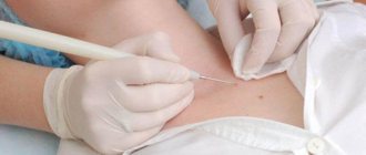
Among the advantages of laser removal of red spots are the non-invasiveness of the procedure, it is carried out under local anesthesia, a short period of postoperative rehabilitation, as well as a minimal cosmetic defect remaining after removal.
Any procedures using a laser require preliminary examination for the presence of contraindications. Thus, laser surgery is not used if the patient has:
- problems of the cardiovascular system and rheological properties of blood;
- oncological neoplasms;
- hypertension and atherosclerosis;
- disorders of the kidneys and liver;
- nervous system disorders;
- elevated body temperature.
Laser surgery is also not used during pregnancy and lactation. If there are no contraindications, postoperative rehabilitation takes up to two weeks, during which redness and swelling may be observed in the affected area. They do not require any additional treatment and soon go away on their own.
Red moles in newborns
During the neonatal period and infancy, red moles in children are very noticeable, but they often disappear by the age of 5.
Such benign tumors do not pose a danger if they:
- They do not itch, do not hurt, do not cause any concern;
- They do not become larger, for example, in a few months they do not increase in size by 3 times or more;
- Not located under the eye, on the nose, on the face or on the genitals.
It is known that hemangiomas can rapidly grow along the periphery, this is especially true for the first months of a child’s life. Therefore, in 10-12% of cases they are removed, since there are indications for this. The fact is that as the mole grows, it will destroy nearby tissues, causing defects not only cosmetic, but also functional. This is relevant in the case when hemangiomas are located on the ears, under the eyes, etc. Normal functioning of organs becomes impossible due to compression by the overgrown formation.
Reasons for appearance
Angiomas occur in newborns. They are formed due to viral and bacterial diseases suffered by a woman during pregnancy. For example, ARVI, influenza, pyelonephritis. The formations have different sizes, but do not exceed 1 cm. Up to 7 years of age, they most often resolve on their own. Adults are characterized by hormonal changes, after which red moles appear.
There are no reliable data on the nature of the formation. There are only hypotheses.
- Changes in hormonal levels. For example, adolescence, pregnancy, lactation, menopause.
- Damage to skin and blood vessels. They must be chronic. For example, frequent damage after shaving.
- Hypovitaminosis. If there is a lack of vitamins K, C, E, D, the vascular endothelium begins to become thinner and damaged.
- Digestive disorders. Moles can form due to frequent inflammatory conditions. For example, gastritis, enteritis, colitis.
- Liver pathologies. Angiomas take on a bright red hue.
- Cardiovascular diseases leading to fragility of blood vessels. This is anemia, hemophilia.
- Lipid imbalance, autoimmune lesions.
- Unfavorable heredity.
- Bad habits. These include frequent consumption of alcohol and nicotine. Visiting a solarium is also harmful.
People at risk should have their moles examined periodically by a dermatologist.
Features of red moles in adults
Red moles, as primary formations, do not appear in adulthood. They arise from previously undetected vascular proliferations. Most often, they are treated before the child enters school, so if a hemangioma is present in adulthood, this means that it was either not treated, or the tumor is located on internal organs.
The most dangerous location of a red mole is considered to be the vertebral body. As the tumor grows, the structures of the spine will weaken, which contributes to the occurrence of fractures.
Surgical removal of tumors
An operation to remove hemangiomas surgically is indicated for large tumors, if the surface of the mole is damaged and inflammation appears, if there is a risk of damage to the tumor by friction of clothing or during hygiene procedures. The surgical method is used in the treatment of angiomas on the arms, legs, chest, abdomen, i.e. in almost any area except the face. Such operations are performed by a vascular surgeon.
Removal of red dots using cryodestruction, sclerotherapy, radiation and microwave therapy is strictly contraindicated. Such treatment can lead to worse consequences than the problem itself.
Surgical removal of hemangioma is most often prescribed in cases where its growth is associated with a risk to vital organs, for example, proliferation (growth) in the area of the larynx, duodenum, internal genital organs; if the neoplasm is deep or combined. Depending on the nature of the tumor, the operation can be performed under local or general anesthesia and take from several tens of minutes to several hours. But in any case, the surgeon must be highly qualified to avoid the risk of blood loss.
Classification of red moles
Morphology of hemangiomas
Red moles can be capillary or cavernous. Capillary hemangiomas are represented by layers or a group of vessels that are closely pressed to each other. Each of the vessels has a wall made of a membrane, one or several layers of cells, similar in structure to epithelial cells. The lumens of these small fused vessels are filled with blood cells. Sometimes fused capillaries form lobules separated by thin stroma.
As for cavernous moles, they are formed by cavities of different sizes and shapes. The cavities are represented by a layer of endothelial cells, which in their structure resembles the endothelium of capillaries. A rupture of the cavity septum and the formation of papillae in the resulting lumen are possible.
Location of red moles
- Simple hemangiomas are located on the surface of the skin.
- Cavernous or cavernous hemangiomas are located under the skin.
- Combined hemangiomas have the cutaneous and subcutaneous parts.
- Mixed moles contain elements of other tissues, for example, nervous or connective. These are hemangiomas such as angiofibromas, hemlymphangiomas, etc.
Origin of red moles
- Acquired moles are those that appear during a person’s life. However, they can have exclusively percutaneous localization. Complex forms of hemangiomas cannot be acquired. Often discovered during surgery, they are present in the human body from birth, but have not been diagnosed.
- Congenital red moles are detected either during the newborn period or in infancy.
Course of the pathology
Simple moles do not pose a threat in terms of disrupting the functioning of certain organs.
In turn, complex moles can affect the following organs and systems:
- On the organs of vision, on the brain, on the ears;
- On the vertebrae, but it is difficult to remove such formations, since they are located in hard-to-reach places;
- On large vessels or on vascular nodes.
Removal of angiomas in Moscow
Listratenkov Kirill Viktorovich is a highly qualified surgeon who has more than 12 years of experience. He can remove a red mole on the body without any problems. By conducting the procedure under sterile conditions using reliable equipment, safety and effectiveness are guaranteed.
For the convenience of clients, Kirill Viktorovich practices online consultations: on the website you can ask a question with an attached photo. He will give an answer soon. Despite the high quality of the services provided, the pricing policy is loyal. To make an initial appointment, just call the number provided or fill out a standard form with contact information.
Features of red moles
Hemangiomas are somewhat different from other formations. Features of red moles are as follows:
- The first 90 days after the baby is born will be characterized by accelerated growth of the mole;
- In premature babies, hemangiomas grow 2 or 3 times faster than in children born on time;
- Small red moles may disappear on their own in the first five years of a child’s life;
- Only simple hemangiomas can resolve spontaneously; other forms of moles require intervention;
- It is impossible to predict what will happen to the mole after it has regressed or after growth has stopped.
Causeless itching
Probably each of us periodically feels the desire to scratch one or another area of the skin. This can happen up to several times a day, for no apparent reason. For example, a person simply scratched his nose, but after that, no thoughts about possible diseases of the nasal cavity (sinusitis, rhinitis) arise. By the way, according to research, a person’s nose itches more often when he knows that he is not telling the truth.
The same can happen with a mole - it is not the mole itself that itches, but the area of skin around or next to it.
Symptoms of red moles
- A simple mole. A simple red mole most often looks like a red spot; its size can vary. The spot rises somewhat above the skin. If you press on the formation from the edge, while capturing a healthy area of skin, the mole will become lighter and smaller. When the pressure stops, the shape, color and size return to the same. In the first 4 months after birth, the mole will grow around the periphery. You can understand this if you make a stencil of it on paper and apply it again after 2-3 weeks.
- Cavernous hemangioma. A cavernous red mole is located under the skin. The skin above it remains unchanged. A mole can be in a capsule or without it. If there is no capsule, then the boundaries of the formation will not be clear. The color of the mole is blue; the vessels that feed the mole may appear through the skin. Just like a simple hemangioma, the cavernous formation becomes smaller in size when pressure is applied to it. Sometimes the skin that is located above the mole has a higher temperature. Education does not pulsate. When palpated, you can feel the lobules of which it consists. Cavernous formations on the neck, near the ears, and on the head are dangerous, as they grow quickly and are capable of penetrating into nearby tissues.
- Combined hemangioma. A combined red mole has both a subcutaneous and a supracutaneous part. Moreover, the portion that is located under the skin is most often larger compared to the supercutaneous part.
- Mixed red mole. A mixed formation has elements not only of vascular tissue, but also of other tissues - lymphoid, nervous, connective.
Signs of hemangioma and pathogenesis
Hemangioma is a red spot, in the center of which there is a point, and from it comes a network of small vessels.
Usually the neoplasm is smooth and can protrude 1-2 cm above the skin. When pressed, it turns pale, but quickly returns to its normal appearance. A characteristic symptom is temperature unevenness: upon palpation, the hemangioma is noticeably warmer than the surrounding areas of the body. It may not manifest itself clinically and may be discovered by chance during examination. The disease occurs in phases: growth, stabilization, and optionally spontaneous regression. Complications are possible during the growth and stabilization phases of hemangioma. If a neoplasm grows near a functional organ, its function may deteriorate - for example, in the periorbital zone it can contribute to decreased vision, and on the laryngeal mucosa - obstruction of breathing.
If the hemangioma is localized near the nerve ending, as it grows, weakness and numbness of the limbs, disruption of the bladder and gastrointestinal tract are possible. Large spinal hemangiomas can cause pain and increase the risk of compression fractures. New growths are easily injured, so they can cause bleeding.
Spontaneous problem resolution
Red moles go away on their own if they are superficial or simple. This is possible in no more than 15% of cases. Most often, formations that are localized on closed parts of the body regress.
At first, the tumor becomes less bright and decreases in size. After six months, the mole becomes a pale pink spot located at skin level. In this case, the skin over it atrophies. After another 3-4 years, only a small depigmented area remains on the skin.
Formation of nevi during pregnancy
Moles on the chest and other parts of the body during pregnancy appear against the background of active hormonal activity and its restructuring.
In particular, the appearance of new moles is influenced by an increase in progesterone, a hormone necessary to maintain pregnancy.
It is quite difficult to judge whether the appearance of new nevi is accompanied by a pathological process.
It will be helpful to see a dermatologist regardless of when new moles appear, during pregnancy or after it.
It is possible that many moles appear during pregnancy.
As a rule, they are not dangerous.
But dermatologists recommend that expectant mothers pay attention to formations and monitor their development.
At the first incomprehensible changes, contact a specialist.
Complications
Hemangiomas (red moles) can disrupt the functioning of organs that are located next to them. This is especially dangerous if they grow in the liver, near the eyes, or in the brain.
- Inflammation and the appearance of ulcers on the mole are possible. As the pathological process fades, spontaneous disappearance of the mole is not excluded.
- Bleeding from a mole as a result of its traumatization. Bleeding is dangerous if there is a large mole, if it is a cavernous or combined formation localized on the internal organs.
- Infection.
Briefly about the main thing:
If your mole becomes inflamed, red, or painful without any external influence, do not panic. Apply compresses according to the above scheme and inflammation should subside within 3-5 days. If this does not happen, see an oncologist immediately.
If you don’t have the mental strength to wait 5 days, you can get my consultation online right now. There is also the opportunity to make an in-person appointment with me at the clinic in St. Petersburg (Ushinsky 2k.1, Globus Medical Center).


