A child has many moles on his body - this is not a cause for concern. Nevi are benign formations that arise due to increased production of the melanin pigment. When the formation does not cause concern, it is not necessary to remove it. It is important to monitor the condition and prevent injury; moles rarely turn into malignant tumors.
The mechanism of the appearance of moles in a child
Moles are formed during the period of intrauterine development; they may not form immediately. As a rule, age spots appear in babies under 1 year of age.
The formation mechanism depends on the individual characteristics of the organism and the type of spot.
| Type of education | Peculiarities |
| Red spots immediately after childbirth | They arise due to pressure on the baby’s skull. They disappear on their own during the first year of a child’s life. |
| Flat nevi of burgundy color | The result of vascular pathology. |
| Flat brown nevi | The spot may have different shades of brown. Does not require removal in the absence of alarming symptoms. |
| Strawberry hemangioma | A bright red spot, soft on palpation. Congenital pathology manifests itself in the first month of a baby’s life. In the absence of indications, removal is not recommended. |
| Cavernous hemangioma | Formations in the form of blood vessels with blurred boundaries. Spots that appear during puberty. Destruction is not required, they go away on their own. |
Reasons for the growth of raised moles in children
The source of the appearance, degeneration, and growth of dark convex spots are the following factors:
- Reduced efficiency of the immune system. Immunity is one of the main defenders against attacks on the body. If the system is weak, the person will constantly be susceptible to disease.
- Genetic predisposition to the appearance of nevi. Inherited spots are often a family pride, so don't worry about their growth.
- Excessive exposure to sunlight and x-rays. It is not recommended to scan internal organs more than once every six months. The rays increase the amount of pigment.
- Changes in hormonal levels. If a raised mole appears due to age-related changes, do not worry.
- Mechanical damage to the skin. Accidental pinching of parts of the skin, blows, abrasions - all are the reasons for the appearance and growth of marks.
Development factors lead to the formation of benign pigmented formations. Cancer is unlikely. It is recommended to consult a pediatrician.
What may the appearance of a large number of moles indicate?
A large number of age spots may indicate:
- frequent exposure of the baby to the sun;
- hormonal surge (typical in adolescence);
- exposure to x-ray radiation;
- consequences of past illnesses and injuries.
The location of moles can be inherited.
The following are more likely to form birthmarks:
- children with light skin color;
- girls (5 times more than boys);
- premature babies.
Frequent age periods of children that are characterized by the appearance of pigmentation: 0.5-2 years, 5-6 years, 12-16 years.
Is the appearance of a large number of nevi dangerous?
A baby has many moles on his body or face - this does not mean that he has melanoma. Evenly colored brownish spots are not a cause for concern.
The indication for contacting a dermatologist is the modification of formations. You should visit a doctor if you have the following signs of degeneration:
- itching, burning, peeling;
- a sharp increase in size of the formation;
- color change;
- asymmetry, uneven edges;
- change in shape (it was flat, it became convex);
- change in structure;
- hair loss from a mole;
- injury.
What symptoms and sizes are safe for children?
When a convex nevus grows, regular monitoring of the process is necessary. Preventing the development of melanoma, a malignant tumor, is the result of responsible parental control over the child’s health.
We recommend reading
- Do I need to get tested before having a mole removed?
- Why moles may appear after childbirth
- Rules for caring for watery moles
Symptoms that are not dangerous:
- the form does not go beyond symmetry;
- A healthy mark has smooth, clear, and not blurry edges;
- the size should not exceed 5 millimeters in diameter;
- color and uniform coloring are a sign of normal condition. The opposite situation includes a changing tone - red, dark, black - interspersed;
- if the hair at the site of the convex nevus does not fall out, this is a good sign.
Moles have their own limit point - the tetrad cell. The value can be 1 mm, in a baby – 3. The normal interval is from 1 to 5 mm.
Removal methods
Birthmarks need to be removed when:
- localization in a place of increased trauma;
- bleeding, pain;
- changing size, color, shape, structure.
It is necessary to postpone the removal of nevi in children under the following conditions:
- hyperthermia of unknown cause;
- herpes;
- infectious diseases;
- inflammatory process on the skin;
- immunodeficiency.
Nevi are removed after preliminary diagnosis by a dermatologist.
The doctor collects anamnesis, examines the mole with a dermatoscope, and issues a referral for laboratory diagnostics (general and extensive blood tests). If there are no indications, it is better not to remove the formation.
- The medicinal method includes the prescription of cauterizing drugs (cryopharma, stefalin, lapis pencil). Use is allowed only after consultation with a doctor.
- Hardware methods. The choice of removal technique depends on the size and location of the tumor.
| Name | Peculiarities |
| Radio wave | Exposure to the Surgitron apparatus using high frequency waves (up to 4 MHz). Radioknife is used for small moles located on the surface of the skin. The advantage of the procedure is the absence of scars and scars. |
| Cryodestruction | For fresh birthmarks, removal is carried out with a cotton swab moistened with liquid nitrogen. Nevi located in the deep layers of the skin are removed with a cryodestructor. The disadvantage of this method is the danger of damaging healthy cells. |
| Diathermoelectrocoagulation | Removal occurs by high frequency electric current. The advantages of the method are the absence of bleeding and the ability to conduct histology of cut biological material. It is allowed to use electrocoagulation to remove moles on the face, head, and neck. Complete healing of the wound occurs after 7-10 days. |
| Laser | Non-contact and bloodless removal using laser. Impact on the problem area of the skin. |
The procedures are performed under local anesthesia.
- Surgical excision is performed in a hospital setting. The birthmark is removed with a scalpel. Surgery is resorted to in difficult cases when the nevus degenerates into a malignant tumor. The damaged area takes a long time to heal, and there is a possibility of infection of the wound.
- Traditional methods include lightening nevi with chalk mixtures, celandine ointment, and chamomile. Methods help to temporarily disguise a cosmetic skin defect. It is not recommended to use cauterizing substances (acetic essence, table vinegar, iodine) on moles. Uncontrolled use of cauterizing agents poses a health hazard and leads to burns.
You cannot remove the formation yourself (by tying it with hair). It is not recommended to carry out the procedure in a beauty salon. A doctor should remove the nevus. After disposal, the biological material is sent for histology.
After excision of moles, you must adhere to the following rules:
- Avoid contact of the damaged area with water and cosmetics.
- Avoid direct sunlight.
- Do not take wound healing medications without a doctor's prescription.
Types of moles
The following types of birthmarks are found in newborns:
Pigmented birthmarks. Vascular birthmarks.
Pigmented birthmarks
Pigmented moles are usually brown, yellowish, or gray in color. They form in areas where there is a higher concentration of cells responsible for skin coloring.
Pigmented birthmarks include:
"Coffee" stains. Cutaneous melanocytosis. Nevi (warts).
"Coffee" stains
These are oval, yellowish or light brown (cafe-au-lait) spots that may be present on a child's skin from birth or appear during the first few years of life.
If there are only a few coffee stains, there is no reason to worry. However, if your baby has several spots larger than a 25-cent coin (about 25 millimeters) on his body, it could be a sign of neurofibromatosis, a genetic disorder that causes abnormal growth of nerve cells.
A large “coffee” spot with jagged edges sometimes indicates the presence of a rare hereditary disease called McCune-Albright-Braitsev syndrome.
At the slightest suspicion, we advise you to show the baby’s moles to a pediatrician and, possibly, a dermatologist and geneticist.
Cutaneous melanocytosis (“Mongolian” spots)
The so-called “Mongoloid” spots are flat moles of various sizes, brown, gray and even bluish in color.
Areas of cutaneous melanocytosis are usually located in the lower back or on the baby’s buttocks. Such birthmarks are very common in dark-skinned, Mongoloid babies. Usually these spots fade before school age.
Nevi
Nevi can be either congenital or acquired. Nevi can grow with the child.
The color of nevi varies from yellowish to black. This is a relatively common occurrence - about 1 percent of all newborns are born with nevi.
As a rule, nevi are not dangerous and are benign skin formations, but sometimes after a long enough time they can turn into a malignant skin tumor (melanoma). It is recommended to periodically show the baby's pigment elements to the pediatrician. If the size of the wart has changed, be sure to tell your doctor about it.
In extremely rare cases (1 in 20,000 children), a nevus can grow to a size of over 20 centimeters in diameter. Such nevi can be either flat or convex, and hair can grow on them. Consult a doctor - he will conduct an examination and advise what to do in this case.
Vascular birthmarks
Vascular birthmarks are collections of blood vessels. For this reason, such spots may be red or even purple.
These spots can be either flat or convex in shape. The following types of vascular birthmarks are distinguished:
Infantile hemangioma. Simple nevus. "Wine" stains.
Infantile hemangioma (“Strawberry” spots)
So-called “strawberry” spots, or hemangiomas, are collections of small blood vessels under the skin.
Usually these spots are not visible immediately after birth. In the first weeks they may be flesh-colored or even white, and only turn red later. "Strawberry" spots occur in 10 percent of babies aged 2 months, most often appearing by 2-3 weeks.
Such spots can appear on any part of the skin, but, as a rule, they are concentrated on the head or neck. They are more common in Caucasians, female babies, twins, premature babies and low birth weight babies.
Hemangiomas often grow with the baby and reach their largest size by three or four months of age. These spots gradually disappear by school age.
If your baby’s hemangioma worries you, grows too quickly, begins to bleed, or bothers your child, then let a doctor examine it. Typically, hemangiomas do not require treatment and are self-necrotic, but they are sometimes removed if they are causing serious problems, such as being located near a child's eye, in the mouth or throat. They are also removed if there is bleeding or infection.
Most often, hemangioma is single. In rare cases, a child may have several hundred small hemangiomas throughout the body. Sometimes hemangioma lies in the deep layers of the skin.
In the latter case, local treatment, oral medications, injections, laser treatment or surgery are prescribed. Discuss all your options with your doctor.
Simple nevus
Popularly, these pink spots on a child's neck, forehead, upper eyelids, nose or upper lip are called "salmon" spots, "stork bites" and "angel kisses".
Such spots are very common - in more than 80 percent of babies, most often in fair-skinned children. As a rule, they merge with the skin after a few years.
Wine stains
These are large, flat, uneven patches of dark red or purple color on a child's face or neck. They appear due to disturbances in the development of blood vessels under a specific area of the skin. Such spots do not disappear, but, on the contrary, darken over time. Port-wine stains do not go away on their own and require treatment.
To reduce the likelihood of darkening of port-wine stains during adolescence and adulthood, pulsed dye laser treatment is sometimes prescribed to young children. We recommend discussing all possible treatment options with your doctor.
If a port-wine stain is located near the eye, forehead, or scalp, it may indicate an eye and brain disorder called Sturge-Weber disease. This very rare neurological disease causes glaucoma, seizures, developmental delays and learning delays.
The pediatrician will be able to make the correct diagnosis and prescribe the appropriate treatment, including laser lightening of the spot.
Precautions and possible complications
Precautions to take if you have multiple birthmarks:
- If possible, avoid staying outside during periods of solar activity. If exposure to the sun cannot be avoided, the child should use sunscreen.
- Clothing made from natural fabrics is recommended. Avoid overheating and excessive sweating.
- If the nevus is located in places with an increased risk of injury (armpits, groin area), it is preferable to immediately carry out destruction rather than wait until mechanical damage occurs.
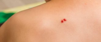
- In case of accidental injury to a mole, it is necessary to treat the damaged area with an antiseptic (hydrogen peroxide) and consult a doctor.
Possible complications in the presence of multiple moles:
- degeneration of benign cells into a malignant tumor;
- After the removal procedure, consequences are possible: long healing, dangerous scars and scars forming, secondary infection of the body.
In order to prevent the formation of melanoma, it is necessary to regularly examine the baby’s skin for the presence of pigment spots. To avoid tanning and sunburn, keep an eye on your child. Sunscreens reduce the likelihood of sunburn, but do not provide 100% protection against skin cancer.
Until what age is the growth of moles normal?
The appearance of moles on the skin is a natural process.
Melanin – protection against sunburn: the population of hot countries has dark skin.
The pigment reduces the negative effects of ultraviolet rays.
Nevi appear as a result of hormonal changes. The growth of dark marks as a person grows older does not affect health. Each person’s body is unique - there is no exact age at which formations appear.
Approximate intervals for a child to grow up with the formation of a favorable nevus:
- early infancy - from 6 months to 2 years, nevi do not grow until six months of age;
- eve of adolescence - 6-7 years;
- Beginning of puberty – 12-14 years.
During these periods, the skin structure increases melanocytes - cells that produce dark pigment.
Growth and increase in bulge without changes in hormonal levels is a deviation from the norm and a reason to visit a doctor.