Lentigo - what is it?
Here we are not talking about freckles, which appear in some people with increasing solar activity.
Lentigo is a slight pigmentation of the skin that does not disappear all year round. Sometimes it appears spontaneously.
This phenomenon is classified as a disease, which is a type of benign skin lesion. Develops against the background of excess melanin in the dermis.
Solar lentigines or lentigines are very common in people over 40 years of age. Sometimes they are also called “age spots” or “senile freckles”.
Dermatologist Fatima Ahmed
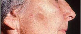
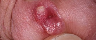
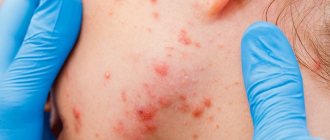
The key symptom of lentigo is the sudden appearance of small brown spots on the skin, with a diameter of 1.5 to 3 cm. Moreover, they can be localized on the face, neck, back, arms and other parts of the body (see photo) in both women and men . It is also possible to damage the mucous membranes.
Lentigo is conventionally divided into congenital and acquired. If there are too many spots, then the diagnosis is lentiginosis.
The danger of this dysfunction is that it can develop from benign pigmentation to malignant. According to statistics, this happens in 4%.
The risk of such degeneration increases due to a number of facts:
- Getting more than three sunburns in your lifetime.
- Combination of lentigo with freckles.
- The presence on the epidermis of more than three spots of atypical size and shape.
The exact causes of lentigo are not known, except for a form that is inherited. However, certain provoking factors have been identified:
- excessive sensitivity of the epidermis to ultraviolet radiation;
- fanatical addiction to sunbathing and repeated sunburn (especially in childhood);
- fair skin and red hair;
- advanced age;
- frequent visits to the solarium;
- suppressed immunity due to the use of depressants, cyostatics and glucocorticoids, as well as due to poor nutrition;
- papilloma virus infection;
- endocrine changes during pregnancy, during puberty and the onset of menopause;
- disruptions in the gastrointestinal tract;
- injuries of various origins;
- severe poisoning;
- inflammatory processes of a dermatological nature.
To accurately diagnose the disease itself and find out the cause of its development, you should go to the clinic for a medical examination.
Etiology and pathogenesis of lentigo
The main cause of solar lentigo is chronic exposure solar radiation . Changes do not occur immediately - they accumulate over many years of ultraviolet irradiation of exposed areas of the face and body. The likelihood of lentigo increases if a person has light skin of Fitzpatrick phototype I or II. Solar lentigo can also occur after long periods of visiting a solarium .
Quite often, lentigo appears after PUVA therapy (psoralen (P) + ultraviolet A (UVA) = PUVA or PUVA) - therapeutic exposure of the skin to ultraviolet A (UVA) with preliminary intake of a photosensitizer (psoralen). These pigmented lesions are called PUVA lentigo, and they are a side effect of phototherapy for psoriasis.
Microscopy of solar lentigo lesions reveals elongation of the epidermal ridges (lentiginous hyperplasia), an increase in the number of melanocytes that produce excess pigment, as well as an increase in the number of melanophages - macrophages that have absorbed the pigment and turned dark. Atypical melanocytes are not normally detected in solar lentigo.
In addition to solar and PUVA lentigo, simple lentigo (lentigo simplex), the causes of which have not been established, as well as radiation lentigo (radiation lentigo) after high-dose radiotherapy can occur on the skin. In the future, any of these neoplasms can transform into lentigo maligna, so they should be treated with special attention. Primary differential diagnosis between ordinary and malignant lentigo is carried out with high efficiency using FotoFinder .
In some cases, lentigo is a sign of a hereditary disease - for example, LEOPARD syndrome. LEOPARD is an acronym whose letters describe the symptoms of the disease: L (lentigo), E (cardiac conduction disorder), O (ocular hypertelorism), P (pulmonary stenosis), A (genital anomalies), R (slow growth), D (deafness).
What are the types of lentigo (5 options)
There is the following classification of lentigo:
- Solar – occurs as a result of prolonged exposure to the sun. People with fair and sensitive skin types are more susceptible to it. The appearance of this type of pigmentation is perceived as a primary sign of photoaging. The spots appear mainly on the neck, arms and face. This type of lentigo is not considered a disease, but rather a cosmetic defect.
- Simple (adolescent or juvenile) - it has nothing to do with ultraviolet radiation and skin aging. Most often it makes itself felt in adolescence or immediately after childbirth in women. Lentigines appear on the skin and mucous membranes in the form of spots up to 10 mm in size.
- Senile - they are also called liver spots. They are dark brown washouts (up to 1 cm in diameter) with uneven contours that appear on the face, neck and arms. People over 50 years of age are more susceptible to this disease if they previously did not particularly adhere to sun protection.
- Melanotic - these are ink-colored spots that appear randomly on the skin, which is more exposed to direct sunlight. They have a teardrop shape.
- PUVA - appears six months after undergoing therapy against psoriasis. The spots have unclear boundaries and resemble freckles in appearance. The torso is more susceptible to damage.
Chloasma also occurs. Women often encounter it during pregnancy or menopause.
Dermatologist Michelle Green
Lentignosis of the mucous components is characterized by a scattering of flat pigment spots of a light cream color and with smooth contours.
Such a disease can be acquired at the genetic level by inheritance or due to certain third-party factors.
Even knowing the signs of each type of lentigo, you should not make a diagnosis yourself. This is only within the competence of the relevant specialist. Therefore, there is no need to postpone your visit to him.
What types of hyperpigmentation exist?
Freckles (ephylides ) are a genetically determined skin condition. These are small round spots, 1-2mm in diameter. As a rule, they are characteristic of blondes, brown-haired and red-haired people and appear or intensify when exposed to ultraviolet radiation on the skin of the face, hands, and body unprotected from UV rays. In winter, they may become less bright or disappear altogether.
Lentigines are oval-shaped spots, slightly protruding above the skin with a diameter of 2 to 20 mm. Typically located where the skin is most exposed to UV radiation
- Face
- Hands
- Neckline and shoulder area
- Upper back
What is the clinical picture
When pigmentation is detected on the skin, most people do not associate it with any disease. Moreover, rarely does anyone know about lentigo and what it is.
This is due to the fact that the spots do not cause discomfort. The only thing the patient notes is a change in their color from light to darker (as in the photo).
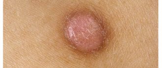
Sometimes the pigmented areas may thicken slightly or become bulging.
Such changes signal the degeneration of this neoplasm from benign to malignant. Then you need to urgently consult a doctor for diagnosis.
If your dermatologist has looked at the spots and determined that they are benign (not cancerous) and cannot turn into skin cancer, then he can begin to treat them. Sometimes we simply freeze the lesions with liquid nitrogen on the day of presentation. Laser treatment can make dark skin even darker if the wrong type of laser was used.
Dermatological surgeon Amy Paul
Contraindications
It is not dangerous to remove lentigo with a laser, although there are a number of contraindications:
- oncological diseases;
- pregnancy and breastfeeding;
- blood diseases;
- diabetes;
- skin damage in areas of increased pigmentation.
Diseases from the list may not be absolute contraindications, but consultation with your doctor is required. During pregnancy and lactation, laser lightening is not recommended, because the spots that arise often go away on their own after the baby finishes feeding, as hormonal levels change.
How is lentigo diagnosed (4 procedures)
Establishing a diagnosis does not require any complex laboratory procedures. An examination of the patient by a dermatologist and an anamnesis is sufficient.
What is done during diagnosis:
- Dermatoscopy – a photo of the affected areas is taken in high resolution and in good lighting.
- Biopsy - a piece of tissue is taken from the spot for laboratory testing. This procedure is prescribed if there is doubt about the benignity of the tumors.
- A general blood test and protein tumor markers are necessary to exclude the possibility that hyperpigmentation is malignant.
- Confocal laser scanning microscopy (CLSM ) - this means an instrumental examination of a skin section in layers.
There are no contraindications to performing all these procedures. They are completely safe.
Every time you have a suspicious spot or mole, you should go to a dermatologist for diagnosis.
Dermatological surgeon Mitchell Schwartz
Diagnosis of nodular melanoma
Nodular melanoma is diagnosed by oncologists at the Yusupov Hospital taking into account the patient’s complaints, history of the disease, visual examination data, blood tests for tumor markers and the results of luminescent dermatoscopy. When making a diagnosis, the following factors are taken into account:
- Increase in planar dimensions;
- Change in the shape, outline and color of a pigment formation within a short period of time;
- The presence of unusual sensations (itching, burning, itching, bloating), increased bleeding;
- The appearance of peeling and ulceration on the surface of nodular melanoma.
If oncologists suspect regional metastasis, the patient undergoes an ultrasound examination of the lymph nodes and a biopsy. To exclude hematogenous secondary lesions, the following examination is carried out:
- Chest X-ray;
- Ultrasound examination of the abdominal organs;
- Computed and magnetic resonance tomography;
- Scintigraphy of skeletal bones.
Doctors at the Oncology Clinic carry out differential diagnosis of nodular melanoma with other types of melanoma, telangiectatic granuloma, hemangioma, pigmented form of basal cell skin cancer and blue nevus. The final diagnosis is made based on the results of histological examination of the removed tumor.
Is it possible to cure this disease and how?
It is necessary to treat lentigo when the spot is too large, is constantly exposed to mechanical and solar influence and spoils the appearance with its presence.
Typically treatment comes down to the following steps:
- regular whitening of pigmented areas on the skin with special medications;
- constant application of protective creams against UV radiation to the skin;
- superficial exfoliation of the epidermis using various cosmetic procedures.
Don't worry if the spots disappear and then return after a few days or weeks. The pigment usually consists of several layers, so peeling occurs layer by layer.
Anesthesiologist Laura Rome
As a last resort, they resort to surgical intervention. Then the lentigo is cut out from the skin surface.
During the operation, the specialist captures part of the healthy tissue so that the stain does not appear again in the future. The remaining scar is easily removed plastically.
Arbutin, azelic acid and licorice extract have a good effect as whitening agents.
They contain ascorbic and kojic acid, retinoids and hydroquinone. The latter component is present in modern drugs, but in minute quantities. All other components are absolutely harmless and easily tolerated.
Laser destruction
For those who want to get rid of lentigo without subsequent scars, laser therapy is recommended.
With it, the beam captures and destroys only the neoplasm, and does not affect healthy tissue in the neighborhood. This procedure is painless and is performed without local anesthesia.
If you have persistent solar lentigo/sun spots, the best combination is the Fraxel Dual laser and Yag laser.
Dermatologist Michelle Green
During the session, the patient may feel a slight tingling sensation on the skin.
Immediately after pigmentation has been reduced, redness may appear in the areas treated with the laser. If you are prone to allergies, slight darkening may appear in these places.
Before laser therapy, the patient must undergo a series of examinations to exclude existing contraindications.
Phototherapy
If the diagnosis of malignant pigmentation is excluded in the patient, then a phototherapy method may be offered.
This means exposing problem areas to a light beam. This stimulates the natural synthesis of collagen, thereby increasing the regenerative capabilities of tissues and rejuvenating them at the cellular level.
I have better results with the Q-swithced laser. I have used the Cutera Laser Genesis with great success. For patients who want a more conservative approach, a combination of hydroquinone and retin-A creams is suitable. IPL phototherapy is effective in some patients, but it is still superior for treating melasma.
Plastic surgeon Temp Patterson
Peeling
People with light age spots are recommended to resort to chemical peeling of the skin. The fact is that, in addition to removing darkened areas, adjacent healthy skin is also affected.
The following types of peelings are often used:
- retinoic;
- TCA peel;
- ABR peeling;
- pyruvic;
- azelaic.
This procedure is performed with local anesthesia. This is followed by a short recovery period of 10-12 days. As a result, the skin structure is completely restored and pigmentation disappears without a trace.
However, in addition to the beauty treatment, you should also follow a proper skin care regimen to brighten your skin.
Plastic surgeon Grant Stevens
There are many different types of peelings, but experts often praise these options:
- Peeling Cosmelan. The peeling itself is brown in color and has a thick consistency. Contains azelaic, phytic, kojic, ascorbic acids, as well as arbutin. The product brightens the skin well. First, in the clinic, the patient is applied a mixture of Cosmelan 1, which should remain on the skin for about 8-12 hours. At home, the mask is removed with cleansing cosmetics. 2 days will pass, after which the patient independently applies the Cosmelan 2 mixture every day. This must be done throughout the year.
- Jessner peeling. This name is given to a patented product that contains salicylic, lactic acids and resorcinol. This is a medium peel. The effect is amazing. The product will not only help remove pigmentation, but will also eliminate many other problems (acne, wrinkles, etc.).
Lentigo – treatment with the M22 device
Modern methods of getting rid of lentigo include photothermolysis - treatment with IPL light or laser radiation. The essence of this method is the pulsed effect of light beams on the skin in the affected area, during which excess melanin is destroyed without affecting healthy areas of the skin.
It is generally accepted that laser copes with this problem better. However, modern light devices, for example, the M22 multi-module platform, demonstrate a high rate of removal of various age spots. For each type of hyperpigmentation, including lentigo, a special program is provided, which significantly increases the effectiveness of M22 compared to other photosystems. M22 rays penetrate the skin, destroy excess melanocytes and keratinized cells, cause the synthesis of collagen and elastin fibers and other active substances. These processes promote rejuvenation and rapid regeneration of damaged areas, lightening of spots and smoothing of the relief. After just a few sessions, the spots cease to be noticeable and the skin acquires a healthy color.
Question answer
Yes, but initially it is recommended to consult a dermatologist. He will prescribe suitable medications. For example, whitening creams with hydroquinone 4% or retinol have worked well. You can supplement therapy with traditional methods, but they will not be able to fully cope with the problem. You should also understand that treatment with such creams will take a lot of time.
Yes it is possible. Therefore, go to the doctor for diagnosis in a timely manner. Degeneration is observed in 4% of patients. A malignant formation usually has smooth edges and is dark in color. The size usually grows and can reach up to 20 cm.
In the table of classification of pathologies, lentigo was included in the group of diseases associated with increased levels of melanin and pigmentation disorders. Assigned number L81.
What determines the success of treatment
The result of hyperpigmentation treatment depends on how deep the pigment lies.
Based on the depth of the pigment, all types of hyperpigmentation can be divided into two large groups. It is this classification that allows us to understand whether our treatment will be successful or not.
Epidemal hyperpigmentation is pigmentation with clear boundaries, sometimes protruding above the surface of the skin, usually dark brown in color. She is the one who responds well to treatment
Dermal hyperpigmentation is a dull light brown pigmentation, sometimes with a grayish tint, that does not have clear boundaries. This type of pigmentation is practically untreatable.
Mixed hyperpigmentation - pigmentation has areas of dark and light shades at the same time. Treatment in this case has a partial positive effect.
Lentigo therapy at home
You can treat lentigo yourself at home if it is clearly established that the pathology is benign.
All folk recipes are based on the use of natural lightening agents:
- lemon juice;
- hydrogen peroxide;
- ammonia;
- rowan;
- calendula;
- black currant;
- parsley;
- almonds;
- honey
Here are some productive recipes for homemade masks:
- The stems, leaves and roots of parsley are finely chopped to a paste. Then add honey. The resulting composition is applied to problem areas and, after leaving for half an hour, washed off with warm water.
- Place the lemon peel in boiling water and keep it warm for about 30 minutes. Then let the water sit and wipe the stains up to 3 times a day.
- Grind almonds in a coffee grinder and mix them in equal proportions with oatmeal. Add 5-7 g of milk powder and flavor with a small amount of water to form a mass similar to sour cream. Use this thickener to scrub the affected areas for about 5 minutes and wash off any remaining residue. Do this no more than 2 times a week.
- Dilute dry white clay (1 tbsp) with warm water until smooth and add a little lemon juice. Apply the composition to age spots and wash off after 20-25 minutes. This mask is used every day.
The suggested masks should be done in the evening, when you are not planning to go outside. This is due to the fact that after the procedure you cannot expose the skin to UV exposure.
As a treatment for lentigo, it is recommended to use pharmaceutical ointments with a whitening effect:
- zinc;
- sulfuric;
- Clotrimozole;
- salicylic.
Among the creams, the following brands are distinguished: Skinoren-gel, Melanativ, Clearvin. They should be applied to the skin twice a day.
I recommend using a combination of whitening cream (hydroquinone 4%) and tretinoin (vitamin A). But it will take months of treatment to achieve results. The laser is much faster.
Plastic surgeon Sam Naficy
Melanoma: stages of development
The effectiveness of therapy depends on the stage of development of the disease. In other words, the earlier the diagnosis is made, the more likely a positive prognosis is. The following stages of melanoma development are distinguished:
- "0". The malignant process affects exclusively the epidermis. Melanoma is rarely diagnosed at this stage.
- "I". The thickness of the neoplasm does not exceed 1 mm. There are no ulcers on the surface, no bleeding or peeling. After surgical removal of melanoma, the disease can be completely overcome.
- "II". Education begins to grow deeper. At the same time, its thickness increases to 2 mm - 4 mm. The surface begins to peel and bleed. The lymph nodes at this stage are not affected, so metastases to distant organs do not occur. Treatment also involves surgical removal of the formation.
- "III". At this stage, cancer cells travel to the lymph nodes. Melanoma begins to actively penetrate into the deeper layers. In addition to surgical treatment, chemotherapy and radiation therapy are indicated.
- "IV". This is the last stage of melanoma development. It is characterized by the occurrence of metastases on internal organs. Treatment involves an individual approach and long-term complex therapy.
Preventive measures (5 tips)
To minimize the risk of lentigo, you should adhere to certain preventive measures:
- before each exit to fresh air, lubricate the skin with cream with an SPF value of at least 30;
- people over 50 years old should try to spend less time in the sun and not visit the solarium;
- in the summer, always wear a hat and sunglasses;
- regularly undergo examination by a dermatologist to detect atypical moles or other pigmentation;
- immediately go to the doctor if dark spots of unknown etiology are detected on the body.
These spots usually appear due to constant sun exposure, so daily use of sunscreen is important for prevention.
Dermatological surgeon Mitchell Schwartz
If a stain on the skin does not cause discomfort, but is constantly subject to friction from clothing, then it is advisable to remove it.
Technique
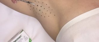
First, make-up or cream residues are carefully removed from the surface of the skin. Then a special gel is applied to the pigmented areas to improve the conductivity of light beams. The patient is required to wear special glasses to protect his eyes from the effects of the device.
The procedure begins with test flashes, since it is necessary to make sure that the depth of penetration of the wave and the intensity of the impact are selected correctly for the specific skin type and color of the spots. The duration of the impulse is also individual.
During laser treatment on sensitive areas such as the lips, there may be some discomfort due to the heat absorbed by the melanin. Therefore, before the procedure begins, the specialist is interested in the pain threshold and suggests applying an anesthetic.
Immediately after laser removal of lentigo on the face is completed, the treated lesions may become red. They are lubricated with regenerating cream. After 3-5 days, in some cases the skin begins to peel off. You need to try not to injure these areas of the skin so that rehabilitation goes quickly.
Depending on the size of the spots, the intensity of the color and the reaction to laser exposure, a decision is made whether repeated removal sessions are necessary.
Expert opinion
- Cosmetologist
- Surgeon
Anna Avaliani
practicing cosmetologist
I recommend paying attention to Q-Switch technologies (VersaPulse C laser or ConBio laser). Either of these two options should be better than IPL phototherapy, peels, or retinol. I would recommend starting with QS532 if the lentigines are superficial, or QS755 if they are deeper lesions. Usually 1-3 procedures are required.
Aisha Baron
plastic surgeon
Take a comprehensive approach to the problem.
Go to a dermatologist who will select the appropriate procedure. Indeed, laser technologies have proven themselves to be the best. Use creams recommended by a specialist. In addition, don't forget to protect your skin from the sun. Now it is more or less clear what lentigo is. But this pathology was considered only in relation to humans.
It turns out that this disease is common in dogs and cats. You can find out more about this on veterinary forums.
Summarizing all that has been said, the conclusion suggests itself that timely diagnosis of the disease is the key to successful treatment and a guarantee of safety from oncology.
Clinical manifestations of lentiginosis
Lentigenic lesions appear as flat or slightly concave round spots of varying diameters and colors ranging from light brown to dark brown. They usually appear on open areas of the skin—the face, the backs of the arms and hands, and the upper half of the torso ( Fig. 1 ).
Immediately after their appearance, their size does not exceed 5 mm, but as they “age”, solar lentigines become larger, darker and gradually merge into large spots. They may contain areas of normal skin or be separated by fine wrinkles. Skin affected by lentigo appears loose and inelastic—in general, it looks worse than it should for its age ( Fig. 2 ). The epidermis becomes thinner and dry, and the skin color becomes unevenly patchy.
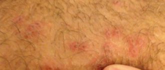
Lentigo can occur after a long course of PUVA therapy for psoriasis - from six months or more. In this case, pigment spots are similar to solar lentigo, but have irregular contours - sometimes they can be confused with freckles. After cessation of phototherapy sessions, small PUVA lentigoes usually remain on the skin for about 3-6 months, while large star-shaped lesions last up to 2 years.
Solar lentigo can appear after long and/or multiple courses of visiting a solarium - usually such pigmentation occurs in women prone to abuse of artificial ultraviolet radiation. Externally, these lesions are similar to PUVA lentigo and may be present in areas not naturally tanned (for example, in the armpit).
In some people, solar lentigo transforms from smooth spots into rough lesions, which can later develop into malignant tumors (for example, lentigo maligna). And although the likelihood of such an event is small, the problem is relevant and requires attention from both the patient himself and his attending physician.
A special type of benign neoplasm is macular nevus . It is both a lentigo and a melanocytic nevus, where multiple dark spots are located in the area of a large and lighter congenital or acquired spot ( Fig. 3 ). A spotted nevus can transform into melanoma, which is something to keep in mind.
Rice. 1. A lesion of solar lentigo on a woman’s cheek (www.medscape.com)
Rice. 2. Solar (senile) lentigo on aging skin of the hands (Danish national service on dermato-venereology)
https://www.danderm-pdv.is.kkh.dk/atlas/7-130-2.html
Rice . 3. Spotted nevus (Danish national service on dermato-venereology)
https://www.danderm-pdv.is.kkh.dk/atlas/7-150.html
Treatment methods
Melanoma, the survival prognosis of which depends on timely diagnosis, is successfully treated by surgical removal in the initial stages of development. After surgery, antitumor drugs are additionally prescribed. If the disease has affected the lymph nodes or metastases are diagnosed, chemotherapy is indicated. Radiation therapy is prescribed to exclude relapses.
Immunotherapy, which is aimed at enhancing natural protective reactions, is considered effective. In this case, the body is actively involved in the fight against malignant cells, which makes it possible to defeat the disease.
After treatment of melanoma in the initial stages, the patient must be observed for 5 years. In other cases, condition monitoring must be ensured for 10 years. After this, if there are no relapses, a traditional medical examination can be performed annually.
Risk groups and factors predisposing to the development of skin melanoma.
Individuals at increased risk of developing melanoma include:
- with white skin, red hair, blue, gray or green eyes;
- constantly sunburned;
- those who have suffered sunburn and have been exposed to the sun for a long time, especially under the age of 20;
- having close relatives with melanoma (other skin cancer);
- having more than 100 moles on the body or more than 50 before the age of 20;
- having Dubreuil's melanosis (precancerous skin disease).
There are well-known cases of melanoma being diagnosed after injury to a pigmented nevus of the skin (mole). Melanoma often occurs after accidental or intentional (cutting, burning) injury to a mole. Sometimes 1-2 injuries are enough for melanoma to occur.
Trauma to the nevus can be chronic and occur unnoticed. For example, a well-starched shirt collar can injure moles on the neck, and bras can injure moles on the torso. Moles localized on the soles of the feet, palms, and perineum are chronically injured.
The influence of certain hormones on the development and clinical course of melanoma is assumed. Puberty, pregnancy and menopausal changes are regarded as risk factors for the development of melanoma from pigmented nevi.
Genetic predisposition plays a role (familial cases of melanoma).
Diagnosis of the disease
All changes that occur on the surface of the skin, first of all, should not be ignored by the patient himself. The appearance of suspicious lesions in the form of rashes, spots, swellings requires the use of a biopsy (diagnostic method) to determine the causes and the presence of malignant melanocytes. When performing a biopsy, the following information must be included in the report:
- Place of defeat.
- Diagnosis.
- Engaging research boundaries.
- Breslau level.
Additional research methods for biopsy are:
- A morphological method of research in which a fragment of infected tissue taken by excision is studied.
- Dermatoscuopia. This involves studying the pigment spot under an electron microscope.
- Calculation of tumor marker levels.
During the study, the condition of the patient’s entire body, the diseases that exist or previously existed, and the area of skin (a layer is removed) where the tumor is present are studied. Blood is taken for analysis to identify the presence of material in it that can create all the necessary conditions for the formation of a tumor in the cell. If the result is positive, after a biopsy, specialists prescribe a course of complex treatment with chemotherapy, radiation therapy, and, if necessary, surgery with the use of cytostatics.