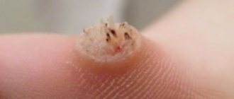
Dermatofibroma is a benign formation of the skin and connective tissue, similar in appearance to a mole. In most cases, they have a round shape and a smooth surface, much less often - keratinized or warty. Part of the dermatofibroma, a photo of which you can easily find, is located in the bulk of the skin, while the other rises above the surface of the dermis. If you touch such a mole, it seems that there is a small pebble stuck under the skin.
Dermafibroma of the skin occurs as a single formation; in rare cases, doctors diagnose several moles at once in one place. Dermafibromas themselves consist of fibrous tissue and contain a certain number of histiocytes and fibroblasts. As soon as a small grayish or pinkish dot appears on your skin, you need to consult a doctor. If treatment is not started in time, such a formation will turn black or dark brown and grow up to one centimeter. According to MBK-10, dermatofibroma is a response to any damage.
Causes of dermatofibroma
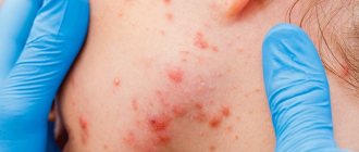
Modern specialists have still not been able to determine the exact reasons why dermatofibroma of the skin occurs. In most cases, doctors consider trauma to the skin of various etymologies as the main factor in the development of this disease: insect bites, frequent injections in one area, infectious diseases that affect the condition of the skin. The hereditary factor also plays a significant role here: if one of your relatives had problems of this kind, they will probably overtake you too. In addition, dermotofibroma on the leg or face often appears in individuals who suffer from acne and liver disease. The following causes of this disease are identified:
- Frequent skin injuries.
- Gender: statistics have shown that women suffer from such formations much more often than men.
- Negative environmental influences.
- Infectious diseases.
- Genetic predisposition.
- Age – older people are more affected by dermotofibroma.
Knowing the cause of this disease will help determine the effective and correct treatment. It is highly not recommended to treat dermotofibroma using traditional methods, as there is a high probability of developing serious complications. Disturbances in the functioning of internal organs, in particular the gastrointestinal tract, can contribute to the development of the disease.
Causes of appearance and clinical picture
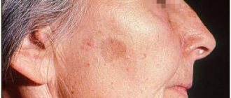
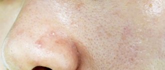
Medical science does not have a clear answer to the question: “Why does dermatofibroma appear.” Among the main causes of this skin disease, doctors consider heredity and negative external factors that can trigger the appearance of growths. For example, dermatofibroma can appear at the site of microtrauma, an insect bite, acne, or after an infectious dermatological disease. Growths due to liver dysfunction are also detected. A formation on the face can be a consequence of age-related skin changes.
Based on clinical manifestations, the pathology is easily identified by a dermatologist. The bulk of the tumor is “buried” in the skin. Only a small part of the “pea” protrudes above the surface of the epidermis.
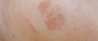
Important: Most often, this type of neoplasm is single, that is, one node forms on the skin. But in some cases there may be two or more manifestations. The color of the growths varies. The formations can be natural flesh-colored, gray, pink, brown and even purple.
Specific symptoms of the disease include a dense or soft consistency. Education remains fluid. When pressed, it easily moves to the side with the skin fold. When injured, the growth may begin to grow; damage to the tumor causes pain and itching.
Symptoms of dermatofibroma
Recognizing dermatofibroma of the skin, a photo of which you can easily find, is quite simple even by external signs. Such formations are round in shape, often smooth, protrude slightly beyond the skin, and are localized singly. Their diameter in rare cases exceeds 1 centimeter, most often varying from 0.5 to 0.7 millimeters. If several dermatofibromas appear on the body, they are scattered chaotically and are in no way tied to one particular part.
Dermatofibroma is a formation that can occur on any part of the body. In the vast majority of cases, it is located on the lower back, shoulders, back or lower leg. In rare cases, doctors diagnose such lesions on the palms and soles. The color of such formations varies from red-brown to grayish. In rare cases, dermatofibroma mimics the surrounding tissues and does not manifest itself in anything other than compaction. The edges of the formation are dark, becoming lighter towards the center. On palpation, a serious compaction is felt, there is a sensation of a foreign body under the skin.
General information
Dermatofibroma is a nodule-like formation on the skin that is one centimeter in diameter. This growth on the skin consists of fibrous fibrous tissue, which contains fibroblasts and histiocytes .
Also in medicine there are other names for dermatofibroma - sclerosing hemangioma , histiocytoma . This formation is benign. As a rule, dermatofibroma appears on the skin of the legs. Sometimes for some time a person does not even notice that he is developing sclerosing hemangioma. Hemangioma therapy is performed only if the formation causes discomfort to the patient.
Treatment
Dermatofibroma rarely goes away on its own without some intervention. Many people live with such an education all their lives: once it appears, it may not manifest itself in any way and may not grow anymore. However, there is always a risk that the tumor will sooner or later become malignant, which is why it needs to be gotten rid of as quickly as possible. Removing dermatofibroma surgically can provoke relapses, which is why many doctors have long abandoned this method in favor of using a laser.
At the Healthy Skin Center, experienced specialists will provide you with a detailed consultation, thanks to which you will be able to determine how to treat dermatofibroma on your leg. Typically, doctors send their patients for a procedure using the Surgitron radio wave therapy device. Under the influence of high frequencies, the affected cells seem to evaporate, while the healthy ones remain in place. Using this method ensures that dermatofibroma will never appear on the treated area of the skin again.
Prevention
Due to the unclear etiology of the disease, it is difficult to formulate specific preventive measures. We only know what is needed:
- avoid damage to the skin;
- monitor your diet, get the required amount of vitamins and microelements from food;
- avoid overwork, decreased immunity;
- It is not recommended to expose the body to excessive sun exposure, as this leads to a decrease in immunity.
If you have the slightest doubt or discomfort, do not hesitate to contact a specialist - a dermatovenerologist. Only he will be able to make an accurate diagnosis and prescribe adequate treatment. Any self-medication is always fraught with bad consequences.
Important points when contacting a dermatologist if you have a hair disease.How to care for braces
- ADVANTAGES OF TREATMENT ABROAD
Also, it is necessary to remember that this formation in itself does not pose a particular threat and you can calmly live with it all your life. So, the main thing is not to panic.
Removal of dermatofibroma
- Dermatofibroma
Dermatofibroma is a deep-lying benign tumor of dark brown connective tissue, has the appearance of a retracted or hemispherical protruding formation, up to 1-2 cm in diameter, with a dense consistency.
As a rule, it is located on the skin of the upper and lower extremities. Not to be confused with botryomycoma and atheroma. Diagnosis of dermatofibroma: ultrasound of the skin, advanced dermatoscopy.
Kinds
Dermatofibroma is classified as follows:
1) Lenticular appearance, when dense formations are formed from small nodes of 1 centimeter (diameter), and the predominant color is red;
2) Solid appearance, consisting of indurated and red-flesh size up to 2 centimeters (diameter), which occurs in any lesions of the skin. Such formations can be either multiple or single;
3) The soft type, the inside of which is soft lobules of a flesh-bluish tint, usually such formations appear on the torso, face or neck.
Treatment of dermatofibroma
Removal using radiosurgery.
In our center, tumor removal is performed by a dermato-oncologist using radio wave surgery under local anesthesia.
In order to remove the formation more efficiently, the doctor uses a dermato-oncological magnifying glass, which allows for more detailed and complete removal of skin tumors without leaving pathological cells in the wound. Therefore, after removal, we can say with complete confidence that the formation has been completely removed and you will no longer have it. Read more about tumor removal in our center...
The cost of removing dermatofibroma is from 2300 rubles.
List of sources
- Kurbanova A.A., Skin diseases: a guide for doctors and students of medical universities / A.A. Kurbanova. - M.: GEOTAR-Medicine, 1998;
- Skripkin Yu.K., Skin and venereal diseases: a guide for doctors and students of medical universities / Yu.K. Skripkin. - M.: Triada-farm, 2001;
- Paltsev M.A., Potekaev N.N., Kazantseva I.A., Lysenko A.I., Lysenko L.V., Chervonnaya L.V. Clinical and morphological diagnosis of skin diseases: Atlas. M.: Medicine; 2004;
- Fitzpatrick T., Elling D.L. Secrets of dermatology. Per. with English-M.S-Pb-Bean, Nevsky dialect.-1999.
How is skin pathology diagnosed?
First of all, at an appointment with a dermatologist, the doctor conducts a visual examination. If the data obtained is not enough to make a diagnosis, a hardware diagnostic method is used - dermatoscopy.
Interesting: Using a special device - a dermatoscope - a specialist examines the skin formation, including its part hidden deep in the skin layers. A multiple increase in growth is displayed on the screen and stored in the database. This allows you to monitor the condition of the tumor and monitor its growth during subsequent visits.
The doctor may also prescribe a biopsy and histology of the tumor. Laboratory examination of tissues allows us to identify fibrous fibers, blood vessels, pathogenic cells, and lipids.