There are contraindications. Specialist consultation is required.
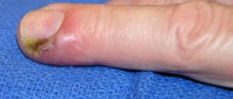
A furuncle is a purulent inflammation of the hair follicle. It develops due to staphylococcal infection entering the sebaceous glands due to microtraumas, non-compliance with hygiene rules, excessive sweating, allergies, and decreased immunity. By the nature of the rashes there are single and recurrent, in form single and multiple (furunculosis).
Anatomy of the rectum and perianal region, functions of the obturator apparatus
The rectum and anal canal are the final sections of the digestive tract. The immediate tasks of the rectum are the accumulation, formation and excretion of intestinal contents and gases. The functions of the anal canal are unique: a system of nervous reflexes and a complex muscle complex provide one of the most important functions of the body - control of bowel movements, as well as differentiation of the composition of intestinal contents without the need for conscious assessment and subsequent “smart” control of evacuation. In other words, a healthy person most of the time does not think about what kind of contents are in the rectum, whether it is necessary and whether it is possible to retain it in a given situation. All this is controlled unconsciously, thanks to multi-level neuromuscular self-regulation of the holding process.
The approximate boundary between the rectum and the anal canal on the side of the mucous membrane (lumen) of the intestine is formed by the so-called dentate line. There are vertical folds on it, alternating with depressions - crypts. The ducts of the anal glands open into the crypts. The mucus produced by these glands facilitates the sliding of intestinal contents as they pass through the anal canal. Circumstances such as trauma, swelling of the mucous membrane due to defecation disorders, chronic intestinal diseases can lead to inflammatory changes in the crypts and anal glands.
Outside the mucous membrane there is a complex of muscles - these are the sphincter muscles mentioned above, which provide the function of retention and excretion. There are internal and external sphincters. The internal sphincter forms the so-called resting tone, uninterruptedly ensuring the tightness of the anal canal. Its regulation and reduction occur unconsciously, that is, without our will. The external sphincter surrounds the internal one and consists of several layers (portions). Its reduction occurs due to our volitional effort (Fig. 1).
Figure 1. Schematic representation of the rectal sphincters
What is a fistula
A fistula is a pathological, normally non-existent, passage in the form of a tube connecting the lumen of a hollow organ with the external environment or the lumen of another organ. A rectal fistula, a kind of “tunnel,” connects the lumen of the rectum with the skin of the perineum, buttocks or (rarely) other hollow organs, sharply reducing the patient’s quality of life. This pathological process can involve a large portion of sphincter muscles, which initially characterizes such a fistula as complex and does not allow for “minimally invasive” treatment. In such a situation, a careful assessment of the degree of involvement of muscle structures and planning of special techniques for economical excision of the fistula with subsequent plastic closure of the resulting defect are required.
Important! Such operations must be performed by expert surgeons with extensive experience in such interventions, otherwise the likelihood of relapse (recurrence of the disease) increases several times.
Fistulas have the following structure:
- internal opening: it is usually the affected anal crypt (that is, the most terminal part of the gland duct). The crypt is connected by a duct to the anal gland, located in the space between the sphincters. It is the inflammation of this gland that in the vast majority of cases leads to the development of the disease (Fig. 2).
Figure 2. Stages of fistula formation: a) - anal gland with an excretory duct, b) - inflammatory process in the gland, with a forming abscess (abscess) c) - formed fistula tract.
- fistula tract, which can be tortuous, have cavities and branches
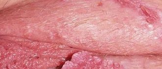
- external opening: but most often located on the skin of the perineum near the anus (Fig. 3), sometimes in the vagina or urethra (urethra).
Important! If you have the slightest suspicion of a rectal fistula, you should immediately consult a specialist. If treatment is untimely or inadequate, the disease can be complicated by acute inflammation and additional purulent leaks, which significantly worsens the prognosis!
However, it also happens that the fistula tract ends blindly in the pararectal (peri-rectal) tissue, tissues of the perineum, buttocks, that is, it has only an internal opening of the fistula, which opens from the mucous membrane of the rectum and has no “exit” to the outside.
Figure 3. External opening of the fistula
How common is rectal fistula?
The incidence in European countries is about 10-25 people per 100 thousand population. Mostly people of working age suffer from rectal fistulas. Men get sick 2-3 times more often than women.
Important! This disease, being benign, can dramatically reduce a person’s quality of life. A serious and most common complication of inadequate treatment of perianal fistulas is impaired holding function due to damage to the muscular apparatus of the anal canal.
Rehabilitation period
After 1-2 hours the patient is discharged from the clinic. If the drainage is removed, it must be removed by a surgeon after 1-2 days. It is recommended to limit physical activity for 2-3 days after the procedure. For 2 weeks, it is necessary to go for examinations and dressings at the frequency specified by the surgeon, as well as ensure a balanced diet and proper rest. The tissues heal completely within 10-14 days.
To consult, clarify prices and remove a boil in St. Petersburg, call SM-Clinic.
Causes of fistulas
If you experience symptoms characteristic of a rectal fistula, in the vast majority of cases the cause for this was an inflammatory process in the anal gland (Fig. 2). In turn, inflammation of the gland is usually preceded by microtrauma of the anal canal. The cause of such injury is most often excessive irritation and/or trauma to the anal canal when too hard stool passes through the anal canal during constipation or, conversely, during diarrhea. Relatively rare causes of so-called specific fistulas of the anal canal and rectum are inflammatory bowel diseases (Crohn's disease, ulcerative colitis), tuberculosis, rectal tumors, and the consequences of radiation therapy of the pelvic organs. Inflammation of the gland is accompanied by the development of an abscess (ulcer) in the soft tissues of the perineum and is usually characterized by severe pain, which forces the patient to consult a doctor. Timely and correctly performed opening of the abscess leads to rapid relief of the condition, however, according to modern data, about half of all abscesses are accompanied by the subsequent formation of a fistula and require repeated intervention.
It is this repeated intervention that is aimed at radically getting rid of the fistula and requires not only an extremely thoughtful approach to the choice of treatment tactics, but also extensive surgical experience on the part of the surgeon.
Important! Opening an abscess is a relatively simple operation, but if performed incorrectly, it is at this stage that the muscular complex of the anal canal is most often damaged, which leads to disruption of the function of controlling intestinal contents!
If your visit to the doctor is postponed, the abscess may open on its own with the release of pus, after which your health usually improves and the pain in the perineal area decreases sharply. Symptoms may subside or disappear completely for a period of several weeks or months to many years. Despite the improvement in general well-being, spontaneous opening of an abscess is a relatively unfavorable scenario, because in this case, there is no complete evacuation of the contents of the purulent cavity, but only part of the purulent discharge occurs through the formed hole, which can soon close again. Proper treatment of an abscess consists of creating adequate drainage by creating a sufficient hole, evacuating the contents, rinsing the abscess cavity with antiseptic solutions and, if possible, removing the inflamed gland that is causing the inflammation.
Important! Sometimes, in the hands of an expert surgeon, with the relative simplicity of the fistula tract, radical excision of the fistula at the stage of surgical opening of the abscess is possible, but performing this intervention is extremely dangerous in the hands of an insufficiently experienced specialist and today is not recommended in most international recommendations.
When an abscess spontaneously opens, the likelihood of a return of the acute disease increases and the process becomes chronic with the formation of a fistula. A long course of the disease with periodic exacerbation leads to scarring of the surrounding tissues and, most importantly, the sphincter muscles responsible for the holding function. In addition, in conditions of constant inflammation, a relatively simple fistula can give rise to additional passages and leaks, which significantly complicates subsequent treatment.
Timely consultation with a doctor at an early stage of the disease can reduce the likelihood of developing a fistula and such dangerous complications of the inflammatory process as pelvic phlegmon. Therefore, even minor symptoms that do not significantly affect the quality of life require attention and evaluation by a qualified specialist.
How is the treatment carried out?
Treatment of eyelid abscess occurs only by surgical intervention.
In this case, the abscess is opened , and such a procedure is performed only in a hospital setting, although, as an exception, such a procedure can also be performed in a clinic.
After opening the abscess, its contents are removed , and then the cavity is washed using antibacterial and antiseptic solutions .
The operation usually does not take more than 15 minutes and is considered a relatively easy procedure in ophthalmic practice.
After opening, no cosmetic defects remain in the area of the abscess.
But experts recommend not only going to the hospital in time if abscesses occur, but also taking a number of measures that will help prevent the onset of the disease . In particular, it is advisable to carry out procedures such as systemic antibiotic therapy, ultraviolet irradiation of the blood, and autohemotherapy .
In the latter case, a course of treatment is performed, which involves 12 procedures of administering drugs to the gluteal region to help strengthen the patient’s immunity.
Note! Ultraviolet irradiation of blood is a process of ultraviolet irradiation, which occurs through a special needle equipped with an emitter.
Such a needle is inserted into the patient’s vein, and all harmful and opportunistic microorganisms present in the body die. Ten procedures are enough - and the risk of formation of an abscess of the eyelid and other purulent inflammatory diseases is reduced.
What types of fistulas are there?
Traditionally, rectal fistulas are classified according to their location relative to the sphincter muscles. In accordance with this, intrasphincteric, subcutaneous-submucosal, intersphincteric, transsphincteric, suprasphincteric, extrasphincteric fistulas are distinguished (Fig. 4).
Figure 4. Types of fistulas
- Extrasphincteric fistula
- Low transsphincteric fistula
- Subcutaneous submucosal fistula
- Suprasphincteric fistula
- Intersphincteric fistula
The fistula tract can be located at a short distance from the edge of the anus and occupy no more than 1/3 of the lower part of the sphincter muscles - then they speak of a low (simple) fistula, or go high up, crossing a significant portion of the muscle complex - these are high (complex) fistulas. The complexity of the fistula is determined not only by the degree of muscle involvement, but also by the presence of swelling, active inflammation, and the presence of scarring around the fistula.
Subcutaneous-submucosal fistulas are the simplest fistulas. Simple fistulas also include intra- and intersphincteric fistulas. The fistulous tract in this form is usually straight, without streaks or cavities, and involves a minimal number of muscle fibers, thus not disrupting the function of the sphincter. Intersphincteric (intrasphincteric) fistulas pass through the internal sphincter into the intersphincteric space and then extend to the perineum. The external sphincter is not involved. As a rule, such fistulas are also simple and do not pose any difficulties for surgical treatment.
Transsphincteric fistulas of the rectum are more common than extrasphincteric fistulas and can affect more extensive layers (portions) of the sphincter complex. In this case, the fistula tract involves both external and internal sphincters.
Important! Tortuous fistula tracts, purulent cavities in the peri-rectal tissue, scarring in the surrounding tissues (including the sphincter) are more common if the fistula is located high in relation to the sphincter.
Suprasphincteric (from supra - above (lat.)) fistulas, starting in the intersphincteric space, rise upward and cross the muscular structures of the anal canal at its highest point, thus involving the entire obturator apparatus.
With such fistulas, high blind branches can form, which can sometimes be identified through the wall of the rectum.
In 3-5% of cases, extrasphincteric fistulas occur, in which the fistula tract goes around the external sphincter, and the internal opening of the fistula is low, in the area of the crypts. These fistulas are characterized by the formation of a tortuous course, streaks, and cicatricial changes.
In addition, additional fistula tracts may extend from the main fistula tract in various directions, and along the fistula, cavities with accumulation of mucus, pus, and necrotic (destroyed) tissues may form.
If the patient has already undergone surgery to eliminate a rectal fistula, and the symptoms of the disease resume after some time, the fistula is called recurrent.
Important! Treatment of recurrent and postoperative fistulas should be carried out only in specialized clinics and only by experienced surgeons!
Causes
The main cause of an abscess is streptococci and staphylococci entering the eye , but these bacteria can enter the body in different ways:
- During diseases such as boils, barley and blepharitis in a complicated form, the infection can spread to the eyelid.
- Sometimes the source of infection is teeth with caries .
- When the general immunity of the body decreases, infections can spread throughout the body, including the eyelid area.
- When squeezing pimples and pustules, there is a risk of infection in the eyelid.
Important! Decreased immunity can lead to hematogenous transmission of infection (transition from other organs within the body through the blood).
Symptoms of rectal abscess
- the presence of a small, painless wound - an external fistula opening on the skin of the perineum or buttocks.
- purulent, mucous or bloody discharge from the fistula opening
- itching, burning, feeling of fullness and discomfort in the fistula area.
- the presence of an internal opening of the fistula, detected upon examination of the mucous membrane of the anal canal (in approximately 30% it is not possible to find the internal opening during examination)
With an incomplete fistula, skin manifestations may be blurred or absent, and symptoms can vary from mild discomfort, a feeling of tightness or fullness, the presence of a lump in the anus, to discharge from the rectum.
When the disease worsens, a painful thickening is observed in the area of the external opening on the skin, the pain is often pulsating, bursting in nature. This may be accompanied by an increase in temperature and general weakness - these are the symptoms of acute abscess or paraproctitis.
Important! Acute paraproctitis should be opened as early as possible, in a surgical hospital!
Sometimes patients mistake the painful process for an exacerbation of hemorrhoidal disease and begin self-medication, which in no case should be done until the correct diagnosis is established!
Which doctor should you contact if symptoms occur?
If you or your loved ones have the described complaints, you should contact a coloproctologist as soon as possible, who, after consultation and examination, will establish a preliminary diagnosis and determine further tactics and urgency of treatment, or, if in doubt about the diagnosis, a list of necessary additional studies.
If surgery is required, you will be offered hospitalization in a hospital, where, after assessing the results of the studies, which, as a rule, do not take much time and are easy to perform, the operation will be performed.
Is it possible to cure an abscess at home?
Patients with acute conditions are admitted to dentistry without a queue. Therefore, if your health worsens, you should not postpone your visit.
The first question that a patient has is: why does pus appear in the gums and how to treat the pathology? The cause of infiltration is a weakened immune system and non-compliance with dental care measures. At the first symptoms of inflammation, you should immediately consult a dentist. Self-opening of a flux neoplasm threatens the addition of a secondary infection.
It is strictly forbidden to heat the inflamed area. Heat activates purulent-necrotic processes and accelerates the spread of infectious agents.
Timely sanitation of the inflammatory focus in a clinical setting prevents complications.
What studies should be performed in preparation for surgical treatment?
Before the operation, you need to perform a number of standard tests, however, if the disease worsens with the formation of an abscess, the operation is performed for emergency reasons, so waiting for test results should not delay surgical treatment.
In addition to general clinical standard laboratory tests, if a complex fistula or deep-seated abscess is suspected, a number of additional diagnostic procedures may be needed: MRI of the pelvis, ultrasound examination of the pelvis and perineal tissue.
As a rule, the external fistula opening is detected during examination, the internal one - during digital examination of the rectum or anoscopy. However, in some cases - with recurrent, complex fistulas - there is a need to trace the fistula tract along its entire length, evaluate its relationship with the structures of the perineum, and determine the presence of additional tracts or leaks. Today, only MRI of the pelvic organs has this capability, due to its high specificity and sensitivity. That is why this study is the main method for diagnosing complex rectal fistulas.
To objectively assess the functional state of the sphincter (that is, how well it contracts and performs its function), in some cases it is necessary to perform anorectal manometry. The indications and need for it in your case will be determined by your attending physician.
In addition, the patient is usually asked to fill out a special questionnaire, the results of which can be used to determine the degree of continentness (retention of solid and liquid stools and gases) before surgery.
In women, it is often necessary to conduct a vaginal examination to exclude communication between the fistula tract and the vagina when the fistula is located anteriorly.
To exclude other diseases of the colon (in patients over 45 years of age, in cases of colon cancer in relatives, in the presence of two or more fistula tracts), a colonoscopy may be necessary.
Treatment of rectal fistula
The only radical treatment for rectal fistula is surgery. The purpose of the operation is to eliminate the fistula and simultaneously preserve the function of the sphincters. These are two mandatory conditions that must be met during treatment. Once the diagnosis is established, your attending physician will tell you about all treatment options for this disease, their risks and the effectiveness of each method, and will also indicate which type of treatment is optimal in your case.
The type of fistula and the degree of involvement of the sphincter in the pathological process determine the choice and extent of surgical intervention.
In case of a low location of the fistula tract, affecting no more than 1/3 of the height of the sphincter apparatus (subcutaneous-submucosal, intersphincteric, low transsphincteric fistulas without additional leaks), the fistula tract is removed within healthy tissues with dissection of a small volume of sphincter fibers, which are then sutured (Fig. 5). For the most low-lying fistulas, an effective method is simple dissection of the fistula.
Figure 5. View of the postoperative wound after excision of the fistula with suturing of the sphincter. Marked stitches on the muscles
With this type of surgical treatment, after excision, a virtually painless wound defect is formed, which heals with the formation of a barely noticeable scar. When using this surgical intervention, cure occurs in 92-97% of cases.
For high or recurrent fistulas, the use of the above method of dissecting the fistula is unacceptable due to the risk of developing incontinence of intestinal contents or gases as a result of damage to a large volume of muscle mass. Instead, the fistula tract is cut out in the form of a tube within healthy tissue. In this case, a defect is formed in the wall of the rectum, which is closed with a small section of the mucous-submucous membrane of the intestine, the so-called plastic surgery with a displaced flap (Fig. 6).
Figure 6. Schematic representation of the operation using a repositioned flap.
Another relatively low-traumatic method for the sphincteric apparatus is the method of ligation of the fistula tract in the intersphincteric layer - LIFT (ligation of intersphincteric fistula tract), proposed in 2007 by the Thai surgeon A. Rojanasakul. During this operation, the area of the fistula is excised through a small access in the projection of the intersphincteric space, the remaining areas of the fistula are bandaged. With this technique, there is no damage to the sphincter, the soft tissue defect is insignificant, and the wound heals faster. However, only the author of this technique was able to achieve outstanding treatment results with a cure rate of up to 94%. Modern world statistics indicate the effectiveness of this operation at the level of 60-70%.
Treatment of complex fistulas should be divided into 2 stages.
At the first stage, a drainage ligature is installed into the fistula tract - a thin, almost invisible suture material (Fig. 7). The main goals of this stage of treatment are: the formation of a direct fistula tract around the ligature, drainage of possible leaks, reduction of the inflammatory process, prevention of self-closure of the external opening for constant drainage of inflammatory and/or purulent discharge.
Figure 7. Drainage ligature
At the second stage, after the acute inflammation subsides (4-6 weeks), the main surgical treatment is performed - excision of the fistula.
Important! Two-stage surgical treatment provides conditions under which radical surgery becomes possible with minimal complications.
Currently, in the treatment of rectal fistulas, various biological materials based on connective tissue (Permacol), fibrin glue, and collagen paste are widely used, which are introduced into the fistula tract, tamponing it. These methods have a lesser effect on the rectal closure apparatus; their use does not lead to the formation of a massive wound defect. However, the rate of disease recurrence is significantly higher than radical surgery.
Promising modern methods for treating fistulas are endoscopic (VAAFT) and laser (FiLaC) technologies for removal of affected tissue. These technologies practically eliminate the possibility of damage to the sphincter apparatus, however, today, the effectiveness in leading clinics in the world does not exceed 50-60%.
Anesthesia and pain relief after surgery
The vast majority of surgical interventions for rectal fistulas are performed under subarachnoid or, as it is also called, spinal anesthesia. It refers to regional anesthesia methods that block the transmission of nerve impulses in a specific part of the body. A distinctive feature of any method of regional anesthesia is the minimum number of complications, in particular from the cardiovascular system, respiratory system, and brain. At the same time, you are conscious or, at your request, are in a superficial medicated sleep (sedation).
In the case of subarachnoid anesthesia during perineal surgery, the blockade of the nerve impulse occurs at the lumbosacral level. In our clinic, anesthesiologists and resuscitators prefer to perform a “saddle” block, which provides ideal anesthesia of the perineum with virtually no motor block, i.e. with preservation of motor function. If necessary, anesthesia can be supplemented with intravenous sedation. All this ensures maximum comfort for the patient and ideal working conditions for the surgeon.
If you are constantly taking a number of medications, for example, antihypertensives, those that affect blood clotting, or others, be sure to inform your anesthesiologist-resuscitator about this a few days before surgery.
Surgical interventions on the perineum can also be performed under general anesthesia.
After the operation, you will be prescribed pain relief as planned for several days, which can be easily done at home using tablets. These include local anesthetic drugs, intravenous analgesics and, in case of severe pain, potent drugs.
How to behave after surgery?
After the operation, you will remain in the hospital for 1 to 5 days, depending on the complexity of the operation. You will be prescribed, if necessary, antibacterial therapy and dressings with wound-healing ointments will be performed. After surgery, you may need to hold stool to allow the wound to heal.
Your doctor will teach you how to care for the wound, and it is extremely important to follow these recommendations strictly. One of the important recommendations is to irrigate the wound with a stream of water 3-4 times a day for the purpose of mechanical cleaning and periodic medical monitoring of proper healing in the direction “from the bottom of the wound.” After training, as a rule, you will be able to perform dressings yourself at home.
Stages of boil development and symptoms
As the boil matures, it sequentially goes through 3 stages of development. Each of them is characterized by a specific clinical picture.
The first is the infiltration stage . After staphylococcus enters the hair follicle, inflammation gradually develops in a small limited area. The affected area turns red, hardens and swells.
At the second stage - suppuration and necrosis - the focus of inflammation increases to several centimeters in diameter. Pain in the affected tissue appears, increasing with palpation.
, the purulent-necrotic stage develops . A small, raised, cone-shaped pustule forms in the center of the infiltrate. A necrotic core of pus and dead cells forms inside the abscess. The process is often accompanied by intoxication fever, general weakness and severe pain.
After some time, the pustule bursts, pus flows out of it and the necrotic core comes off. Swelling subsides, pain subsides. The patient's condition is improving.
The last stage is healing . After cleansing the wound, a deep cavity remains at the site of the infiltration. Gradually it fills with granulation tissue. The skin is restored. Scars form in place of extensive boils.
What complications can there be after surgery?
Currently, with the availability of standardized technology and an integrated approach to the diagnosis and treatment of rectal fistulas, unpleasant consequences are minimized. However, it should be noted that the risk of complications always exists.
The most serious complication after surgery for rectal fistulas is the development of anal incontinence. The risk of its occurrence is especially high during repeated interventions, when the anatomy is significantly changed and the holding function may be initially compromised. It is worth noting that when operations are performed by an experienced specialist who performs this type of intervention on an ongoing basis, the risk of incontinence is practically absent.
In addition, bleeding may develop both in the early postoperative period and several days after surgery. The nature and severity of the complication are determined only after examination. Stopping bleeding is usually possible in a dressing room. In some cases, repeat surgery may be required.
In addition, when treating complex and recurrent fistulas, due to pronounced scar changes, the sutures fixing the mucomuscular flap may diverge, which leads to inflammation in the wound and requires repeated interventions.
Opening abscesses at the Miracle Doctor clinic in Moscow
- Safe
All procedures in the clinic are performed by experienced surgeons under sterile conditions. - Painless
Opening and drainage of the abscess is performed on the day of treatment under local anesthesia. - Effective
If you follow the doctor’s recommendations, the inflammatory process goes away quickly.
Causes of abscesses
- damage to the skin (pricks, scratches, abrasions);
- inflammatory diseases of the skin and subcutaneous tissue (carbuncles, boils);
- exposure of the skin to aggressive chemicals that cause its death (acids, alkalis);
- carrying out therapeutic injections in violation of hygiene rules, etc.
Symptoms of an abscess and indications for its removal
The main sign of a purulent inflammatory process is the appearance of a painful lump under the skin. The pain intensifies when pressing on the affected area. Gradually, this area may turn red and increase in size up to several centimeters. After a few days, the lump will become soft and a white or yellow abscess will form in its center. At this stage, patients’ general health deteriorates: body temperature rises (sometimes up to 40 ºС), headache and general weakness occur - general intoxication of the body begins.
You cannot wait for the abscess to go away on its own, since in each person the inflammatory process can occur with varying intensity and lead to dangerous consequences. Prices for opening abscesses are affordable for all patients, so you should not waste time and risk your health.
It is imperative to contact a medical facility if:
- new foci of inflammation form next to the abscess;
- localization of inflammation - the area of the nasolabial triangle, spine or buttocks;
- ulcers appear in patients with diabetes.
What is the likelihood of the disease returning?
The risk of disease relapse with complex, high fistulas is slightly higher than with simple intrasphincteric fistulas and depends on the type of fistula, its location, and the presence of previous surgical interventions. Treatment of recurrent fistulas is a particularly difficult task for a coloproctologist. Previous operations on the anal canal create gross cicatricial deformities, changing the anatomy and significantly impairing the function of retention, and a long course of the disease or inadequate previous treatment can lead to the formation of additional streaks and passages. In such conditions, planning of surgical intervention should be carried out after a full examination.
The success of the upcoming surgical intervention largely depends on a comprehensive assessment of the available data using various diagnostic methods and a mandatory conversation with the surgeon. An important factor is also the implementation of surgical intervention in a specialized hospital, where this type of treatment is based on world research data, their own many years of experience and is carried out by specialists who have successfully completed training and courses to improve their skills in treating this disease.
Important! The skill of a coloproctologist is judged all over the world by the ability to correctly operate anorectal fistulas, since since the existence of proctology, the treatment of fistulas has been and is now perhaps the most difficult section of diseases of the anorectal zone!
The correct treatment for most boils is surgery.
Self-medication with ichthyol, Vishnevsky ointment, compresses can provoke uncontrolled spread and generalization of the purulent process, therefore only a doctor determines the treatment tactics for a boil. If the abscess is not properly opened and drained, antibiotics, anti-inflammatory and other drugs cannot effectively act on the tissue affected by pus.
Opening and draining the boil
If the purulent process extends beyond the hair follicle, mandatory surgical intervention is required. It is performed using classical techniques or using a laser. Laser removal of a boil is accompanied by additional disinfection of the affected tissues of the boil. The most optimal intervention technique in each specific clinical case is determined by the doctor.
The surgeon also determines the time for opening in such a way that the rod is fully formed - otherwise a relapse of the disease is possible. The procedure is performed under local anesthesia. After opening, pus and necrosis are removed. Drainage is removed according to indications. The tissues are treated with an antiseptic. An antibiotic ointment and a sterile dressing are applied to the wound.
Why do you choose us?
Part of the Perm State Medical University named after. I.M. Sechenova Clinic of Coloproctology and Minimally Invasive Surgery is an example of a new generation of medicine, harmoniously combining the deepest fundamental knowledge, honed skills, a modern multidisciplinary approach and attentive attitude to any category of patients.
KKMH is a guarantee that the surgical interventions performed within our walls correspond to the most current ideas about colorectal surgery. This includes our own department of anesthesiology and intensive care, whose employees ensure a smooth course of the operation and the early postoperative period and treat seriously ill patients. This is an “open intensive care unit”, where you can not only find out the most complete information about the condition of a loved one, but also be with him during a difficult time for him.
The doors of our Clinic are open to patients who were denied treatment in other hospitals due to the complexity of the surgical intervention or the neglect of the process. Elderly patients, patients with a “bouquet” of concomitant diseases (so-called comorbid) are an area of our special interest. The presence in the hospital of such highly professional specialists as a cardiologist, pulmonologist, urologist, etc. allows us to treat patients of any age and with any concomitant diseases.
KKMH is a dynamically developing team of specialists who sincerely and deeply love their work, who are continuously learning and teaching others, who are interested in keeping you healthy.
Technique for opening a purulent abscess
Diagnostics To determine an abscess, the doctor only needs to examine the patient externally. The surgeon examines the area of inflammation and decides on the need to open it and drain it.
Carrying out outpatient surgery. The doctor disinfects the site of inflammation and treats it with an anesthetic; if necessary, gives several painkiller injections around the abscess. After this, a small incision is made above the swelling, through which the purulent contents are removed. The opened cavity is washed and drained using special materials. The incision site is covered with a sterile gauze bandage.
Recovery period. The doctor provides the patient with recommendations regarding further treatment, changing bandages, and observing personal hygiene rules. Depending on the severity of the inflammatory process, a specialist may prescribe antibiotics. After 1–2 days, the patient needs to visit the doctor again to have the drain removed. If you follow the doctor's recommendations, the incision will heal completely within 2-3 weeks.
FAQ
Is it possible to cure a subcutaneous abscess without surgery?
If the patient began taking antibiotics in a timely manner and does not have complications in the form of intoxication of the body, then this is possible. In any case, such treatment must be carried out under the supervision of a doctor to prevent complications.
Is there a chance that I will be admitted to hospital due to an abscess?
In most cases, abscess opening is performed on an outpatient basis, with the patient returning home immediately after the procedure. At the same time, there are situations when people come to us with advanced pathology and complications. If the doctor comes to the conclusion that the patient requires treatment in a hospital (for example, due to a deep-seated abscess, the development of phlegmon, lymphangitis, etc.), then we can offer this option.
Is it true that patients with diabetes require hospitalization when abscesses appear?
Yes it's true. In patients with this diagnosis, purulent processes in the body tend to progress very quickly and spread to surrounding tissues. For this reason, diabetes mellitus requires hospital treatment. The patient will be discharged as soon as the blood glucose level has stabilized and the threat of complications has been eliminated.
Will there be a scar after removing the abscess?
This depends on the individual characteristics of the patient’s body, as well as on the scale and location of inflammation. The main goal of the procedure is to remove the purulent contents of the cavity in the tissues. For some, the inflammation may be minor, in which case a gentle incision is made, which leaves a slight mark on the skin. Someone comes to us with an advanced abscess, and then more extensive intervention may be required.