Potassium permanganate, potassium permanganate or potassium salt of permanganate acid, is well known to each of us under the name potassium permanganate. Acquaintance with this chemical compound occurs in early childhood, when a newborn baby is bathed in a weak solution to protect delicate skin from various bacteria.
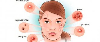
With age, a more saturated solution is used as an antiseptic for various skin inflammations with pustular formations. It is thanks to its high antiseptic effect that potassium permanganate is an excellent remedy for acne, and since there is a bottle with these crystals in almost every home, it is quite simple to try this method of struggle, but the effectiveness and safety of the drug is another question .
Mechanisms of action
Skin treatment with potassium permanganate differs from treatment with drugs that contain antibiotics, due to the fact that antibiotics tend to destroy both pathogenic and beneficial microflora, and it is the pathogenic flora that often develops resistance to antibiotics.
But potassium permanganate, like any natural antiseptic , although it does not have a strong effect on microflora, but along with the low-aggressive effect it has on pathogenic bacteria, it does not completely destroy safe microflora.
At the same time, its pronounced therapeutic effect is fully present, and the fact that potassium permanganate is not addictive makes its long-term use possible. Potassium permanganate has oxidative properties, to which the biological mechanisms of adaptation of bacteria do not work, this makes it possible to use a solution of potassium permanganate even in such a branch of medicine as purulent surgery.
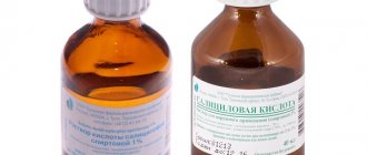
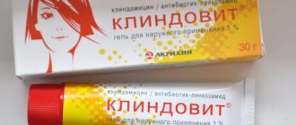
Another drug for external use, the main properties of which are antiseptics and disinfection, is cindol.
This drug is based on zinc; it is also popularly called chatterbox (due to the fact that it must be shaken thoroughly before use).
In cosmetology , potassium permanganate has been used for acne for a long time , but when treating with this remedy, it is important not to forget that the potassium permanganate solution dries out the skin and can react with other cosmetics; although its effect is effective, it requires caution to avoid chemical burns.
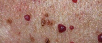
At home, without spending a lot of time and money, you can get rid of acne by preparing a solution of potassium permanganate, which can be stored for a long period of time in the refrigerator. Every person has the opportunity to fight acne on the back, chest and other parts of the body using this simple remedy.
The effectiveness of potassium permanganate solution for acne on the back
The effectiveness of using a solution of potassium permanganate against acne on different areas of the skin (on the back and not only) has been known and proven for a long time, because the product not only has a drying effect, but also prevents the appearance of new rashes , so regular use of the solution leads to a significant reduction in the number of acne on skin.
This happens due to the fact that potassium permanganate has the following properties:
- disinfection;
- fight inflammation;
- lack of addiction from pathogenic pathogenic flora;
- reduction.
For the back, this treatment method is considered almost the most ideal.
It can be used for 7 days or more, until recovery, and this area of the body is least exposed to sunlight and external pollution, since it can be completely covered under clothing.
Types of wounds
They can be:
- Operating: applied deliberately for the purpose of performing an operation.
- Accidental: as a rule, these are household damages.
Depending on the nature and conditions of the damage, wounds are distinguished:
- chopped;
- I stab;
- bruised;
- torn;
- crushed;
- chopped;
- bitten;
- mixed;
- firearms.
In children , cut, puncture, bitten, mixed, and also superficial (abrasions, scratches, etc.) predominate. Particular attention is paid to bites that result from an animal or human bite (for example, another child), as they are the most infected. This is due to the large number of harmful bacteria in the oral cavity. They are most often complicated by infection.
According to the degree of infection there are:
- Freshly infected. As a rule, this damage occurs within 3 days from the date of its application. Defects with soil contamination are especially infected.
- Purulent. An infectious process develops in these lesions. Microbes, when multiplying, lead to increased inflammation, necrosis, and also contribute to the appearance of pus. Children with purulent manifestations develop symptoms of general intoxication: nausea, headache, dizziness, weakness, lethargy.
Contraindications for this drug
Of course, the procedure has a number of contraindications that should not be forgotten during the treatment process; this primarily concerns the fact that the chemical composition of potassium permanganate can react with other substances and drugs that are applied to the skin.
Therefore, when using the solution, do not forget about these rules:
- You should not apply creams or cosmetic scrubs to the skin before the procedure, because this will not allow potassium permanganate to act fully and will cause an unpredictable reaction even with the natural components of the cosmetic product.
- There is no need to squeeze out pimples before starting the procedure , even despite the antimicrobial effect that potassium permanganate has on rashes, if the solution is applied to a fresh skin lesion, this most often contributes to the further spread of the rash over the skin. Also carefully use washcloths on the affected area of the skin; if there are wounds or scratches near the pimple, treat them first and only then use a solution of potassium permanganate. This is necessary because as a result of tissue damage, the inflammatory reaction increases, which means the effect of the procedure is lost.
- It is not advisable to use this method to treat rashes on the face , with the exception of only one pimple, which can be gotten rid of in just one night by applying the solution to the problem area. But use is permissible only for a single rash, because after treatment with potassium permanganate, quite unaesthetic stains often remain that need to be additionally removed.
If you suffer from rashes on your face, we recommend that you pay attention to the so-called “liquid gold” - olive oil. It contains antioxidants and a full range of vitamins, is suitable for any skin type, and can be used daily for facial skin care.
Mycoses of smooth skin
Among the widespread fungal diseases today, the most common are mycoses of smooth skin, such as microsporia, trichophytosis, lichen versicolor, mycosis of the feet (hands), and candidiasis. Sources of infection can be sick animals (cats, dogs, mouse-like rodents, cattle, etc.), as well as humans. In recent years, there has been an increase in the number of diseases caused by opportunistic fungi, among which superficial forms of candidiasis are most often recorded. Such a wide prevalence of these mycoses can be explained by the massive use of modern therapeutic agents, the environmental situation and other factors that reduce the body's defenses. One of the reasons for the significant prevalence of mycoses is the weakening of sanitary educational work in recent years. Due to insufficient awareness about the sources and ways of spreading infection, as well as adequate preventive measures, patients turn to the doctor late, and therefore mycoses become chronic, including in children suffering from mycoses of the scalp and smooth skin.
Microsporia is a fungal disease caused by various types of fungi of the genus Microsporum. In Russia, microsporia, which has spread over the last 50 years, is caused by a zoophilic fungus - fluffy microsporum (Microsporum canis), which parasitizes the skin of cats, dogs, and less often other animals. Infection from a sick person is observed in 2% of cases.
Epidemiology . Infection in 80-85% of cases occurs as a result of direct contact with a sick animal or through objects contaminated with the fur of these animals. Infection of children can also occur after playing in the sandbox, since the causative agent of microsporia is highly resistant to environmental factors and can remain viable in infected scales and hair for up to 7-10 years. Children often suffer from microsporia.
Clinic . After 5-7 days from the moment of infection, lesions appear on smooth skin, which can be observed on both open and closed parts of the body (children love to pick up animals and put them in bed with them). The lesions are round or oval in shape, pink or red, with clear boundaries, a raised ridge along the periphery, covered with blisters and thin crusts, with peeling in the center. The lesions are usually small, from 1 to 2 cm in diameter, single or multiple, sometimes merging. In 85-90% of patients, vellus hair is affected.
Treatment . If there are single foci of microsporia on smooth skin without damage to vellus hair, you can limit yourself to only external antifungal agents. The lesions should be lubricated with alcohol tincture of iodine (2-5%) in the morning, and sulfur-salicylic ointment (10% and 3%, respectively) should be rubbed in in the evening. You can rub the following antimycotics 2 times a day: mycozolon, mycoseptin, travogen or 1 time a day in the evening - mifungar cream, mycospor - until the clinical manifestations resolve. In case of multiple lesions of smooth skin and single lesions (up to 3) involving vellus hair, it is recommended to prescribe the antifungal antibiotic griseofulvin at the rate of 22 mg per 1 kg of the child’s body weight, in 3 doses after meals, in combination with exfoliating the stratum corneum of the epidermis in keratolytic lesions means (salicylic acid 3.0, lactic or benzoic acid 3.0, collodion up to 30.0). The lesions are lubricated with one of these products 2 times a day for 3–4 days, then 2% salicylic ointment is applied under compress paper for 24 hours, the detached scales of the stratum corneum of the epidermis are removed with tweezers and vellus hair is epilated. If during a control study carried out using a fluorescent lamp or microscope, affected hair is detected, the procedure is repeated. Detachment of the stratum corneum of the epidermis and manual hair removal of vellus hair can be carried out after using the “sealing” method. The lesions are sealed in a tile-like manner with strips of adhesive plaster for 2-3 days, this causes an aggravation of the process, which, in turn, facilitates hair removal.
The results of treatment for smooth skin microsporia are monitored using a fluorescent lamp or microscopic examination for fungi. The first control study is done after the resolution of clinical manifestations, then 3-4 days before the first negative test, and then after 3 days. The criteria for cure are resolution of the lesions, absence of luminescence and three negative tests on microscopic examination.
During the treatment, bed and underwear are disinfected: boiling in a soap-soda solution (1%) for 15 minutes (10 g of laundry soap and 10 g of caustic soda per 1 liter of water); ironing outerwear, furniture covers, and bedding five times with a hot iron through damp cloth.
Prevention. The main measure to prevent microsporia is compliance with sanitary and hygienic rules (you cannot use other people’s underwear, clothes, etc.; after playing with animals, you must wash your hands).
Trichophytosis is a fungal disease caused by various types of fungi of the genus Trichophyton. Trichophytons can be anthropophilic, parasitic on humans, and zoophilic, whose carriers are animals. Anthropophilic trichophytons include Trichophyton (Tr.) tonsuraus and Tr. violaceum, to zoophiles - Tr. mentagrophytes var gypseum and Tr. verrucosum.
Epidemiology. With superficial trichophytosis, caused by anthropophilic fungi, infection occurs through close contact with a sick person or indirectly through household items. Often children become infected from their mother, grandchildren from grandmothers suffering from a chronic form of the disease. The incubation period lasts up to a week. In zooanthroponotic trichophytosis, the sources of infection are sick animals: cattle, rodents. The highest incidence of this type of trichophytosis is recorded in the fall, which is associated with field work: it is at this time that the likelihood of infection through hay and straw increases. The incubation period ranges from 1–2 weeks to 2 months.
Clinic. On smooth skin with superficial trichophytosis, lesions can appear on any part of the skin - face, neck, chest, forearms. They have clear boundaries of a round or oval shape, with a raised ridge along the periphery of a bright red color; they are larger in size than in microsporia. The lesions are reddish-bluish in color, with peeling, nodules on the surface; in the chronic form, they develop on the skin of the buttocks, knee joints, forearms, less often the back of the hands and other parts of the body; the lesions do not have clear boundaries. Lamellar peeling is observed on the skin of the palms and soles. Vellus hairs are often affected.
With trichophytosis, caused by zoophilic fungi, the disease on the skin can occur in three forms: superficial, infiltrative and suppurative. Lesions are usually located on open areas of the skin. With a superficial form, they are round or oval in shape, with clear boundaries, a raised ridge along the periphery, on which bubbles, crusts, a pink center, and a bright red ridge are visible. The lesions are larger in size than with microsporia. Sometimes they are located around natural openings - eyes, mouth, nose. In the infiltrative form, the lesions rise above the skin level and are accompanied by inflammatory phenomena - infiltration. The suppurative form is characterized by the development of tumor-like formations, bright red in color, covered with purulent crusts due to the addition of a bacterial infection. When the lesion is compressed, pus is released from the hair follicles and pain is noted. The disease is accompanied by a violation of the general condition, sometimes the temperature rises. After the resolution of clinical manifestations, cicatricial atrophy of the skin remains at the site of former lesions. Clinical forms of zooanthroponotic trichophytosis can transform into one another.
Diagnostics. The diagnosis of trichophytosis is established on the basis of the clinic and when the fungus is detected during microscopy of pathological material, and the type of pathogen is determined using cultural examination.
Treatment. Treatment is carried out with antimycotics for external use. The lesions are lubricated with tincture of iodine (2-5%) during the day, and sulfur-salicylic ointment (10% and 3%, respectively) or mycoseptin is rubbed in in the evening. You can carry out monotherapy with ointment or cream (kanison, mifungar, mycozoral, mycospor (bifosin), exoderil, mycozoral, etc. In the infiltrative form, 10% sulfur-tar ointment is prescribed to resolve infiltration 2 times a day. Treatment of the suppurative form of trichophytosis begins with removing crusts from the affected area using bandages with 2% salicylic ointment, which are applied for several hours. After removing the crusts, vellus hair is epilated. Then apply lotions with solutions that have a disinfectant and anti-inflammatory effect (furacilin 1:5000, rivanol 1:1000 , potassium permanganate 1:6000, ichthyol solution (10%), etc.). As a result of this treatment, the hair follicles are freed from pus, inflammatory phenomena are reduced. Next, sulfur-tar ointment (5-10%) is prescribed for resorption of the infiltrate (5-10%) in the form of rubbing or under wax paper.After the infiltrate has resolved, antimycotics are used for external use (see superficial form of trichophytosis).In cases where vellus hair is affected in lesions on smooth skin, the stratum corneum of the epidermis is detached, followed by hair removal. To do this, you can use salicylic collodion (10-15%), milky-salicylic-resorcinol collodion (15%). If there is no effect, griseofulvin is prescribed orally at a daily dose of 18 mg per 1 kg of body weight, in 3 divided doses after meals daily until a negative test for fungi, then every other day. As an alternative method, terbinafine (Lamisil, Exifin) can be prescribed to adults 250 mg (1 tablet) once a day after meals every day, children weighing up to 20 kg - 62.5 mg, from 20 to 40 kg - 125 mg , over 40 kg - 250 mg in combination with antimycotics for external use.
The criteria for cure for trichophytosis are resolution of clinical manifestations and three negative fungal test results at three-day intervals.
Prevention. Prevention of trichophytosis depends on the type of pathogen. With superficial trichophytosis caused by anthropophilic fungi, the main preventive measure is to identify the source of infection, and it can be children suffering from superficial trichophytosis, or adults suffering from a chronic form of the lesion. In recent years, cases of chronic trichophytosis have been observed in middle-aged and older children. For suppurative trichophytosis, preventive measures are carried out jointly by medical workers, epidemiologists and veterinary services.
Mycosis of the smooth skin of the feet (hands). In a number of countries, up to 50% of the population suffers from mycosis of the feet. This disease is more common in adults, but in recent years it has often been observed in children, even infants.
Etiology. The main causative agents of mycosis of the feet are the fungus Trichophyton rubrum (T. rubrum), which is isolated in almost 90% of cases, and T. mentagrophytes var. interdigitale (T. interdigitale). Damage to the interdigital folds, which may be caused by yeast-like fungi, is recorded in 2-5% of cases. The anthropophilic fungus Epidermophyton floccosum is rarely isolated in our country.
Epidemiology. Infection with mycosis of the feet can occur in the family through close contact with a patient or through household items, as well as in a bathhouse, sauna, gym, or when using someone else's shoes and clothes.
Pathogenesis. The penetration of fungi into the skin is facilitated by cracks and abrasions in the interdigital folds caused by sweating or dry skin, abrasion, poor drying after water procedures, narrowness of the interdigital folds, flat feet, etc.
Clinic. Clinical manifestations on the skin depend on the type of pathogen and the general condition of the patient. The T.rubrum fungus can cause damage to the skin of all interdigital folds, soles, palms, dorsum of the feet and hands, legs, thighs, inguinal-femoral, intergluteal folds, under the mammary glands and axillary area, trunk, face, and rarely the scalp. The process may involve vellus and long hair, nail plates of the feet and hands. When the skin of the feet is affected, there are 3 clinical forms: squamous, intertriginous, squamous-hyperkeratotic.
The squamous form is characterized by the presence of peeling on the skin of the interdigital folds, soles, and palms. It can be flour-shaped, ring-shaped, lamellar. In the area of the arches of the feet and palms, an increase in the skin pattern is observed.
The intertriginous form is the most common and is characterized by slight redness and peeling on the lateral contact surfaces of the fingers or maceration, the presence of erosions, superficial or deep cracks in all folds of the feet. This form can transform into dyshidrotic, in which vesicles or blisters form in the area of the arches, along the outer and inner edges of the feet and in the interdigital folds. Superficial blisters open with the formation of erosions, which can merge, resulting in the formation of lesions with clear boundaries and oozing. When a bacterial infection is attached, pustules, lymphadenitis and lymphangitis occur. In the dyshidrotic form of mycosis, secondary allergic rashes are observed on the lateral and palmar surfaces of the fingers, palms, forearms, and shins. Sometimes the disease becomes chronic with an exacerbation in spring and summer.
The squamous-hyperkeratotic form is characterized by the development of foci of hyperkeratosis against the background of peeling. The skin of the soles (palms) becomes reddish-bluish in color, and pityriasis-like peeling is noted in the skin grooves, which extends to the plantar and palmar surfaces of the fingers. Pronounced ring-shaped and lamellar peeling may be detected on the palms and soles. In some patients, it is insignificant due to frequent hand washing.
In children, lesions of smooth skin on the feet are characterized by fine-plate peeling on the inner surface of the terminal phalanges of the toes, usually 3 and 4, or there are superficial, less often deep cracks in the interdigital folds or under the toes, hyperemia and maceration. On the soles, the skin may not be changed or the skin pattern may be enhanced; sometimes ring-shaped peeling is observed. Subjectively, patients are bothered by itching. In children, more often than in adults, exudative forms of lesions occur with the formation of blisters and weeping eczema-like lesions. They appear not only on the feet, but also on the hands.
Rubrophytia of smooth skin of large folds and other areas of the skin is characterized by the development of lesions with clear boundaries, irregular outlines, with an intermittent ridge along the periphery, consisting of merging pink nodules, scales and crusts, with a bluish tint (the color is bluish-pink in the center) . On the extensor surface of the forearms and shins, rashes can be located in the form of open rings. Lesions with nodular and nodular elements are often observed. The disease sometimes occurs as an infiltrative-suppurative trichophytosis (more often in men when localized in the chin area and above the upper lip). Foci of rubrophytosis on smooth skin can resemble psoriasis, lupus erythematosus, eczema and other dermatoses.
The fungus T. interdigitale affects the skin of the 3rd and 4th interdigital folds, the upper third of the sole, the lateral surfaces of the foot and toes, and the arch of the foot. This mushroom has pronounced allergenic properties. With mycosis of the feet caused by T. interdigitale, the same clinical forms of damage are observed as with rubrophytosis, but the disease is often accompanied by more pronounced inflammatory phenomena. In the dyshidrotic, less often intertriginous form, large blisters may appear on the skin of the soles and fingers, along with small blisters; in the case of bacterial flora, with purulent contents. The foot becomes edematous, swollen, and pain appears when walking. The disease is accompanied by an increase in temperature, deterioration of health, development of allergic rashes on the skin of the upper and lower extremities, torso, face, enlargement of the inguinal lymph nodes; the clinical picture is similar to that observed with eczema.
Diagnosis. The diagnosis is established on the basis of clinical manifestations, detection of the fungus by microscopic examination of skin flakes and identification of the type of pathogen by cultural examination.
Treatment. Treatment of mycosis of the smooth skin of the feet and other localizations is carried out with antifungal agents for external use. For squamous and intertriginous forms of lesions on the feet and other areas of the skin, medications are used in the form of a cream, ointment, solution, spray; you can combine a cream or ointment with a solution, alternating their use. Currently, the following medications are used to treat this disease: exifin cream, mycozoral cream, nizoral cream, canizon cream and solution, mycozon cream, mycospor (bifosin) cream, mifungar cream, lamisil cream and spray, mycoterbin cream. These drugs are applied to cleansed and dried skin once a day, the average duration of treatment is no more than 2 weeks. Antimycotics such as travogen, ekalin, batrafen, mycoseptin, mycozolon are used 2 times a day until clinical manifestations resolve, then treatment is continued for another 1-2 weeks, but once a day - to prevent relapse. In nodular and nodular forms of rubrophytosis, after relieving acute inflammatory phenomena using one of these ointments, sulfur-tar ointment (5-10%) is prescribed in order to further resolve the clinical manifestations. For intertriginous and dyshidrotic forms (the presence of only small blisters) of mycosis of the feet, drugs with a combined effect are used, which, along with an antifungal agent, include a corticosteroid, for example mycozolon, travocort, or a corticosteroid and an antibacterial drug - triderm, pimafucort.
In case of acute inflammatory phenomena (wetting, the presence of blisters) and severe itching, treatment is carried out as for eczema: desensitizing agents (intravenous or intramuscular administration of calcium chloride solution (10%), sodium thiosulfate solution (30%), calcium gluconate solution (10%) or calcium pantothenate orally; antihistamines. For external medications, at the first stage of therapy, lotions are used (2% boric acid solution, potassium permanganate solution 1:6000, 0.5% resorcinol solution), 1-2% aqueous solutions of methylene blue or brilliant green, fucorcin. Then they switch to pastes - boron-naphthalan, ichthyol-naphthalan, ACD paste - F3 with naphthalan, if complicated by bacterial flora - lincomycin (2%). At the 2nd stage of treatment after resolution of acute inflammatory phenomena, the above-mentioned antimycotic agents are used.
A drug such as Triderm, which contains, in addition to an antimycotic (clotrimazole 1%), a broad-spectrum antibiotic (gentamicin sulfate 0.1%) and a corticosteroid (betamethasone dipropionate 0), can quickly and effectively eliminate the symptoms of inflammation and itching in the presence of both fungal and bacterial infections. .05%). The presence of Triderm in 2 dosage forms - ointment and cream - makes it possible to use it for different types and at different stages of the pathological process.
If external therapy is ineffective, systemic antimycotics are prescribed: itraconazole in a continuous regimen of 200 mg per day for 7 days, then 100 mg for 1-2 weeks; terbinafine (Lamisil, Exifin) 250 mg once a day every day for 3-4 weeks; fluconazole (150 mg once a week for at least 4 weeks).
Prevention. To prevent foot mycosis, it is necessary to observe, first of all, the rules of personal hygiene in the family, as well as when visiting a bathhouse, sauna, swimming pool, gym, etc.; disinfect shoes (gloves) and linen during the treatment period. After visiting a bathhouse, swimming pool, sauna, to prevent mycosis of the feet, apply Daktarin spray powder to the skin of the interdigital folds and soles.
Tinea versicolor is a fungal disease caused by Malassezia furfur (Pityrosporum orbiculare), a yeast fungus. Lichen versicolor is quite widespread in all countries; young and middle-aged people suffer from it.
Etiology. Malassezia furfur as a saprophyte is found on human skin and, under favorable conditions, causes clinical manifestations.
Pathogenesis. Factors contributing to the development of the disease have not yet been precisely established, however, lichen versicolor is more common in people suffering from excessive sweating, changes in the chemical composition of sweat, diseases of the gastrointestinal tract, endocrine pathology, vegetative-vascular disorders, as well as immune deficiency .
Clinic. The disease is characterized by the presence of small spots on the skin of the chest, neck, back, abdomen, less often the upper and lower extremities, axillary and inguinal-femoral areas, on the head; the spots are initially pink in color and then become light and dark brown; Slight peeling is also observed, sometimes it can be hidden and can only be revealed by scraping. The rashes often merge, forming large areas of damage. After tanning, as a rule, white spots remain as a result of increased flaking. The disease is characterized by a long course with frequent exacerbations.
Diagnosis. The diagnosis is made on the basis of clinical manifestations, when the pathogen is detected in skin flakes during a microscopic examination and in the presence of a characteristic yellow or brown glow under a Wood's fluorescent lamp, as well as a positive iodine test.
Treatment. Currently, there is a sufficient selection of antifungal drugs for topical use that have a pronounced antifungal effect against the causative agent of lichen versicolor. These include imidazole and triazole derivatives, allylamine compounds. During the treatment of the disease, the following is used: exifin cream (applied to cleansed and dried skin in the affected areas 2 times a day for 7-14 days, if necessary, after a 2-week break, the course of treatment can be repeated), nizoral cream, mycozoral ointment, cream and canizon solution, mycozon cream, mifungar cream (prescribed once a day, duration of treatment is 2-3 weeks); lamisil cream and spray; nizoral shampoo (for three days, apply to the affected areas of the skin for 3-5 minutes and wash off in the shower). For common, often recurrent forms of lichen versicolor, systemic antimycotics are more effective: itraconazole (prescribed 100 mg once a day for two weeks, then take a two-week break, repeat the course of treatment if necessary), fluconazole (150 mg once a week within 4-8 weeks). During treatment, it is necessary to disinfect the patient’s clothes, hats, underwear and bed linen by boiling in a 2% soap-soda solution and ironing with a hot iron while wet. Family members of the patient should also be examined.
Prevention. To prevent recurrence of mycosis, it is necessary to use nizoral shampoo. Treatment should be carried out from March to May once a month for 3 days in a row.
Smooth skin candidiasis is a fungal disease caused by yeast-like fungi of the genus Candida.
Etiology. The pathogens are opportunistic fungi that are widely distributed in the environment. They can also be found on the skin and mucous membrane of the mouth, digestive tract, and genitals of a healthy person.
Epidemiology. Infection from the external environment can occur with constant fractional or massive infection with fungi.
Pathogenesis. Both endogenous and exogenous factors can contribute to the occurrence of candidiasis. Endogenous factors include endocrine disorders (usually diabetes mellitus), immune deficiency, severe somatic diseases and a number of others. The development of the disease is possible after the use of a number of modern medications: broad-spectrum antibiotics, immunosuppressive and hormonal drugs. The occurrence of candidiasis in the interdigital folds of the hands is facilitated by frequent contact with water, as this develops maceration of the skin, which is a favorable environment for the introduction of the pathogen from the external environment.
Clinic. On smooth skin, small folds on the hands and feet are more often affected, less often - large ones (inguinal-femoral, axillary, under the mammary glands, intergluteal). Lesions outside the folds are located mainly in patients suffering from diabetes mellitus, severe general diseases, and in infants.
In some patients, the disease begins in small folds of the skin with the formation of small, barely noticeable blisters on the lateral contacting surfaces of hyperemic skin, the process gradually spreads to the area of the fold, then peeling, maceration appears, or immediately shiny eroded surfaces of a deep red color with clear boundaries appear, with peeling of the stratum corneum of the epidermis along the periphery. The 3rd and 4th interdigital folds on one or both hands are most often affected. The disease is accompanied by itching, burning, and sometimes pain. The course is chronic, with frequent relapses.
In large folds, the lesions are dark red in color, shiny, with a moist surface, with a strip of exfoliating stratum corneum of the epidermis, occupying a significant surface, having clear boundaries and irregular outlines. New small erosions appear around large foci. In children, the process of large folds can spread to the skin of the thighs, buttocks, abdomen, and torso. Painful cracks sometimes form deep in the folds.
Candidiasis of smooth skin outside the folds has a similar clinical picture.
Diagnosis. The diagnosis is made on the basis of a typical clinic when a fungus is detected in scrapings from skin flakes during a microscopic examination.
Treatment. Limited and sometimes widespread acute forms of smooth skin lesions, especially those that developed during therapy with antibacterial drugs, as a rule, are easily treated with local antimycotic agents in the form of a solution, cream, ointment and can resolve even without treatment after discontinuation of antibiotics.
For candidiasis of smooth skin of large folds with acute inflammatory phenomena, treatment should begin with the use of an aqueous solution of methylene blue or brilliant green (1-2%) in combination with indifferent powder and continue for 2-3 days, then antifungal drugs are used until clinical resolution manifestations.
Among the antimycotic agents for candidiasis of smooth skin, the following are used: Canison solution and cream, Mycoson cream, Mifungar cream, Candida cream and solution, Triderm ointment and cream, Pimafucort, Pimafucin, Travocort, Travogen, Nizoral cream, Mycozoral ointment, Ekalin.
For common skin processes and in case of ineffectiveness of local therapy, systemic antimycotics are prescribed: fluconazole (Diflucan, Forkan, Mycosist) - adults at a dose of 100-200 mg, children at a dose of 3-5 mg per kg of body weight, itraconazole (100-200 mg), nizoral (adults 200 mg, children weighing up to 30 kg - 100 mg, over 30 kg - 200 mg) once a day daily, as well as the polyene antibiotic natamycin (adults 100 mg 4 times a day, children 50 mg 2–4 times a day). The duration of treatment is 2–4 weeks.
Prevention. Prevention of smooth skin candidiasis in adults and children consists of preventing its development in people suffering from underlying diseases, as well as in people receiving long-term antibacterial, corticosteroid, and immunosuppressive therapy. To prevent the development of candida infection in children hospitalized in somatic departments and receiving broad-spectrum antibiotics, it is necessary to prescribe fluconazole at the rate of 3 mg per kg of body weight once a day, treatment is carried out during the entire main course of therapy. Patients with intestinal candidiasis are prescribed nystatin 2–4 million units per day or natamycin 50 mg for children and 100 mg for adults 2 times a day for 15 days.
Zh.V. Stepanova, Doctor of Medical Sciences, TsNIIKV
Note!
- In recent years, there has been an increase in the number of diseases caused by opportunistic fungi, among which superficial forms of candidiasis are most often recorded.
- Due to insufficient awareness about the sources and ways of spreading infection, as well as adequate preventive measures, patients turn to the doctor late, and therefore mycoses become chronic
- 50% of the population suffers from mycosis of the feet. Adults get sick more often. Recently, there has been an increase in incidence in children, even infants.
- Treatment of mycosis of the smooth skin of the feet and other localizations is carried out with antifungal agents for external use.
- If external therapy is ineffective, systemic antimycotics are prescribed.
Tips for use
If you have had skin problems for a long time and you want to try treatment with a solution of potassium permanganate, be sure to consult with your dermatologist or cosmetologist; only a doctor can assess how safe this treatment method is.
In order for potassium permanganate to be effective in treatment, you need to use a concentrated solution, which is prepared as follows:
We take a container that can be tightly closed, because the prepared solution needs to be stored only in this form and in the refrigerator. In this case, the container must be clean and dry, made of glass. Simple kitchen utensils that are used for food will not work; for this it is better to take a small pharmacy container , approximately 100 ml. This amount of solution is enough for treatment for a period of 7 days. Crystals of potassium permanganate are dissolved in hot (but not boiling water) distilled or purified water.
It is necessary to dissolve the crystals by gradually adding the dry product carefully; during the preparation of the solution, it is constantly stirred, adding new portions. This process must be carried out until the crystals completely cease to be visible in the water. But if several of them remain at the bottom of the dish undissolved, then this is not a problem, the main thing is that they do not get on the skin . The solution, cooled to a temperature of +20 degrees, should look like an opaque liquid of a rich purple color. When applied to the skin, a specific smell of ozone appears, and it immediately dries out any pimple.
Before applying the solution to your shoulders, back or other area of skin, you must take a shower with simple cosmetic or antibacterial soap to remove sweat, dust and other contaminants from the surface of the skin. Next, apply the solution to the pimples point by point; for this you can use a cotton swab or cotton swab. But the impact should not last longer than 5 seconds . Often, when there is a large area of skin damage, it is recommended to take a bath with a diluted potassium permanganate concentrate; the water should be pink, and the bath can be taken for 5-7 minutes.
After using the solution, a feeling of tightness appears on the skin, but it is strictly forbidden to smear the skin with cream , so as not to worsen the therapeutic effect. Spot treatment or lubrication of groups of pimples is carried out up to 2 times a day, and a bath is taken once a day. After treatment, do not use any emollient creams or ointments.
Due to the fact that the solution stains the skin brown, it is important not to wear light-colored clothing and use the solution carefully when treating facial skin.
Products for treating wounds in children
Below we list the most popular and affordable means for treating superficial wounds in children:
- Iodine. Suitable for superficial abrasions, but not for deeper injuries.
- Diamond green.
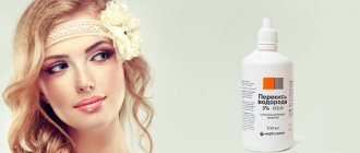
- Hydrogen peroxide. It is the drug of choice for first aid in the treatment of superficial abrasions in children. Allows you to clean abrasions and scratches well. An equally important property is stopping bleeding.
- Furacilin. To prepare a solution for topical use, dissolve the tablet in water.
- Miramistin. It is also an antiseptic for topical use. Allows you to prevent suppuration. If a purulent process occurs, it allows you to fight it. A pleasant bonus will be the possibility of using this remedy for acute respiratory viral infections, sore throats and pharyngitis in children.
- Chlorhexidine.
An antiseptic helps prevent suppuration.
To speed up healing, ointments and creams can be used such as:
- Levomekol;
- Betadine;
- Vishnevsky ointment;
- cream Bepanten plus.
Why can’t potassium permanganate be used to treat acne on the face?
On the face, the solution is used only for a single rash and extremely rarely, so that the facial skin does not turn brown. Under no circumstances apply the solution to your face when taking a bath and make sure that potassium permanganate does not get into your eyes, otherwise, in addition to a brown tint to the skin, an eye burn may occur, which will require serious treatment and can lead to loss of vision.
In addition, it is the face that is most exposed to the sun, dust and other contaminants, which reduce the effectiveness of treatment with this method.
How to remove potassium permanganate stains?
This type of acne treatment is quite effective, but the most unpleasant moment may be the presence of stains that the solution leaves. It is because of this that it is best to use a solution of potassium permanganate for treatment before the weekend or at night. But if you need to quickly remove stains from potassium permanganate on the face, neck, chest or other noticeable place, then this can be done using various drugs and substances.
Such means include:
- ascorbic acid;
- running water;
- lemon juice;
- citric acid solution;
- alcohol solution;
- White Spirit.
Running water
It is clear that it is best to rinse damaged areas of skin with copious amounts of running water.
But hands or clothes contaminated during the treatment process can be rubbed with one of the above substances, and then rinsed thoroughly in water. So, when treating with ascorbic acid, it is enough to apply a damp cotton swab with powder to the stain/stains for up to 10 seconds, and then rinse with water.
Lemon juice
When using lemon, you can take freshly squeezed juice of one citrus or even a slice of this fruit.
Rub the contaminated area and then rinse with water. A solution of citric acid is prepared as follows : dissolve a packet of citric acid in a glass of water (250 ml), then either wash your hands in it or apply the solution with a swab to the contaminated area. By rinsing your hands or cloth with water, you will see how the stains disappear almost before your eyes.
Alcohol solution
The alcohol solution must also be diluted with water and under no circumstances should you wipe the skin with pure alcohol , because this way the skin will receive a severe burn, which does not heal for a long time and can leave scars.
White Spirit
White spirit is a universal solvent; it will help you deal with potassium permanganate on your hands and clothes without unnecessary problems. If the treatment did not give the desired effect the first time, then after a while you can repeat the procedure several times .
If you used a very saturated solution of potassium permanganate, the stains may take several days to disappear, be patient and do not use an alcohol solution or white spirit very often, because this can cause a chemical burn to the skin. It is best to use potassium permanganate with medical gloves to avoid stains.
| For those who struggle with acne for a long period of time, potassium permanganate can be a remedy that will help restore health and beauty to the skin. It is important not to self-medicate and consult a doctor before starting treatment . Often, it is potassium permanganate (potassium permanganate) that becomes the remedy that will help overcome acne, acne vulgaris and furunculosis, especially if the treatment is prescribed by a doctor and it is comprehensive. Maintain the concentration and carefully ensure that undissolved crystals do not get on the skin or mucous membranes; it is also important to dilute the concentrate before use. | |
Wound healing and factors influencing this process
The wound healing process can be disrupted by many factors.
Healing is the process of repairing damaged tissue and restoring its integrity. During the healing process, certain processes occur. We will not dwell on them in more detail, but will only indicate those factors that may disrupt the healing process:
- Age. As a rule, this applies more to older people. The child's body has great regenerative capabilities. This is due to the high level of anabolic (construction) processes in the body.
- Attachment of infection.
- Decreased immunity.
- Poor circulation in the wound area.
- Chronic diseases (diseases of the respiratory and cardiovascular systems, diabetes, tumors and others).
Healing complications
- Attachment of infection . Most often, a nonspecific purulent infection develops. The threat is tetanus, rabies, diphtheria. Therefore, injuries that have been bitten and contaminated with soil or old metal objects should be immediately examined by a surgeon with a number of preventive measures taken (administration of anti-tetanus serum, rabies vaccine).
- Bleeding.
- Divergence of defect edges.
- In some cases, healing may be complicated by a hypertrophic scar or keloid . They are an abnormal scar that can spread beyond the defect area and also contribute to the development of complications.