Why do benign formations appear on the skin?
Cosmetologists and dermatologists do not know the exact mechanism of their formation. Most often the cause is:
- injuries;
- viruses;
- systemic diseases of the body, for example xanthomas, occur due to an excess of fat in the blood;
- long-term skin diseases;
- exposure to aggressive substances;
- excessive exposure to ultraviolet radiation;
- x-rays;
- heredity (for example, seborrheic dermatosis).
Most skin lesions are benign
Benign and malignant neoplasms on the skin: what are the differences?
Benign pathologies do not pose a threat to human life. If they reach large sizes, they can interfere with the adequate functioning of various body systems. In contrast, malignant ones grow quickly and aggressively, penetrate into surrounding tissues, and form metastases over time. Some damage vital organs and cause death.
Sometimes benign skin tumors change due to external or hereditary causes. They acquire the ability to degenerate into malignant pathologies. Such conditions are called borderline or precancerous. They pose a great danger to health and life, although they do not always have pronounced symptoms.
2.Classification
Keratomas are classified according to a number of characteristics:
- senile keratoma (gray-white growth on an open area of the body, prone to inflammation);
- seborrheic keratoma (the most dangerous type of growth, which turns brown over time, causes itching and peeling);
- horny keratoma (usually a strongly protruding, small diameter, but tall, dark-colored growth that can degenerate);
- solar keratoma (multifocal keratoma, most often affecting exposed skin in men over 40 years of age, dangerous for asymptomatic degeneration into cancer);
- follicular keratoma (growth on the scalp or upper lip, more common in women).
Visit our Dermatology page
What is the structure of benign neoplasms
The growths consist of cells that have partially retained their original functions and are capable of growing slowly. They are similar in structure to the tissues from which they originated. They can put pressure on nearby tissues, but do not penetrate them, since they have a capsule in their structure. They respond well to hardware and surgical treatment and, as a rule, do not cause relapses.
There are always congenital formations on the skin - moles or warts, as well as acquired ones. The latter are formed on the surface or in the subcutaneous layer as a result of metabolic disorders, decreased immunity, or under the influence of a virus.
Stages
In its development, amelanotic melanoma can go through 4 stages. Each of them, except the fourth, is divided into several substages.
The stages are determined according to three criteria:
- thickness of the tumor (depth of its germination into the skin);
- metastases to regional lymph nodes;
- metastases to internal organs.
| Stage | Tumor thickness | Metastases to regional lymph nodes | Metastases to internal organs |
| 0 | within the epidermis | No | No |
| I.A. | to the papillary layer of the dermis, reticular (deep) does not affect, no ulcers | No | No |
| I.B. | penetrates into the deep layer of the dermis, possibly into the subcutaneous tissue (thickness - from 1 to 2 mm), no ulcers | No | No |
| II A | tumor thickness - from 1 to 2 mm, there is ulceration or thickness from 2 to 4 mm, no ulcers | No | No |
| II B | thickness from 2 to 4 mm, with ulcerations or thickness above 4 mm, without ulcers | No | No |
| II C | thickness more than 4 mm, with ulcerations | No | No |
| III A | any thickness, no ulcers yet | there are micrometastases in 1-3 regional lymph nodes | No |
| III B | any thickness, without ulceration | or macrometastases in 1-3 regional lymph nodes or metastases to the skin near the tumor, but without metastases in regional lymph nodes | No |
| any thickness, with the formation of ulcers | or micrometastases in 1-3 regional lymph nodes or metastases to the skin near the tumor, but without metastases in regional lymph nodes | No | |
| III C | any thickness, with ulceration | or micro- or macrometastases in 2-3 regional lymph nodes | No |
| any | more than 3 regional lymph nodes are affected by metastases, or there are several affected lymph nodes fused together, or there are metastases in the skin near the tumor, despite the fact that they are also present in the regional lymph nodes | No | |
| IV | any size | any quantity | There is |
Types of benign formations
- Warts and papillomas
Papilloma is a small tumor on a stalk or broad base, which has clear boundaries. Flint is visible on an uneven, grainy surface. The growth is painted in any color - from white to dark brown. It is found both individually and in large quantities.
Both papillomas and warts appear as a result of the papilloma virus entering the body, live in different parts of the body, and reach a diameter of several centimeters. They are activated on the skin as a result of nervous tension, stress, decreased immunity or vegetative disorders.
Papillomas and warts are not dangerous if they are not injured
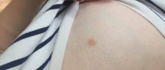
- Birthmarks
They are not prone to degeneration, but such cases do occur, so it is necessary to pay attention to changes in contour, color, and size, especially if the area is constantly injured.
- Lipoma
A round, soft-to-the-touch tumor of adipose tissue that does not disrupt the functions of the body, but only creates a cosmetic defect. Wen appears in various areas where there is a fat layer. Reaching large sizes, they grow into tissue and approach bone surfaces, often spreading to muscles and blood vessels.
- Atheroma
Atheroma is a residual cyst caused by blockage of the ducts of the sebaceous glands. It contains products produced by the sebaceous glands in the form of an odorless curd mass. The pathology has clear contours and a dense consistency. Superficial atheromas occur mainly on the back, head, neck, face, and limbs - due to poor hygiene, metabolic disorders and unprofessional depilation.
When subjected to mechanical action, the atheroma becomes inflamed, swells and becomes red. When an infection gets inside, pus is formed and a greasy consistency is erupted. There is a risk of degeneration into liposarcoma, so it is recommended to remove this pathology.
A seemingly harmless atheroma can degenerate into liposarcoma
- Nevus
More often than others, melanoma degenerates into a malignant formation, especially with prolonged exposure to negative external factors or hereditary predisposition.
- Lymphangioma
The pathology is mainly congenital, developing from lymph nodes in the skin, but can also spread to fiber or muscles. It is most often localized on the head, face and upper body. The blue tumor-like growth rises above the skin, has a dense consistency and clear boundaries, dimensions are 1-5 mm. Since it puts pressure on vital organs (lungs, larynx, trachea), surgical removal is resorted to.
- Hemangioma
It is formed on the basis of blood vessel cells in the subcutaneous layer, rises above the surface, and is characterized by rapid spontaneous growth. When pressed, the growth decreases. It is mainly localized in the head and neck area, mainly in young children. Cavernous hemangioma is colored blue and resembles a node, located deep in the tissues. Capillary - in the epithelium, has a red or blue color. If it becomes inflamed, it can provoke open bleeding, and if it is close to vital organs, it disrupts their functions.
Hemangiomas are diagnosed more often in children under three years of age.
- Fibroma (dermafibroma)
A tumor of dense consistency, light pink in color, is formed from connective tissue. Mild varieties occur more often on the neck and chest, in the groin folds and armpits in women. Hard - in different areas near the upper layers of the skin. Dermatologists recommend removing fibromas, since under favorable conditions they can transform into fibrosarcoma.
- Keratoma
This skin tumor is formed from keratinocytes that make up the stratum corneum. A growth in the form of a spot or node is formed from dead cells and is localized on the back, head, face, and limbs. Such a benign neoplasm requires urgent treatment.
- Neurofibroma
Formed from nerve sheath cells, it looks like a hard tubercle up to three centimeters in diameter. Causes discomfort and pain because it puts pressure on nerve endings. Most often located on the face, back, abdomen, arms and legs. If there are many formations, they talk about neurofibromatosis - a disease that is mainly inherited.
Molluscum contagiosum or infectious molluscum
Molluscum contagiosum or infectious molluscum is a skin tumor of a viral nature, benign in nature. She's contagious. The main routes of transmission from a sick baby to a healthy one are through direct close contact. The second option for infection may be the use of shared hygiene items.
Another great place to catch this virus is swimming pools - humid air, skin contact and a large number of people contribute to this.
Not everyone is equally susceptible to the virus - usually children from the group of frequently ill people, allergy sufferers, and digestive problems are more likely to suffer. Children with skin problems are more likely to get sick when there are areas of damage - abrasions, scratches, dermatitis - then it is easier for the virus to penetrate the thickness of the skin.
Rashes can appear on any part of the body - face, arms, neck, legs, shoulders, abdomen and even genitals - they cannot only appear on the palms and soles, which is why they differ significantly from papillomas. Usually they are not complicated - but if the child picks or combs them, microbes can appear.
Of course, in the case of a typical course and manifestation in several children, there is no doubt, but sometimes the mollusk is difficult to recognize. Therefore, let a pediatric dermatologist make an accurate diagnosis and treatment.
Treatment of this viral disease consists of removing the tumors by a pediatric dermatologist (or a pediatric ophthalmologist, if the molluscum is on the eyelid) using anesthetics. Further, it is possible to prescribe antiviral agents and certain treatment of skin areas where there were previously nodules, to exclude the possible reappearance of tumors. The main preventive measures are timely diagnosis of this disease and its treatment; therefore, it is necessary to conduct preventive examinations.
Removal of benign skin tumors
Dermatologists are convinced: it is necessary to get rid of benign formations, with the exception of small scatterings of moles or other minor defects throughout the body. This is especially true for the face, because the growth attracts attention, spoils the overall impression and gives a lot of unpleasant emotions to its owner.
There are several methods for removing benign skin tumors:
- Electrocoagulation
Under local anesthesia, the growth is cut off with a special surgical coagulator, which creates a high-frequency current. Simultaneously with removal, the tissues are soldered together, which avoids bleeding and infection. The crust at the treatment site disappears after 7-10 days, sometimes leaving a slightly noticeable scar. The method is effective if the defect is small.
- Cryodestruction
Liquid nitrogen can only be applied to formations in the upper layers of the epidermis. If it is flat, apply applications with liquid nitrogen. In case of deeper occurrence, a cryodestructor is used. After the procedure, the body begins a reaction of rejection of the treated defective tissue. The resulting crust disappears and heals within a month and a half.
- Laser removal
A powerful beam of light destroys neoplasm cells and evaporates them from the skin. The laser does not affect healthy neighboring tissues. The manipulation is carried out under local anesthesia, it is low-traumatic and bloodless. Laser treatment parameters are selected individually. Damaged tissues are evaporated layer by layer until the beam reaches healthy skin. The formed crust disappears on its own after 1-2 weeks.
- Radio wave method
High frequency radio waves cut and coagulate tissue. The crust disappears after a week. The method is contraindicated in patients with a pacemaker, herpes, or elevated body temperature.
- Surgical removal of benign skin tumors
This method is used when the growths are too large and other technologies cannot cope. The formation is excised with a scalpel and removed, capturing a small area of healthy skin. The scar after surgery takes several weeks to heal, but the wound requires long-term careful care. The description of this traumatic technology is not encouraging, so it is better to choose another method to eliminate defects on the face.
On the face, neoplasms are removed using laser or cryodestruction
Treatment of the disease
At Euroonko, non-pigmented melanoma is treated according to modern protocols, taking into account the stage of the process:
- In stage I of the disease, surgical removal of the tumor and tissue around it is performed.
- In stage II, the tumor is removed and the lymph nodes are additionally checked for cancer cells. If mutated melanocytes are found in the lymph nodes, the nodes are removed. Treatment may include photodynamic therapy and radiation therapy. Here, if there is a mutation in the BRAF gene, treatment is carried out with BRAF inhibitors.
- Stage III is treated by removing the tumor and all nearby lymph nodes. Treatment is complemented by chemotherapy and radiotherapy. Immunotherapy and the introduction of BRAF inhibitors are involved. Therapy is required to improve the condition and relieve symptoms.
- At stage IV, large tumors and metastases are removed, if necessary. Chemotherapy, radiotherapy and palliative treatment are prescribed. Immunotherapy is mandatory.
Modern treatment of melanoma includes such a new and very promising direction as immunotherapy. And if previously only chemotherapy was carried out, which was successful only in 10% of cases, now the concept has changed greatly for the better. Immunotherapy involves the administration of special drugs that bind to certain proteins on the membranes of lymphocytes and “show” them that there are cancer cells in the body. Then the lymphocytes start working again and destroy them - throughout the body, and not locally.
Immunotherapy helps the body fight cancer. Even with advanced forms of malignant melanocytic tumor, patients can count on successful treatment.
For amelanotic melanoma containing a BRAF gene mutation, Euroonko provides targeted therapy with BRAF inhibitor drugs. These drugs suppress tumor progression and enhance immune activation. BRAF inhibitors are used starting from stages IIC-III. They increase long-term survival and are recommended for use even with an inoperable tumor or the presence of distant metastases.
Therapy with BRAF inhibitors is carried out for a long time - 6-9 months.
Prevention of skin tumors
Unfortunately, medicine has not yet learned to prevent the appearance of various formations on the skin. But dermatologists give their patients the following preventive recommendations:
- do not delay contacting a doctor if a tumor appears on the skin;
- remove formations only after a specialist and diagnostics confirm their benign nature;
- avoid excessive exposure to the open sun;
- use sunscreen, especially if you are prone to moles and hyperpigmentation;
- do not come into contact with chemically active and carcinogenic substances;
- do not eat foods that contribute to the development of cancer (smoked meats, sausages, animal fats, meat products with food stabilizers).
Do you have a lot of moles? Forget about tanning in the open sun
Chemotherapy
It is carried out both as an independent method of treatment and in combination with surgical intervention. Prescribing chemotherapy before surgery is intended to reduce the pathological focus. The purpose of this method of treating skin cancer after surgery is designed to completely destroy cancer cells.
The disadvantage of this method is the inability to exclude the effects of drugs on healthy cells. The question of the need for chemotherapy is decided by the attending physician based on the individual characteristics of the development of the tumor process.
Do benign formations hide the danger?
Benign neoplasms are unpredictable structures that can manifest themselves at any time or not at all. The process of their transformation into malignant ones has not been fully studied. There is no clear answer to the question of what exactly activates this process. It is believed that mechanical trauma, excess ultraviolet radiation, metabolic disorders and other factors contribute to degeneration. One way or another, if you have a benign skin lesion, you should not experiment and rely on chance. Moreover, today removal does not cause difficulties.
Diagnostic methods
In addition to standard diagnostic methods (history collection, examination), specialists use additional instrumental and laboratory methods to establish a diagnosis of facial skin basal cell carcinoma. These include:
- A smear-imprint (if there is an erosion or ulcer on the surface of the skin) or a scraping from the surface of a tumor formation (in the absence of ulceration) and subsequent cytological examination.
- Biopsy. It is usually carried out to clarify the diagnosis if basal cell carcinoma is suspected based on the results of a cytological analysis.
The dermoscopic method is also actively used. Dermoscopy is especially informative in the differential diagnosis of the pigmented form of basal cell carcinoma of the facial skin and melanoma.