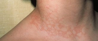
Pictured is Quincke's edema
Angioedema is a condition in which small blood vessels leak fluid into the tissue, causing swelling. The disease has no cure, but symptoms can be controlled with medication and diet. Only in rare cases, the cause of Quincke's edema is an allergy.
This is a fairly common disease. Every tenth person is susceptible to urticaria and of these, every third has Quincke's edema. Without urticaria, the disease is much less common. In most cases it disappears on its own, but may reappear over time.
Angioedema - symptoms
The most affected areas of the body are the face, lips, tongue, throat and genital areas. Swelling in one area usually lasts 1-3 days. Sometimes internal organs such as the esophagus or stomach swell, causing internal pain. Other clinical symptoms of angioedema are itching, tingling or burning. But most often the main symptom is swelling and discomfort. The swelling can be painful if the joints are affected. Signs of angioedema such as swelling can be very large and may not go away for several days.
Relevance
According to statistics from a number of European countries (Germany, England, France, etc.), from 10 to 30% of the population suffers from allergic diseases [1]. One of the manifestations of a human allergic reaction is Quincke's edema (QE) - an acute disease characterized by the appearance of clearly limited swelling of the skin, subcutaneous tissue, and the mucous membrane of various organs and systems of the body. OK was first described by Heinrich Quincke in 1882, but only almost 100 years later (in 1963) the pathogenesis of this disease was determined. For a long time, OK was presented as an exclusively allergic reaction. The progressive development of allergology and immunology has gradually expanded knowledge about OK. This disease is divided into hereditary and acquired forms. It was found that OK can be a manifestation of allergy and pseudo-allergy. It has also been found that this disease can develop as a result of taking angiotensin-converting enzyme inhibitors (ACE inhibitors) (captopril, enalapril). It should be noted that in 45% of patients it is not possible to identify the causative factor for the development of OK [2]. Thus, over time, a number of human pathological conditions with identical clinical manifestations, but with different etiologies and pathogenesis, began to be classified as a separate nosological form. This introduced significant uncertainty into the clinical work of specialists.
Today, according to ICD-10, OK is classified as angioedema (AO) with identification code T78.3. There are: I. Allergic A.O. (giant urticaria); II. Pseudoallergic edema (nonspecific histamine liberation); III. Complement-dependent edema: 1. Hereditary A.O. (HAE): a) HAE with a quantitative deficiency of C1 inhibitor (HAE type I); b) HAE with a functional deficiency of the C1 inhibitor (HAE type II); c) HAE, in which the level of C1 inhibitor and its function in patients are normal (HAE type III); 2. Acquired A.O. (PAO): a) immunocomplex (PAO type I); b) autoimmune (PAO type II); c) when administering drugs that activate the complement system; IV. Idiopathic A.O..
I
.
Allergic AO
. The most common occurrence in everyday clinical practice is A.O. Up to 80% of the causes of AO are the development of an allergic reaction of type I - reagin (IgE-dependent type). Various factors can lead to the development of allergic AO: medications, food products, some dietary supplements, stinging insect venom, latex (gloves, rubber urinary catheters, endotracheal tubes, intravenous catheters), epidermal allergens - saliva, cat and dog dander, cosmetics . Allergic AO is characterized by a clear connection between exposure to the allergen and the development of the reaction, an acute onset - usually the reaction develops 15-30 minutes after contact with the allergen, rapid development of edema, and a combination with urticaria. Swelling has a characteristic appearance - it is usually dense, asymmetrical, painless swelling, may be pale pink in color and not different from unchanged skin. It is localized mainly in places with well-developed subcutaneous fat (on the face these are most often the lips, eyelids, in the oral cavity - the soft palate, tongue, tonsils). Involvement of the mucous membrane of the respiratory system (swelling of the larynx, trachea, bronchi) is especially dangerous due to the threat of asphyxia. It is possible that AO may develop as the onset of a generalized anaphylactic reaction - anaphylactic shock, which is manifested by generalized itching, urticaria, lacrimation, sneezing, bronchospasm, swelling of the tongue, larynx, pharynx, hoarseness, hypersecretion of mucus in the bronchi, nausea, vomiting, cramping abdominal pain, diarrhea, tachycardia, arterial hypotension, cardiac arrhythmia, development of acute cardiovascular failure, seizures, respiratory arrest, coma. In this case, death occurs from swelling of the larynx and cardiac arrhythmia.
II. Pseudoallergic edema
. The clinical manifestations of pseudoallergic edema are in many ways similar to those of allergic edema, however, the release of allergy mediators occurs in a non-immune way. With this mechanism of edema development, an important role is played by foods and medications that change the metabolism of mediators, the formation of excess leukotrienes, bradykinin, dysbacteriosis, namely muscle relaxants (thiopental, tubocurarine), opiates, antibiotics, radiocontrast agents, local anesthetics, plasma substitutes, plasma, albumin, protamine directly affects mast cells, increasing the level of histamine in the blood; fish, cheese, smoked meats, wine, beer, chocolate, nuts, berries (strawberries, raspberries), citrus fruits - contain a large amount of histamine in their composition; non-steroidal anti-inflammatory drugs (NSAIDs), nutritional supplements (tartrazine), salicylates, sulfites, nitrates - lead to the formation of excess leukotrienes D4, C4, E4, which have significant vasodilatory and chemotactic activity; taking ACE inhibitors is a fairly common cause of the development of AO (from 4 to 8% of all AO) due to the accumulation of bradykinin, which increases vascular permeability. It was noted that 0.1-0.7% of all patients taking ACE inhibitors had an episode of AO [3, 4].
III. Hereditary angioedema (HAE)
is an extremely rare disease, its prevalence may vary in different regions and is approximately 1:50,000 people. Such a low prevalence leads to extremely low awareness among doctors about the disease and, as a consequence, to its poor and late detection, as well as to incorrect management tactics and selection of therapy. HAE is a disease whose main clinical manifestations are recurrent swelling of the mucous membranes and deep layers of the dermis of various locations [5, 6].
The development of edema is caused by an increased release of the mediator bradykinin (a low-molecular nanopeptide that causes an increase in the permeability of the vascular wall and extravasation of plasma) due to a deficiency or impairment of the functional activity of the C1-inhibitor (C1-INH). Most often, the development of the disease is based on a mutation in the SERPING1
[7].
In order to diagnose HAE with C1-ING deficiency, indicators of the level of C1-ING and its functional activity, as well as the level of the C4 component of complement in the blood are used. HAE type I (85% of cases) is diagnosed when the C1-ING level is less than 30% of normal. HAE type II (15% of cases) is diagnosed when a decrease in the functional activity of C1-ING is detected by more than 50% of the norm with a normal or increased level of C1-ING. To make a diagnosis, changes must be recorded in at least 2 studies, which must be repeated at intervals of 1-3 months. The level of complement C4 in patients with HAE I and II is usually reduced (<50% of normal), but the low sensitivity and specificity of this test limit its diagnostic value, so this indicator can only be used as a screening marker for selecting patients due to with its availability in routine practice and low price [5] (Table 1).
Table 1. Diagnostic indicators for hereditary angioedema types I and II
Thus, the mechanism for the development of edema in HAE is due to the excessive activity of complement and Hageman factor with the formation of bradykinin and C2-kinin, which increase vascular permeability. This explains the lack of effect in patients from taking antihistamines and glucocorticosteroids (GCS) (Table 2).
Table 2. Differential diagnosis of hereditary and allergic angioedema
Type III HAE was initially described in women and then in men.
The mechanism of its development is unclear. The C1-ING level and its function in patients are normal. It is possible that the disease is associated with increased production of bradykinin and a slowdown in its destruction due to a decrease in ACE (kininase) activity under the influence of estrogens.
Clinical manifestations of HAE.
The main clinical manifestations of HAE are peripheral edema, abdominal attacks (caused by swelling of the intestinal wall) and upper respiratory tract (URT) edema. Almost all patients with HAE suffer from recurrent peripheral edema, the most common clinical manifestation of the disease. A characteristic feature of edema is the absence of urticaria and changes in the skin (redness, temperature) over the edema. There may be tingling, burning, and soreness at the site of swelling. The most common localization is the upper and lower extremities. Unlike histamine edema, edema caused by bradykinin increases more slowly, but also regresses more slowly. On average, swelling persists for about 72 hours [8].
The next most common symptom (traced in more than 50% of patients) is abdominal attacks, which can occur either together with peripheral edema or in isolation, which significantly complicates diagnosis. The clinical picture can vary from mild discomfort to symptoms of an “acute abdomen”, which become the reason for unnecessary surgical interventions. Abdominal ultrasound and CT can detect swelling of the intestinal area and free fluid in the abdominal or pelvic cavity [9].
The greatest threat to the patient's life is edema of the upper respiratory tract: swelling of the larynx, tongue and palate. Clinically, this is manifested by breathing and swallowing disorders, dysphonia, and stridor. The possibility of swallowing movements should be assessed and the upper respiratory tract accessible to examination should be examined. The time from the onset of symptoms of respiratory failure to complete asphyxia is unpredictable and can range from 20 minutes to 14 hours. Edema of the upper respiratory tract may be the first clinical manifestation of the disease [1, 8].
Trigger factors for the development of HAE may include emotional stress, physical stress, mechanical trauma (even the most minor, such as pressure from a belt or shoe), infectious diseases, the use of certain drugs (certain contraceptives, ACE inhibitors or angiotensin II receptor antagonists), insect bites , a number of food products, weather changes [1].
Factors associated with an increased risk of developing urgent edema include many medical procedures: dental procedures, invasive methods of examination and treatment, surgical interventions (especially those associated with the need for intubation anesthesia). Swelling associated with these procedures usually occurs within 2 days after the procedure. It is very important to remember that manipulations performed in the past without complications do not guarantee the safety of each subsequent medical intervention [10-12].
The presence of uncompensated chronic diseases (for example, caries, tonsillitis, cholecystitis) is also a trigger factor for the development of edema, therefore, in the absence of adequate correction of such diseases, a vicious circle is formed that progressively worsens the course of the underlying disease [13]. In this regard, the presence of a clear protocol for patient management before medical intervention is of great importance to minimize the risk of complications and, consequently, to reduce disability and mortality. In addition, the presence of such a plan significantly increases the chance of patients to receive timely, quality medical care. The enormous importance of timely diagnosis and adequate treatment for such conditions is clearly demonstrated in the study by K. Bork et al.: out of 70 examined fatal cases from asphyxia during airway obstruction, 63 patients were not diagnosed, and only 7 patients had the disease verified [ 8].
Recommendations for invasive medical interventions in patients with HAE
According to available data, based on the principles of evidence-based medicine, when carrying out invasive medical interventions, patients with HAE are recommended to follow the following general rules: use a drug for short-term prophylaxis (C1-esterase inhibitor - benert) to prepare for planned and urgent interventions 1-6 hours before procedures. In its absence, fresh frozen plasma and danazol can be used as second-line drugs [1, 5]. Basic therapy should not be interrupted (if the patient is receiving it) [5].
It is recommended to avoid, if possible, intubation anesthesia and give preference to other types of anesthesia [14], to prefer conduction anesthesia to general anesthesia [14], to ensure the possibility of resuscitation measures, in particular to restore airway patency [14].
You should make sure that the patient is not receiving drugs prohibited for HAE: ACE inhibitors, angiotensin II receptor blockers, estrogen-containing drugs.
It is important to remember that since edema in HAE is caused by the action of bradykinin, this group of edema is insensitive to the use of systemic corticosteroids, antihistamines and adrenaline; therefore, it is not advisable to use these drugs for short-term prevention and relief of edema [1, 5].
According to the latest edition of international recommendations for the management of patients with HAE, the only drug for short-term prevention based on the principles of evidence-based medicine (both for planned and urgent medical intervention) is plasma C1-ING concentrate [1]. This is a C1-esterase inhibitor concentrate obtained from donor blood, which affects all stages of pathogenesis in the treatment of HAE. The only C1-esterase inhibitor drug registered in Russia is benert. Berinert confirmed its effectiveness and safety in clinical trials both abroad and in the Russian Federation. Throughout the world, benert is a C1-ING drug with more than 30 years of experience in use (more than 500 thousand courses of treatment). Data on its safe use in all age categories of patients, including children and pregnant women, have been published. Therefore, if there is a choice, this drug should always be preferred, especially if extensive medical intervention is planned or it is not possible to avoid intubation anesthesia [1, 5].
C1-ING concentrate should be used for prophylactic premedication as close as possible to the beginning of the procedure. According to the instructions adopted in the Russian Federation, the prophylactic dose of the drug before an invasive procedure or surgery is 1000 IU for an adult and 15-30 IU/kg for a child. The drug is administered intravenously [5]. Carrying out such premedication allows you to minimize the risk of possible life-threatening edema, however, it must be remembered that even the use of a concentrate of donor C1-ING esterase from a person does not provide an absolute guarantee: there are reports of clinical cases and series of cases where edema developed even after relatively minor operations against the background premedication [1, 14]. Therefore, even when premedicating during surgery, the patient must have with him means to relieve acute edema [1, 5].
Unfortunately, in routine practice in Russia, the use of benert may be limited due to the low supply of patients and medical institutions with this drug. In such a situation, it is necessary to prepare the patient for the planned intervention with the help of second-line drugs: danazol, fresh frozen plasma, antifibrinolytics.
During planned medical interventions, danazol can be used as a short-term prophylactic medication. This drug belongs to the pharmacological group of attenuated androgens. To date, a lot of experience has been accumulated in the use of attenuated androgens in HAE. Danazol is prescribed according to the following regimen: 200 mg/day 7 days before and 4-5 days after surgery (if the patient was on basic therapy with danazol, then the dose is increased 2 times from the original) with further cancellation or transition to the previous dose of basic therapy [5 , 12]. It should be remembered that attenuated androgens have many side effects and there are categories of patients for whom the use of these drugs is contraindicated, despite their effectiveness. However, short courses of danazol, as a rule, do not have clinically significant consequences for the patient’s health [12].
In the absence of danazol and benert, the only available agent that can reduce the risk of edema during medical intervention is native or fresh frozen plasma, which also donors the C1-esterase inhibitor. It is prescribed in a dose of 250.0 ml 1-6 hours before the procedure. In addition, during planned surgery, it is possible to use danazol and plasma together. Since plasma is not standardized for the content of C1-ING and may contain components such as blood coagulation factor XII, prekallikrein, high molecular weight kininogen, its transfusion may not always lead to the expected effect. Cases have been described of not only the lack of effect from plasma administration, but also increased edema. In addition, the safety of plasma in comparison with that of C1-ING drugs is significantly lower, which is associated with a higher risk of transmission of vector-borne infections, as well as allosensitization [1].
The use of antifibrinolytics as short-term prophylactic drugs is not currently recommended, at least as monotherapy [1]. A number of experts believe that it is possible to use aminocaproic acid as an additional method of preventing the development of edema: in addition to danazol, it is recommended to administer aminocaproic acid by intravenous drip of 200.0 ml [5]. Also, before performing dental procedures, rinse the mouth with a 5% solution of aminocaproic acid.
Much more often than with medical interventions, patients encounter everyday trigger factors: these can be stressful situations (exams, moving, personal experiences), menstruation, infectious diseases, insect bites, physical activity. The upcoming effects of some trigger factors are often known in advance, so it is possible to prepare for them. If it is impossible to prevent C1-ING, it is recommended to prescribe danazol (or increase its dose) according to the regimen recommended for preparation for medical procedures. In some patients (with previously proven effectiveness), it is possible to prescribe or increase the dose of tranexamic acid. Prescribing or doubling the dose of the drug for the period of infectious diseases (starting from the prodromal stage) is also considered effective.
Any preventive premedication does not exclude the development of “breakthrough” attacks, so there should not only be a supply of drugs (berinert or icatibant) to relieve edema, but also in case of insufficient response to pharmacotherapy, the possibility of intubation. The patient should be left under the supervision of an anesthesiologist-resuscitator when the first signs of compression of the upper respiratory tract appear [1, 14].
Today, the main problem in the management of patients with HAE in our country is the low awareness of doctors about this disease and, as a consequence, its low detection rate. Patients have been treated for years with incorrect diagnoses, prescribed inappropriate therapy (or not prescribed at all), and denied relevant medical care. In our practice, we often encounter patients in whom the development of edema after surgical procedures is regarded as drug intolerance to local anesthetics; because of this, they undergo many painful procedures without anesthesia (which can also lead to edema) or are denied assistance. Fortunately, today doctors have enough drugs in their arsenal to minimize the risk of complications due to medical manipulation.
Acquired C1-ING deficiency is a rarer pathology than HAE. PAO can occur due to pronounced utilization and consumption of normal C1-ING (PAO type I) or the synthesis of autoantibodies against C1-ING, which impairs its function (PAO type II). PAO occurs in tumors (lymphoproliferative diseases, etc.), autoimmune and infectious diseases [15].
IV. Idiopathic A.O.
The diagnosis of idiopathic AO is made if the cause of AO is not found. This form is characterized by the absence of a family history of the disease. In recent large studies, idiopathic AO accounted for up to 40% of all cases of isolated AO. Idiopathic A.O. characterized by dense pasty whitish swelling, without itching, pain, or hyperemia. The development of idiopathic AO is associated with trauma, sometimes surgical, tissue compression (for example, during a handshake), minor bruise, hypothermia, emotional stress, and the menstrual cycle. Young and middle-aged women are most often affected. The increase in idiopathic AO occurs within 48-72 hours, followed by spontaneous reverse development within 3-4 days. In the absence of exacerbation, patients are practically healthy.
The greatest danger is posed by idiopathic AO of the larynx, which can cause death in patients from asphyxia. Death is most likely between the ages of 30 and 40 years. Possible idiopathic AO of the mucous membrane of the gastrointestinal tract, which is manifested by severe abdominal pain, vomiting bile, and watery diarrhea. This localization of idiopathic AO can simulate the picture of an “acute abdomen”, while there is no rigidity of the abdominal wall, fever and leukocytosis. In such cases, unjustified surgical intervention can cause progression of idiopathic AO up to death [7].
In conclusion, I would like to note that in modern planned ENT surgery, such a complication as angioedema is quite rare. However, given the fatal nature of its consequences, more detailed detailing of the patient’s medical history, timely prediction and verification of the type of angioedema are necessary. This can serve as the key to adequate perioperative prevention and etiopathogenetic therapy, allowing one to avoid such a formidable complication as laryngeal edema, which is the cause of death from asphyxia.
The authors declare no conflict of interest.
The authors declare no conflicts of interest.
Information about authors
Kryukov A.I. — Doctor of Medical Sciences, Prof., Director of the State Budgetary Institution “NIKIO named after. L.I. Sverzhevsky" DZM, head. Department of Otorhinolaryngology "Russian National Research Medical University named after. N.I. Pirogov" Ministry of Health of the Russian Federation, Chief Freelance Otorhinolaryngologist of Moscow, Honored. scientist of the Russian Federation; https://orcid.org/0000-0002-0149-0676
Kunelskaya N.L. - Doctor of Medical Sciences, Prof. Department of Otorhinolaryngology l/f FSBEI HE "RNIMU named after. N.I. Pirogov" Ministry of Health of the Russian Federation, deputy. Director for Scientific Work of the State Budgetary Institution "NIKIO named after. L.I. Sverzhevsky" DZM; e-mail; https://orcid.org/0000-0002-1001-2609
Tsarapkin G.Yu. - Doctor of Medical Sciences, Ved. Researcher, Head of the Department of Pathology of the Upper Respiratory Tract and Aesthetic Rhinofacial Surgery, State Budgetary Institution of Healthcare "NIKIO named after. L.I. Sverzhevsky" DZM; e-mail; https://orcid.org/0000-0003-2349-7438
Tovmasyan A.S. - Candidate of Medical Sciences, Senior Researcher, State Budgetary Institution "NIKIO named after. L.I. Sverzhevsky" DZM, e-mail; https://orcid.org/0000-0002-1214-4939
Lapchenko A.A. - Candidate of Medical Sciences, Head of the Department of Otorhinolaryngology No. 32, City Clinical Hospital No. 1 named after. N.I. Pirogov" DZM; e-mail; https://orcid.org/0000-0001-6729-8815
Kishinevsky A.E. - Junior Researcher, State Budgetary Institution "NIKIO named after. L.I. Sverzhevsky" DZM; e-mail; https://orcid.org/0000-0002-6700-3308
Gorovaya E.V. — otorhinolaryngologist of the KDO, junior researcher in the department of pathology of the upper respiratory tract and aesthetic rhinofacial surgery of the State Budgetary Healthcare Institution “NIKIO named after. L.I. Sverzhevsky" DZM; e-mail; https://orcid.org/0000-0003-2072-5415
Aleksanyan T.A. — Candidate of Medical Sciences, otorhinolaryngologist, plastic surgeon; e-mail; https://orcid.org/0000-0002-9164-6282
How to quote:
Kryukov A.I., Kunelskaya N.L., Tsarapkin G.Yu., Tovmasyan A.S., Lapchenko A.A., Kishinevsky A.E., Gorovaya E.V., Aleksanyan T.A. Angioedema. Classification, diagnosis, prevention, treatment tactics. Bulletin of Otorhinolaryngology
. 2019;84(3):68-73. https://doi.org/10.17116/otorino201984031
Angioedema - causes
Swelling may occur during treatment with drugs that control heart rhythm and blood pressure during the first months. Sometimes appears after increasing the dose. There is such a thing as hereditary angioedema. This is a rare condition that affects 1 in 75,000 people. It is characterized by a low level of an enzyme that helps keep blood vessels in a stable state. An attack may be caused by emotional stress, alcohol, hormonal changes, or trauma (such as dental surgery). Another cause of Quincke's edema is infection. This is the most common cause of symptoms, especially in children. Swelling can also be caused by an allergic reaction to food or medication and usually goes away within 24 hours.
Causes of the disease
Quincke's edema what is it: experts distinguish between allergic and pseudoallergic etiology of the disease.
In the first option, tissue swelling occurs due to direct contact with the allergen. For a reaction to develop, the body must produce antibodies in advance (during a previous encounter with the antigen). Secondary penetration of specific allergens provokes an inflammatory process at the entry point with the appearance of capillary dilation, increased vascular permeability and tissue swelling.
The pathological condition occurs:
- due to food - fish, seafood, eggs, chocolate, citrus fruits, berries;
- medications - antibacterial, painkillers or vaccines (rarely - multivitamin complexes, oral contraceptives), the severity of the reaction depends on the type of administration, and intensifies with injections;
- plant pollen, cosmetics;
- insect bites, waste products or animal undercoat.
Pseudoallergic Quincke's edema is a hereditary disease; patients experience dysfunction of the complement system, which is responsible for activating the reaction to allergens. In this case, the inflammatory process develops under the influence of chemical elements, cold or heat influences, as a response to stress.
The prerequisites for the occurrence of the disease are considered to be certain pathologies, represented by:
- hypothyroidism, helminthiasis;
- chronic lesions of the gastrointestinal tract;
- autoimmune diseases.
These diseases can lead to a recurrent course of angioedema.
Quincke's edema - treatment
First you need to make sure that you have angioedema. Therefore, it is important to see a doctor for tests. For some people, the swelling goes away on its own and never appears again. First of all, avoid the aggravating factors described above. The main treatment for angioedema is antihistamines. They are taken both once when symptoms appear, and for prevention when the disease is regular. To prevent the disease from recurring, a diet prescribed by a nutritionist may be required. To determine exactly how to treat angioedema , be sure to visit your doctor. Only he can find out the cause and prescribe the optimal treatment.
See also: Psoriatic arthritis
We invite you to familiarize yourself with the ESMA line of medical devices.
Features of therapy
When the victim is hospitalized, a laboratory diagnostic examination is carried out to help identify the cause of the reaction and the severity of the condition. In the first days, the patient is transferred to a hypoallergenic diet, with a ban on the consumption of chocolate products, all types of berries, oranges, tangerines, grapefruits and natural honey.
Therapeutic procedures involve the use of medications:
- antihistamine action - for one week the patient is treated with Zyrtec, Cetrin, Fenistil, Loratadine;
- hormonal - several days of therapy with Prednisone, Dexamethasone;
- Polysorb and Enterosgel are prescribed for 72 hours to cleanse the body of specific allergens;
- a decrease in the permeability of blood vessel walls is achieved by using Ascorutin;
- The nervous system is maintained by calcium and ascorbic acid.
For pseudoallergic Quincke's edema, symptomatic medications are used; the choice of appropriate medications remains with the attending physician.
What is tongue swelling, and how dangerous is it?
Edema is an increase in volume of an organ. The tongue may swell and increase in width, and also partially, for example, when a tumor-like lump forms in some place. Such conditions are extremely alarming; they may indicate the progression of a dental disease or an allergic reaction.
Note! Tongue swelling should not be confused with a slight swelling on the tip of the tongue that appears after biting or eating hot, sour foods.
Edema may also be accompanied by the following symptoms of various diseases:
- choking cough and sneezing;
- profuse lacrimation;
- rash on the surface of the face and body;
- feeling of a “lump in the throat”;
- itching in the mouth and sore throat;
- increased body temperature;
- weakness;
- difficulty swallowing;
- speech problems;
- swelling of the tissues of the oral cavity and face;
- feeling of a foreign body in the mouth, etc.
It is important! The most dangerous symptom is swelling of the tongue at its root, when the tissues block the airways, the person feels a sharp suffocation, blushes, and rolls his eyes upward. In such situations, you should urgently call an ambulance or provide emergency first aid to the patient.
Dentistry for those who love to smile
+7
Make an appointment
How to remove allergic swelling from the eyelids
Angioedema develops in a time period from several minutes to several hours after contact with the allergen. It is very important to know what to do with allergic swelling of the eyelids, since it can spread to the respiratory tract and internal organs and cause serious complications.
Doctors recommend following the following sequence of actions:
- Sit or lay the person down so that nothing restrains his movements. Remove tight clothing.
- If the allergen is known, stop exposure to it as quickly as possible. For example, if the cause is pollen from a plant, you need to move away from it and remove clothing that may have particles on it.
- Give the person antihistamines (Claritin, Loratodin, Suprastin, Tavegil, Zodak, etc.). If the patient's condition worsens, glucocorticoids can be administered intravenously.
- If you have a food allergy, you can give the patient laxatives or enterosorbents.
- You can apply ice or a cold compress to the affected area to reduce swelling.
It is important to monitor symptoms closely. The patient may require professional medical attention: if the swelling increases, difficulty breathing, swelling or pain in the abdomen occurs.
After you have removed allergic swelling from the eyelid and eye, you need to be extremely careful over the next 24 hours. You need to wash your face only with warm water, do not use eye makeup or contact lenses, and avoid stressful situations. It will be helpful to apply soothing compresses to the eyes.
There are special medications that cause changes in mast cells. For example, for allergic swelling of the eyelids, drops are sometimes prescribed - Kromhexal, Lecrolin, Alomide. However, you need to keep in mind that they do not have an immediate effect and they must be used in advance, before contact with the allergen - for example, 1-2 weeks before flowering if you are allergic to pollen.
Angioedema of the eyelids can be diagnosed after the attack has passed mainly by allergy tests: you will need to take tests for immunoglobulin E in the blood, eosinophils, as well as a general blood test. Treatment for allergic eyelid edema will include:
- regular use of antihistamines;
- hypoallergenic diet (you need to exclude foods that can cause allergies from the menu);
- keeping a food diary to identify the cause of pathology;
- taking allergy tests for various substances;
- complete exclusion of contact with allergens after determining which substances cause an allergic reaction;
- If you are unable to find out what exactly causes Quincke's edema, you should always carry fast-acting antihistamines with you.