Eventration is the exit of internal organs through a missing or damaged area of the abdominal wall. It manifests itself as a visible tissue defect, prolapse of abdominal organs through it, local and diffuse pain in the abdominal cavity, dyspepsia, and increasing intoxication. Diagnosed using a physical examination, ultrasound of internal organs, electroenterography, thoracic radiography. In case of incomplete eventration, conservative management using a tight bandage is possible; in other cases, reconstructive plastic surgery of the abdominal wall is necessary.
General information
Eventration is a rare surgical pathology in which, due to the formation of a through defect in the peritoneum, muscle-aponeurotic layer, subcutaneous tissue, and skin, the abdominal cavity depressurizes and the internal organs extend beyond its boundaries. Less often, the damage is non-penetrating with the viscera emerging under the skin.
Most eventrations are of traumatic origin, their prevalence especially increases during war - according to military surgeons, the disease is detected in 14-23% of victims with abdominal wounds. Suture dehiscence on days 10-20 after laparotomy is observed in 0.5-3% of patients. The prevalence of eventration during emergency abdominal surgery is 10 times higher. In 49% of cases, the greater omentum comes into the wound, in 29% - the small intestine.
Eventration
Reasons for eventration
Prolapse of abdominal organs has a polyetiological origin. The most common cause of eventration is damage to the anterior abdominal wall - cut, stab wounds, wounds from fragments in explosions, ruptures under the pressure of massive objects, thoracoabdominal injuries. Postlaparotomy prolapse of abdominal organs is promoted by:
- Pathological changes in the wound area. Surgical sutures often come apart when a wound infection develops. Less commonly, eventration complicates the course of postoperative peritonitis or occurs against the background of wound granulomas, seromas, hematomas, or tissue reactions to suture material. The immediate cause of suture dehiscence is the slowdown of regeneration processes, purulent melting of tissues with their cutting through by suture material.
- Errors during the intervention. The risk of eventration increases with improper preoperative preparation, underestimation of the severity of the patient’s condition, and violation of the rules of asepsis and antisepsis. Wound dehiscence is observed with incomplete sanitation, irrational drainage of the abdominal cavity, improper suturing - too infrequent or tight sutures, incorrect use of absorbable material.
- Significant increase in intra-abdominal pressure. Frequent causes of eventration are physical exertion, pressure on the wound from the abdominal cavity during intestinal paresis, flatulence, vomiting, and severe hiccups. The risk group includes patients suffering from diseases with intense cough - whooping cough, ARVI, bronchitis, bronchial asthma, tuberculosis, other bronchopulmonary pathology, mediastinal tumors.
- Exhausted condition of the patient. Tissue regeneration slows down due to intoxication, low levels of serum protein, fibrin, impaired collagen formation, and atrophy of the abdominal muscles. The likelihood of eventration is increased in elderly patients, patients with diabetes mellitus, anemia, liver cirrhosis, atherosclerosis, and cachexia of various origins. In obesity, the key factor is a violation of the supporting function of the abdominal wall.
It is extremely rare that eventration is a congenital anomaly and is caused by the action of various dysontogenetic factors. Prolapse of internal organs is observed in newborns with a defect in the form of underdevelopment of the anterior abdominal wall (gastroschisis), intrauterine rupture of the amniotic membrane, embryonic hernia of the umbilical cord.
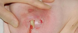
Reasons for the appearance of an abscess on a postoperative suture
In traumatological, orthopedic, and surgical medical practices, it is generally accepted that any wound is considered infected. Inside damaged skin there is a certain amount of bacteria. Signs of suppuration may not appear. For infection of the postoperative area to develop, the presence of one or a combination of provoking factors must be present:
- large depth of defect;
- the presence of foreign bodies and blood in the wound;
- a sufficient number of bacteria (staphylococci, streptococci, E. coli, salmonella, shigella);
- the presence of necrotic tissue in the postoperative area.
The reasons for the development of suppuration are separately identified:
- The appearance of signs of an abscess is associated with non-compliance with the rules of asepsis and sterilization by medical workers. Poorly processed instruments and insufficiently washed surgeon's hands transfer a huge amount of bacteria into the wound. After removal of the pathological focus, some microorganisms remain. They begin to actively multiply, which leads to the appearance of typical symptoms of suppuration in the postoperative area. Often signs are observed after cesarean section or appendectomy.
- A pathological process in a wound can develop if the patient is hypersensitive to surgical materials. In some cases, a reaction of rejection of implants, prostheses, dressings and suture elements occurs. Signs of suppuration appear a couple of days after surgery.
- A high risk of developing an abscess in the postoperative area is observed in people with endocrine diseases. These include diabetes mellitus.
- Signs of wound suppuration appear against the background of a weakened immune system. The body cannot fight the pathogenic bacteria that surround a person. Microorganisms easily penetrate deep into the tissue defect and cause typical symptoms.
- The pathological process develops when harmful microorganisms are transferred from an infected lesion to healthy tissue. During an operation to remove appendicitis, paraproctitis, lung lobes, fallopian tubes, and hemorrhoids, some bacteria are introduced into the area of the tissue incision. In the postoperative wound, suppuration begins.
- Improper application of a ligature suture can cause signs of a purulent process.
The pathological condition requires identification of the etiological factor in order to prescribe adequate therapy.
Pathogenesis
The mechanism of development of eventration is determined by its etiology. In case of mechanical injuries, the integrity of the abdominal cavity, diaphragm is disrupted, and the insides come out into the resulting wound. In some patients, the walls of the organs are also damaged. The pathogenesis of postoperative eventration is associated with a discrepancy between the tensile strength of the wound and the loads acting on it. The created suture is weakened when the regenerative capacity of tissues decreases due to purulent, hyperergic, pathological non-inflammatory processes, insufficiency of protein, fibrin, collagen, and other plastic materials for wound healing.
The provoking factor is often an increase in abdominal pressure, which stretches the edges of the wound from the inside and facilitates their cutting through by suture material. After the wound diverges, the abdominal organs fall into it. The formation of congenital eventration is based on the underdevelopment of individual elements of the abdominal wall: muscles in gastroschisis, all layers in hernia of the umbilical cord. Regardless of the pathogenesis, upon contact with the environment, internal organs are contaminated with infectious agents, dehydration of the outer membranes occurs, and an inflammatory process develops.
Classification
Systematization of the forms of eventration takes into account the etiology of the disease, localization, and the degree of violation of the integrity of the abdominal wall. By origin, the defect can be congenital, traumatic, or postoperative. When the viscera exits into the environment, eventration is considered external; when ectopy enters the pleural cavity, it is considered internal. Taking into account the severity of prolapse, specialists in the field of general surgery and gastroenterology distinguish between the degrees of severity (stages of development) of the post-laparatomy form of the disease:
- Subcutaneous eventration (I degree). There is a divergence of the peritoneum, aponeurosis, and abdominal muscles, but the insides are held in place by a skin suture. Occurs 8-10 days after surgery.
- Partial eventration (II degree). On days 9-11 after the intervention, all layers of the abdominal wall separate. The bottom of the defect is formed by internal organs fixed to the edges of the wound and the peritoneum.
- Complete eventration (III degree). There are no signs of an adhesive process. After the sutures of the surgical wound diverge, it is freely filled with the greater omentum and loops of the small intestine.
- True eventration (IV degree). Evisceration occurs, the insides through the wound opening extend beyond the abdominal cavity. Frequent complications and high mortality are observed.
Symptoms of eventration
With open traumatic injuries, a defect in the abdominal wall is revealed, into which the internal organs extend. Possible internal eventration is indicated by sharp pain in the chest, difficulty breathing, severe shortness of breath, and cough. In patients with injuries, painful and hemorrhagic shock is expressed: confusion, pale skin, tachycardia, and a drop in blood pressure are noted. With congenital eventration, a round or oval defect is found in the peri-umbilical region, through which swollen, swollen, fused intestinal loops prolapse. The newborn may have other developmental anomalies.
Postlaparotomy eventration develops 8-10 days after surgery. When organ prolapse begins, patients complain of moderate dull pain in the suture area. There is asymmetry of the abdomen and local pain in the abdominal wall. As the disease progresses, febrile fever, increased heart rate, a sharp decrease in blood pressure, increased sweating, and chills occur. Complete eventration is characterized by the appearance of copious hemorrhagic or purulent wound discharge, a sharp deterioration in the patient’s condition, widespread unbearable abdominal pain, nausea, vomiting, flatulence, and stool retention. Internal organs are visible in the wound.
Complications
When postoperative sutures diverge or open injuries, secondary infection always occurs with the formation of inflammatory infiltrates, ulcers, peritonitis, and phlegmon of the abdominal cavity. Possible strangulation of prolapsed organs in the wound. In immunocompromised patients with eventration, sepsis may develop due to the generalization of the process. Severe endotoxicosis leads to microcirculation disturbances up to the occurrence of disseminated intravascular coagulation syndrome. In 20-30% of cases the disease is fatal. Long-term consequences of abdominal organ prolapse are the formation of large ventral hernias, trophic ulcers of the scar, adhesive disease, and labiform intestinal fistulas.
Is suppuration dangerous and its course?
Postoperative scar abscess requires early diagnosis of signs and timely initiation of conservative therapy. In a short period of time, with adequate treatment, pus is removed and the proliferation of pathogenic microorganisms is suppressed. Tissue regeneration in a cleaned wound occurs quickly.
The danger of an untreated abscess is the manifestation of signs of postoperative complications. Suppuration can develop into phlegmon or fistula. Both pathologies require surgical intervention. With advanced inflammation of the wound, there is a high probability of developing sepsis. The disease is characterized by the patient's severe condition and severe intoxication. A septic reaction develops when bacteria and their metabolic products enter the bloodstream. The pathological situation can be fatal.
To prevent the appearance of signs of complications, it is necessary to treat suppuration in the early stages of development.
Diagnostics
Due to the clarity of pathological manifestations, diagnosing all forms of eventration, except internal and subcutaneous, is usually not difficult. The diagnostic search is aimed at a comprehensive examination of the abdominal organs to assess the severity of pathological changes. The most informative ones are:
- Physical examination. With a closed form of eventration, a tumor-like formation is palpated in the projection of the suture, and subcutaneous emphysema is possible. In case of complete prolapse, purple-bluish intestinal loops are visible in the wound. Auscultation reveals the absence of bowel sounds.
- Ultrasound of the abdominal cavity. Using sonography, you can assess the condition of the wound walls, detect inflammatory infiltrates, swelling and pathological changes in intestinal loops, and free fluid in the peritoneal cavity. Ultrasound is performed dynamically to assess the effectiveness of treatment.
- Electroenterography. A study of the motor activity of the small and large intestines in cases of suspected eventration is carried out in all operated patients. Characteristic is the absence of peristalsis, indicating a persistent violation of the motor-evacuation function of the gastrointestinal tract.
- Laboratory research. In the general blood test, high leukocytosis (more than 20 G/l) is observed with a shift of the formula to the left, and an increase in ESR. During postoperative eventration, a biochemical blood test may reveal hypoproteinemia, increased levels of creatinine, ALT, AST, alkaline phosphatase, and electrolyte disturbances. Using an immunogram, the degree of decrease in the body's reactivity is assessed. To identify a secondary infection and determine the sensitivity of isolated microorganisms to antibiotics, a bacteriological culture of the discharge from the wound is performed.
- Radiography. To visualize thoracic ectopia of the intestine in case of internal injuries, radiography of the chest organs is prescribed.
Differential diagnosis of eventration is carried out with postoperative peritonitis, wound suppuration, intestinal obstruction, recurrent abdominal hernia, inflammatory infiltrate of the postoperative wound. In addition to observation by a surgeon, the patient is recommended to consult a gastroenterologist, traumatologist, infectious disease specialist, and hematologist.
Post-Covid syndrome and chronic Covid
Currently, it is not possible to distinguish between true post-Covid syndrome and a condition where the virus continues to live in the body (the so-called long-Covid or chronic Covid). The clinical picture is so diverse that it is impossible to identify differential signs of post- or longcovid, and the main diagnostic method - PCR test - allows you to “catch” the virus only at the entrance gates of infection (nasal and oral mucosa). It is not yet possible to prove the presence of the virus elsewhere in the body in mass studies.
Symptoms
Post-Covid syndrome is manifested by a wide range of disorders affecting the cardiovascular, nervous and respiratory systems, and the musculoskeletal system. These are unexplained fatigue, increased anxiety, joint pain, chest pain, headaches, decreased memory, attention and thinking, up to dementia, depression, ringing in the ears, blurred vision, periodic fever, sleep disorders, tachycardia (palpitations), etc.
Development mechanism
Post-Covid syndrome is a complex supra-nosological form, the contribution of which is made by:
- immunological disorders as a trace of past infection,
- a newly emerging autoimmune disease, the impetus for which was a coronavirus infection,
- exacerbation of existing chronic diseases,
- dysfunction of organs and systems that arose during an acute condition and did not return to normal,
- anxiety-depressive and other psychiatric syndromes.
Causes
One of the causes of post-Covid syndrome is autoimmune disorders. After any infectious disease, the body has an increased level of autoantibodies, which are produced against the debris of its own dead cells. Over time, the immune system “calms down” and autoantibodies disappear during normal recovery. If this does not happen, autoimmune reactions occur. The situation is aggravated by endothelial damage (the inner lining of blood vessels) characteristic of Covid, long-term inflammation of the nervous tissue (neuroinflammation), and vascular reactions as a consequence of the bradykinin storm.
How to identify
Diagnosis of post-Covid syndrome is based on clinical signs and a history of an acute episode of COVID-19. From laboratory studies, data from a general blood test, markers of inflammation (C-reactive protein, fibrinogen, ferritin), indicators of blood coagulation disorders (D-dimer), indicators of liver, kidney, thyroid function, and electrolytes are used. The choice of instrumental studies depends on the detected disorders. To assess the condition of the heart, it is advisable to do an electrocardiogram, echocardiogram, and Holter monitoring. To assess the condition of the lungs - an x-ray (but not a computed tomogram) and spirometry. In any case, ultrasound examination of the veins of the lower extremities will be useful.
Treatment
Therapy for post-Covid syndrome is strictly individual and depends on the clinical picture. It includes a variety of non-drug rehabilitation and therapy aimed at eliminating residual inflammation, normalizing immunity and protecting affected organs. Experience shows that over time, post-Covid symptoms weaken and may even go away on their own, but with the active participation of the patient under the guidance of a doctor, the post-Covid rehabilitation process will be much more effective and faster.
Among the general strengthening and fast-acting medicinal methods of treatment, neurometabolic therapy and treatment with the drug LAENNEK can be distinguished.
Treatment of post-Covid syndrome at the ROSA Clinic
At the ROSA Clinic, treatment of post-Covid disorders is carried out on an outpatient basis. In severe cases (development of severe weakness, generalized anxiety, insomnia, depression, etc.), hospitalization may be required. For such cases, we have our own comfortable 24-hour hospital, where we carry out examinations and intensive treatment under the supervision of staff.
For all questions related to the identification and treatment of post-Covid syndrome, please contact us. We will try our best to help you.
Treatment of eventration
Medical tactics depend on the etiology and severity of the prolapse. In case of traumatic pathology, suturing of the wound with a thorough revision of the abdominal cavity, antibacterial and antiseptic treatment, resection of the prolapsed omentum and damaged areas of the intestine is recommended. Victims with internal eventration urgently undergo thoracolaparotomy with reduction or resection of the affected abdominal organs and suturing of the diaphragmatic wound. Surgical correction of congenital forms of the disease is carried out within 3-4 hours after the birth of the child.
In case of incomplete postlaparotomy eventration, conservative therapy is acceptable: the patient is placed on bed rest, intestinal function is regulated, antibiotics and infusion therapy are prescribed. In case of I degree of prolapse, a tight bandage is applied; in case of II degree, the wound is additionally sanitized. Reconstructive plastic surgery with subcutaneous eventration is performed after 2-3 months. Secondary sutures to eliminate tissue defects in case of partial prolapse of organs are applied on days 6-8. In case of complete and true eventration, the intervention (elimination of eventration) is performed urgently, taking into account the presence or absence of purulent-inflammatory processes in the postoperative wound and abdominal cavity:
- During aseptic eventration. Irrigation of prolapsed organs with antibacterial drugs with layer-by-layer blind suturing of the abdominal wall through all layers with special surgical sutures is indicated. To reduce tension and prevent teething, unloading fixation is used using buttons, gauze, or rubber tubes.
- Upon eventration into a purulent wound. It is recommended to clean the wound from pus, wash it with an antiseptic solution, and plastic surgery using unloading rubber tubes. In the absence of intestinal paresis and peritonitis, ointment tamponade of the wound, application of an aseptic dressing and plaster splint, and suturing of the defect after epithelialization are possible.
- In case of recurrent eventration. Allograft placement is most effective. Taking into account the immunological activity of materials, synthetic explants and the use of dura mater are preferred. A similar approach is used when tensioning the edges of the wound or a significant postoperative defect in the abdominal wall.
In the postoperative period, wound care is carried out; antibiotics, immunostimulants, and vitamin therapy are prescribed to improve regenerative processes. Prevention of intestinal obstruction and peritonitis, detoxification and anti-shock therapy with the introduction of colloid and crystalloid solutions to correct metabolic disorders are carried out.
Complications of wounds. Basic principles of treatment
Wounds can be accompanied by a variety of complications, both immediately after the wounds are inflicted and in the long term. Wound complications include:
· The development of traumatic or hemorrhagic shock is the earliest and most serious complication. In the absence of immediate help, it causes an unfavorable outcome.
· Seromas are accumulations of wound exudate in wound cavities, dangerous due to the possibility of suppuration. When seroma develops, it is necessary to ensure the evacuation of fluid from the wound.
· Wound hematomas are formed due to incomplete stopping of bleeding. Hematomas are potential foci of infection; in addition, by squeezing surrounding tissues, they lead to ischemia. They must be removed by puncture or during wound revision.
· Necrosis of surrounding tissues - develops when the blood supply to the corresponding area is disrupted due to tissue trauma during surgery or improper suturing. Wet skin necrosis must be removed due to the risk of deep accumulations of pus. Superficial dry necroses of the skin are not removed, as they perform a protective function.
· Wound infection - its development is facilitated by a high level of contamination and high virulence of the microflora that has entered the wound, the presence of foreign bodies in the wound, necrosis, accumulations of fluid or blood, damage due to injury to bones, nerves, blood vessels, chronic disruption of local blood supply, as well as late surgical treatment and general factors influencing the course of the wound process. Experimental and clinical studies have established that in most cases, for the development of an infectious process in a wound, it is necessary for its contamination to exceed a critical level of 105-106 microorganisms per 1 gram of tissue. Among the general factors contributing to the development of wound infection, significant blood loss, the development of traumatic shock, previous fasting, vitamin deficiencies, overwork, the presence of diabetes mellitus and some other chronic diseases play an important role.
The development of pyogenic infection is caused by staphylococcus, Pseudomonas aeruginosa, Escherichia coli and other pyogenic bacteria, anaerobic infection - by clostridia and non-clostridial anaerobic microflora, erysipelas - by streptococci. When a wound infection generalizes, sepsis develops. Most often, the development of pyogenic wound infection occurs on days 3-5 after injury, less often at a later date - on days 13-15. Anaerobic infection can develop very quickly; in fulminant forms, it is diagnosed a few hours after injury.
If it gets into the wound with soil, dust, foreign bodies, Cl. Tetani may develop tetanus. In the absence of specific prevention, the probability of contracting tetanus in the presence of contaminated wounds reaches 0.8%. The rabies virus can enter the body through bite wounds.
· Dehiscence of wound edges - occurs in the presence of local or general factors that impede healing, as well as when sutures are removed too early. During laparotomy, the divergence of the edges of the wound can be complete - with eventration, that is, with internal organs coming out, incomplete - with preservation of the integrity of the peritoneum, and hidden, when the integrity of the skin is preserved. Dehiscence of the wound edges is eliminated surgically.
· Complications of scars are the formation of hypertrophied scars and keloids. Hypertrophied scars develop when there is a tendency to excessive formation of scar tissue and most often when the wound is located perpendicular to the Langer line. Keloids, unlike hypertrophied scars, have a special structure and spread beyond the boundaries of the wound. Complications of scars lead not only to cosmetic, but also to functional defects, such as impaired walking or upper limb function due to limited range of motion in the joints. Surgical correction is indicated for hypertrophied scars with impaired function, but for keloids it often leads to a deterioration in the treatment outcome.
· Long-term chronic wounds can be complicated by the development of malignancy. The diagnosis is confirmed by a biopsy of wound tissue. Surgical treatment requires radical excision within healthy tissue.
Basic principles of wound care
Treatment for injuries usually takes place in two stages - the first aid stage and the qualified assistance stage.
¨ First aid stage
When providing first aid at the site of injury, two main tasks are solved: stopping bleeding and preventing further microbial contamination. First aid includes the use of available methods to temporarily stop bleeding, pain relief, application of a protective bandage and transport immobilization. At this stage, you should not wash the wound or remove foreign bodies from it.
¨ Qualified assistance stage
At the stage of hospital care, the following tasks are solved:
· prevention and treatment of wound complications;
· acceleration of the healing process;
· restoration of functions of damaged organs and tissues.
Basic principles of wound treatment:
· strict adherence to asepsis at all stages of treatment;
· mandatory surgical treatment;
· active drainage;
· the earliest possible closure of wounds with primary or secondary sutures or using autodermoplasty;
· targeted antibacterial and immunotherapy, correction of systemic disorders.
To select adequate wound treatment tactics, a thorough assessment of its condition is necessary, and the following is assessed:
· Localization, size, depth of the wound, damage to underlying structures such as fascia, muscles, tendons, bones.
· The condition of the edges, walls and bottom of the wound, as well as surrounding tissues, the presence and characteristics of necrotic tissue.
· The quantity and quality of exudate - serous, hemorrhagic, purulent.
· Level of microbial contamination. The critical level is the value of 105 - 106 microbial bodies per 1 g of tissue, at which the development of a wound infection is predicted.
· Time elapsed since injury.
¨ Treatment of contaminated wounds
The risk of developing wound complications in the presence of contaminated wounds is much higher than in aseptic wounds. Treatment of contaminated wounds consists of the following steps:
· In case of possible contact of the wound with the ground (all injuries with a violation of the integrity of the body, frostbite, burns, gangrene and tissue necrosis, out-of-hospital births and abortions, animal bites), measures are necessary to prevent a specific infection - tetanus, and in case of animal bites - rabies.
In order to prevent tetanus, vaccinated patients are administered 0.5 ml of adsorbed tetanus toxoid, unvaccinated patients - 1 ml of toxoid and 3000 IU of tetanus toxoid serum. Due to the danger of developing anaphylactic reactions to protein, the administration of anti-tetanus serum is carried out according to Bezredko: first, 0.1 ml of diluted serum is injected intradermally, if the size of the papule is less than 10 mm, after 20 minutes 0.1 ml of undiluted serum is injected subcutaneously, and only if there is no reaction to subcutaneous administration After 30 minutes, the entire dose is administered subcutaneously.
In case of bites from animals (dogs, foxes, wolves, etc.) suspected of rabies, or their saliva getting on damaged tissue, primary surgical treatment of the wound cannot be performed. The wound is only washed and treated with an antiseptic. There are no stitches. A course of subcutaneous administration of rabies vaccine is required, which is performed in specialized rabies centers, and tetanus prophylaxis. In the presence of superficial injuries (abrasions, scratches) of any location except the head, neck, hands, toes and genitals caused by domestic animals, culture purified concentrated rabies vaccine (COCAV) is administered 1 ml immediately, as well as on 3, 7, 14, 30 and 90 days. But if, when observing the animal, it remains healthy for 10 days, then treatment is stopped after 3 injections.
If animal saliva gets on the mucous membranes, if bites are localized in the head, neck, hands, toes and genitals, as well as with deep and multiple bites and any bites of wild animals, in addition to the administration of COCAB, immediate administration of rabies immunoglobulin (RAI) is necessary. Heterologous AIH is prescribed at a dose of 40 IU per kg of body weight, homologous - at a dose of 20 IU per kg of body weight. Most of the dose should be infiltrated into the tissue surrounding the wound, the rest is administered intramuscularly. If observation of the animal is possible, and it remains healthy for 10 days, then the administration of COCAV is stopped after the 3rd injection.
· In all cases of contaminated wounds, except for minor superficial injuries and cases where there are cosmetic and functional contraindications, primary surgical treatment (PST) with wound dissection, revision of the wound canal, excision of the edges, walls and bottom of the wound is mandatory. The purpose of PSO is the complete removal of non-viable and contaminated tissue. The later PST is performed, the lower the likelihood of preventing infectious wound complications.
PSO is not performed when wounds are localized on the face, as it leads to an increase in cosmetic defect, and good blood supply to this area ensures a low risk of suppuration and active wound healing. With extensive wounds of the scalp, performing PSO in full can lead to the impossibility of matching the edges and closing the wound. Non-penetrating puncture wounds without damage to large vessels and bite wounds with suspected penetration of the rabies virus are also not subject to PSO. PSO can be completed with the application of primary sutures - with suturing tightly or, in the presence of risk factors for wound suppuration, - with drainage left in place.
Preferably, flow-wash drainage of sutured wounds followed by dialysis with effective antiseptics. Flow-flushing drainage is carried out by installing counter perforated drains, one of which is used to administer the drug, and the other is used for outflow. The administration of drugs can be jet and drip, fractional or continuous. In this case, outflow can be carried out in a passive or active way - using vacuum.
This method protects wounds from secondary contamination, promotes more complete removal of discharge, creates conditions for a controlled abacterial environment and favorable conditions for wound healing. When draining, several general principles must be followed. Drainage is installed in sloping areas of the wound cavity, where fluid accumulation is maximum. Removing the drainage tube through the counter-aperture is preferable than through the wound, since the drainage, being a foreign body, interferes with the normal healing of the wound and contributes to its suppuration.
If there is a high risk of developing wound suppuration, for example, in the presence of sudden changes in the surrounding tissues, the application of primary delayed sutures, including provisional ones, is indicated. Like the primary ones, these sutures are placed on the wound before the development of granulation tissue, usually 1-5 days after PSO when the inflammatory process subsides. The healing of such wounds proceeds according to the type of primary intention. Sutures are not applied only after treatment of gunshot wounds and if it is impossible to compare the edges of the wound without tension; in the latter cases, the earliest possible closure of the wound defect using reconstructive surgery is indicated.
· Antibiotic prophylaxis is carried out according to the same scheme as for “dirty” surgical interventions. A 5-7 day course of antibiotics is required.
· Antiseptic prophylaxis involves the use of effective antiseptics at all stages of the operation and when caring for the wound. When treating wounds, chlorhexidine, sodium hypochlorite, dioxidine, lavasept, hydrogen peroxide, potassium permanganate and other antiseptics can be used. Drugs such as furatsilin, rivanol, chloramine are currently not recommended for use in surgical departments, since hospital microflora are resistant to them almost everywhere.
· Wound management after PSO with suturing is similar to the management of surgical wounds. Regular changes of aseptic dressings and drainage care are performed. Treatment of open wounds after PSO is carried out, like the treatment of purulent wounds, in accordance with the phases of the wound process.
¨ Treatment of purulent wounds
Treatment of purulent wounds is complex - surgical and conservative.
· In all cases of infected wounds, when there are no special functional contraindications, secondary surgical debridement (SDT) is performed. It consists of opening a purulent focus and leaks, evacuation of pus, excision of non-viable tissue and mandatory provision of adequate drainage of the wound. If the wound is not sutured after VChO, secondary sutures may be applied in the future. In some cases, with radical excision of an abscess during VCO, primary sutures can be applied with mandatory drainage of the wound. Preferably flow-through drainage. If there are contraindications to carrying out VChO, they are limited to measures to ensure adequate evacuation of exudate.
· Further local treatment of purulent wounds depends on the phase of the wound process.
In the inflammation phase, the main goals of treatment are fighting infection, adequate drainage, accelerating the process of wound cleansing, and reducing systemic manifestations of the inflammatory reaction. The basis is treatment with bandages. For all wounds that heal by secondary intention, wet debridement is considered the standard treatment method. Dry debridement with the application of dry sterile wipes to the wound is used only for temporary covering of wounds and treatment of wounds that heal by primary intention.
Wet treatment uses bandages that create a moist environment in the wound. Osmotically active substances, antiseptics, and water-soluble ointments are used. Fat-soluble ointments are contraindicated as they interfere with the outflow of secretions. It is possible to use modern atraumatic dressings with high absorption capacity that maintain a certain level of moisture and help remove exudate from the wound and firmly retain it in the dressing. Modern combination preparations for local treatment of wounds contain immobilized enzymes - gentacycol, lysosorb, dalcex-trypsin.
Dressings should be changed with adequate pain relief. The frequency of changing dressings depends on the condition of the wound. Typically, 1-2 changes of dressings per day are required, hydroactive dressings such as Hydrosorb can remain on the wound for several days, the need to immediately change the dressing arises in the following cases: the patient complains of pain, a fever has developed, the dressing is wet or dirty, or its fixation is impaired. At each dressing, the wound is cleaned of pus and sequestration, necrosis is excised and washed with antiseptics. Chlorhexidine, sodium hypochlorite, dioxidine, lavacept, hydrogen peroxide, and ozonated solutions can be used to wash the wound. To accelerate necrolysis, proteolytic enzymes, ultrasonic cavitation, vacuum wound treatment, and pulsating jet treatment are used. Physiotherapeutic procedures include ultraviolet irradiation of the wound, electro- and phonophoresis with antibacterial and analgesic substances.
In the regeneration phase, the main goals of treatment are to continue the fight against infection, protect granulation tissue and stimulate repair processes. There is no longer any need for drainage. Dressings applied during the regeneration phase should protect the wound from trauma and infection, not stick to the wound and regulate the humidity of the wound environment, preventing both drying and excess moisture. Dressings with fat-soluble antibacterial ointments, stimulating substances, and modern atraumatic dressings are used.
After complete cleansing of the wound, secondary sutures or adhesive tape are indicated; for large defects, autodermoplasty is indicated. Unlike primary sutures, secondary sutures are applied to granulating wounds after the elimination of the inflammatory process. The goal is to reduce the volume of the wound defect and the entry point for infection. After 21 days, secondary sutures are applied only after excision of the formed scar tissue. In cases where it is impossible to compare the edges to close the defect, autodermoplasty is performed as early as possible - immediately after the inflammatory process has subsided.
In the phase of scar reorganization, the main goal of treatment is to accelerate epithelization and protect the wound from trauma. Since drying causes a crust to form, which slows down epithelization, and epithelial cells die with excess moisture, dressings should still keep the wound in a moderately moist state and protect against injury. Bandages with indifferent and stimulating ointments are applied. Sometimes physiotherapy is used - ultraviolet irradiation, laser, pulsating magnetic field.
· General treatment of purulent wounds includes antibacterial therapy, detoxification, immunotherapy, and symptomatic treatment.
Antibacterial therapy is used in phases 1-2 of the wound process. The drug must be prescribed taking into account the sensitivity of the wound microflora. Systemic administration of antibiotics is indicated; topical administration is not currently recommended. The initial empirical choice of antibacterial therapy, pending sensitivity results, should be directed against typical pathogens, which are staphylococci, streptococci and gram-negative aerobic bacteria.
Amoxiclav, levofloxacin are used, as a reserve - cefuroxime, ciprofloxacin, ofloxacin, and for bites - doxycycline. Treatment of staphylococcal wound infections with pathogen resistance requires the use of vancomycin or linezolid. For erysipelas, penicillins, azithromycin, and lincosomide are indicated. If the infection is caused by Pseudomonas aeruginosa, the drugs of choice are carbenicillin, tazocin, timentin, as well as 3rd generation cephalosporins and fluoroquinolones. In addition to antibiotics, bacteriophages are used in the treatment of purulent wounds.
Detoxification is used in the presence of systemic manifestations of the inflammatory process. Infusions of saline solutions, detoxifying solutions, forced diuresis, and in severe cases, extracorporeal detoxification are used.
Immunocorrective therapy can be specific (vaccines, serums, toxoids) and nonspecific. Tetanus toxoid, antitetanus and antigangrenous serum, antitetanus and antistaphylococcal gamma globulin are often used. Among the means of nonspecific immunotherapy in patients with purulent wounds, only immunomodulators are used, and only in the presence of immune disorders and always in combination with an antimicrobial drug, since they aggravate the course of the infection. Synthetic immunomodulators, such as diocephon and polyoxidonium, are the most promising. Polyoxidonium has the properties not only to restore the impaired immune response, but also to absorb toxins, and is also an antioxidant and membrane stabilizer. Usually prescribed 6 mg 2 times a week, a full course of 5-10 injections.
Symptomatic therapy includes pain relief, correction of organ and system disorders, and correction of homeostasis disorders. For pain relief, non-narcotic analgesics are usually used, however, in the early postoperative period, as well as in case of extensive injuries, narcotic drugs can be used. When the temperature rises above 39° C or fever against the background of severe diseases of the cardiovascular and respiratory systems, the prescription of antipyretic drugs is required.
¨ Prevention of infectious complications of surgical wounds
Surgical wounds are applied under conditions that minimize the risk of wound complications. In addition, before inflicting a wound, it is possible to prevent wound complications. Prevention of complications of surgical wounds includes:
· Preparation for surgery
Before a planned operation, a thorough examination of the patient is carried out, during which existing risk factors for wound complications are identified. When assessing the degree of risk, the patient’s age, nutritional status, immune status, concomitant diseases, homeostasis disorders, previous drug treatment, the condition of the tissue in the area of the proposed incision, and the type and duration of the upcoming surgical intervention are taken into account. The existing disorders are corrected and the patient is directly prepared for surgery, taking into account the requirements of asepsis.
During operations on the colon, as well as during extensive surgical interventions in extremely critically ill patients, selective intestinal decontamination is performed to prevent infectious complications. Selective intestinal decontamination reduces the risk of enterogenous infection resulting from the translocation of intestinal microorganisms. Typically a combination of an aminoglycoside or fluoroquinolone with polymyxin and amphotericin B or fluconazole is used.
With each day of hospital stay, the patient’s contamination with pathogens of hospital infections increases, so the stage of inpatient preoperative preparation should not be delayed unnecessarily.
· Careful adherence to surgical techniques
When performing surgery, careful handling of tissues, careful hemostasis, preservation of blood supply to tissues in the wound area, obliteration of the resulting “dead” space, comparison of the edges of the wound and their suturing without tension are necessary. The sutures should not be ischemic, but should ensure complete closure of the wound edges. Whenever possible, the suture material left in the wound should be absorbable and monofilament. In addition, the duration of the operation plays an important role. As it increases, the degree of wound contamination and tissue susceptibility to wound infection pathogens increases due to tissue drying, impaired blood supply, and reactive edema.
· Antibiotic prophylaxis
Antibiotic prophylaxis of infectious wound complications depends on the type of surgical procedure. In clean operations, it is indicated only in the presence of factors that adversely affect the course of the wound process, such as immunodeficiency states, diabetes mellitus, and taking immunosuppressants. For most clean and conditionally clean operations, as well as for contaminated interventions in the upper gastrointestinal tract, 1-2 generation cephalosporins, such as cefazolin or cefuroxime, can be used for antibiotic prophylaxis. For contaminated operations on the colon, biliary system and internal genital organs, the use of protected aminopenicillins or 1-2 generation cephalosporins in combination with metronidazole is indicated.
During perioperative prophylaxis, average therapeutic doses of antibiotics are used. The first dose of the drug is administered intravenously 30-60 minutes before the skin incision, usually during induction of anesthesia. If the operation lasts more than 2-3 hours, repeated administration of the antibiotic is required to maintain its therapeutic concentration in the tissues throughout the entire surgical procedure. In most cases, the duration of antibiotic administration does not exceed 24 hours, however, the presence of additional risk factors necessitates the need to extend prophylaxis to 3 days. For “dirty” interventions, a full course of antibiotic therapy is indicated, which should begin in the preoperative period.
· Antiseptic prophylaxis
Antiseptic prophylaxis involves the use of effective antiseptics at all stages of the operation, including for treating the skin, washing cavities, and subcutaneous tissue. General requirements for the antiseptics used: wide spectrum of action, high bactericidal activity, toxicological safety. To treat the skin, iodophors, chlorhexidine, and surfactants are usually used; for washing cavities, chlorhexidine, sodium hypochlorite, and dioxidine are used.
· Drainage of surgical wounds
Drainage of surgical wounds is carried out according to certain indications. It is necessary when it is impossible to obliterate the “dead space” formed after surgery, when there is a large area of the wound surface of the subcutaneous fat, when using artificial materials for plastic surgery of the aponeurosis, and in some other cases that create the preconditions for the formation of seromas. Drainage is also mandatory for radical excision of ulcers with suturing of the postoperative wound. Aspiration or flow-wash drainage is preferable, while proper care of the drainage system in the postoperative period is mandatory.
· Proper wound management in the postoperative period
Local cold is prescribed immediately after the operation, adequate pain relief, regular changes of aseptic dressings and drainage care are performed, and, if indicated, dialysis and wound evacuation, physiotherapy and other measures.
¨ Control of wound treatment
The effectiveness of wound treatment is assessed by the dynamics of general and local signs of inflammation. They focus on the subsidence of fever, leukocytosis, pain in the wound area, and normalization of the patient’s general well-being. During dressings, the condition of the sutures, the presence and extent of hyperemia and edema in the wound circumference, necrosis of the wound edges, the type of wound discharge and granulations are visually assessed. To monitor the course of the wound process in the treatment of drained wounds, instrumental research methods can be used.
An endoscopic method of examining the wound is used with simultaneous biopsy of subcutaneous fat for bacteriological examination. In this case, during dressing, an endoscope optical tube with end optics with a diameter of 3-6 mm is inserted through the drainage of the postoperative wound, the presence of wound exudate, areas of necrosis, and fibrin is assessed, then a biopsy is taken. The degree of contamination of wound tissue is determined using express methods, for example, phase-contrast microscopy. After taking a biopsy, the wound channel is filled with physiological solution to assess the correct location of the drains and the direction of the fluid flow during its jet injection.
Favorable endoscopic signs of the course of the wound process and indications for stopping drainage are: the presence of bright pink granulations, the absence of pus, necrosis, a significant amount of fibrin, tissue contamination below critical. Sluggish granulations, the presence of a large amount of exudate and fibrin in the wound, as well as high bacterial contamination require continued dialysis of the wound with antiseptic solutions.
After removal of the drainage systems, an ultrasound scan is indicated to assess the condition of the wound channel and surrounding tissues. Favorable ultrasound signs of the course of the wound process are:
· narrowing of the wound channel the next day after removal of the drainage tubes, its visualization in the form of a heterogeneous echo-negative strip by 3-5 days, absence of dilation and disappearance of the channel by 6-7 days;
· uniform echogenicity of surrounding tissues, absence of additional formations in them.
Unfavorable ultrasound signs of the course of the wound process are dilatation of the drainage channel and increased echogenicity of the surrounding tissues with the appearance of additional formations in them. These symptoms indicate the development of purulent-inflammatory wound complications even before the appearance of their clinical signs.
When treating a purulent wound, daily monitoring of the course of the wound process is necessary. With continued exudation and sluggish granulation, treatment adjustment is required. In addition to visual assessment of the condition of the wound and assessment of the severity of general clinical and laboratory symptoms, various methods are used to monitor the dynamics of the microbial landscape, the level of contamination and regenerative processes in tissues: bacteriological, cytological, modern high-precision gas-liquid chromatography, tests using enzyme systems and others.
Prognosis and prevention
The outcome of eventration depends on its form, timeliness of diagnosis and the general condition of the patient. The prognosis is relatively unfavorable for grade 3-4 disease. In preventing the traumatic variant of the disease, a significant role is played by reducing the level of criminal activity and observing safety precautions when performing work in hazardous conditions. To prevent postoperative eventration, it is necessary to carry out competent premedication, follow the technique of suturing the wound, and prescribe adequate drug therapy to stimulate reparative processes and strengthen the immune system.
You can share your medical history, what helped you in the treatment of eventration.
How is such suppuration treated?
Treatment of postoperative damage begins after additional research methods have been carried out. The patient is prescribed general clinical tests. They help determine the presence of laboratory signs of wound inflammation.
Therapeutic elimination of purulent infiltrate depends on the stage of the disease. At the initial stage of suppuration formation, the surgeon cleans the wound of pathological contents, prescribes anti-inflammatory and antimicrobial drugs, and conducts detoxification therapy.
If signs of postoperative suppuration appear, which corresponds to the second stage, the wound is re-cleaned and drugs are used to heal it. The final period of the disease requires the administration of drugs to stimulate the formation of epithelium.
The main method of treating signs of postoperative suppuration is surgical treatment. The procedure is a low-traumatic type of surgical intervention. After excision of the edges of the wound, the area is carefully examined. Purulent streaks and foreign objects are detected in the diseased area. They can cause an abscess. The surgeon cuts through the leaks and removes necrotic tissue. The procedure ends with the placement of drainage at the location of the infected cavity. There are no stitches.
The pathological area with suppuration is lubricated with local antibiotics, antiseptics, and wound healing preparations. The pain is relieved with analgesics. Severe signs of intoxication postoperative syndrome are treated with antipyretic medications. When performing therapeutic measures, the situation ends with the formation of a small scar.
Need advice from an experienced doctor?
Get a doctor's consultation online. Ask your question right now.
Ask a free question
Traditional methods for eliminating signs of suppuration can be replaced with modern ones. These include vacuum therapy, laser and ozone therapy, cryodestruction, and the introduction of absorbents into the wound.
To prevent the appearance of signs of a bacteroid reaction in the form of suppuration, it is necessary to follow the rules of asepsis/antiseptics, regularly change dressings, apply stitches correctly, and buy effective drugs at the pharmacy. Ligature abscess of a postoperative scar is a reversible wound condition that has a favorable prognosis.