Dermatofibroma refers to benign skin tumors that form from the deep layers of connective tissue. These are small nodules with a diameter of up to 10 mm. They have a smooth surface, color varies from flesh to dark brown, grayish, brown. They are located mainly on the feet. The doctor determines whether it is worth removing dermatofibroma individually for each patient. The risk of degeneration into a malignant tumor is low.
- CAUSES AND SYMPTOMS
- TYPES OF DERMATOPHIBROMA OF THE SKIN
- REMOVAL OF DERMATOPHIBROMA
- POSSIBLE COMPLICATIONS
- TREATMENT AT THE PODOLOGY CLINIC
- COST OF SERVICES
Fibrosarcoma: what is it and what types of this pathology are there?
Fibrosarcoma belongs to malignant neoplasms belonging to the class of sarcomas and developing from immature fibrous connective tissue. Fibrosarcoma is a painless, dense, small-lumpy, round-shaped node, clearly separated from nearby structures. The skin over the tumor remains unchanged. As a rule, the disease harms tissues located in depth (muscles, tendons, fascia). If there have been previous traumatic injuries or exposure to ionizing radiation, pathology can develop in the layers of fatty tissue under the skin.
Among the age group under 5 years, fibrosarcoma forms approximately 50% of the total number of soft tissue cancers. As a rule, the proximal parts are affected (often the lower extremities), but the disease can also be based in other parts of the body. The cost of cancer treatment in Israel depends on the location of the process, the type of tumor, and the stage of the disease.
Based on clinical features and growth patterns, the following types of fibrosarcomas are distinguished:
- fibromyxoid - a tumor of this type with a low level of malignancy most often affects adults. The usual localizations of the pathology are the torso, shoulders, and hips. The formation is characterized by slow growth and rare cases of metastasis;
- infantile - it is diagnosed more often in children under 5 years of age and infants under one year old. Most often, a painless and rapidly growing tumor node is located in the area of the lower extremities, neck, head, and torso. If this formation is identified, then the prognosis is quite optimistic;
- myofibrosarcoma is a rare type of tumor, more often noted in children than in adults. The most characteristic areas of its localization are the head and neck.
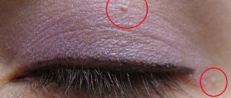
Types of cutaneous dermatofibroma
Depending on the cellular composition of dermatofibroma, there are several types:
- cellular – formed by loose connective tissue, as well as collagen and fibroblasts;
- sclerosing - its peculiarity is that it contains a large number of small vessels;
- fibrous – consists of fibroblasts and collagen, due to which it is not prone to enlargement and growth;
- fibroxanthoma - formed by cells with several nuclei.
If a growth appears on the skin, it is important to consult a doctor as soon as possible. Under no circumstances should you damage or try to remove the tumor yourself, because only a specialist can determine its type and the likelihood of degeneration into a malignant tumor.
About the symptoms of fibrosarcoma
Clinical manifestations are related to the location of the tumor and the extent of the process. When placed in deep layers of soft tissue, fibrosarcoma can occur without symptoms for a long time. The formation is often accidentally discovered during diagnostic procedures for another disease. Many people go to the doctor when fibrosarcoma has reached large proportions, caused deformation of the affected areas, or caused contracture of a nearby joint. Fibrosarcoma of the proximal parts of the extremities can be detected earlier due to the presence of pain caused by compression of the nerves and involvement of the periosteum in the process.
The skin over the surface of the tumor is largely unchanged. With an rapidly growing large fibrosarcoma located on the surface, phenomena such as thinning of the skin, their bluish color, and an expanded network of veins under the skin at the site of the formation may be noted.
Palpation reveals a separate tumor-like neoplasm of an oval or round shape, with a dense consistency. A characteristic feature of all types of sarcomas, as noted by specialists in the treatment of fibrosarcoma in Israel, is a limited node, a “false capsule” or pseudocapsule, which consists of layers of fibrous tissue. As the disease progresses, the false outlines of the tumor become less pronounced.
The level of fibrosarcoma mobility determines the prevalence of the process. Minor local neoplasms may shift (most often in a transverse direction). In the case of germination of nearby structures, fibrosarcoma becomes immobile. When the formation is placed in the space between the muscles, the nodes are perfectly palpable during relaxation, but lose their contours and mobility during muscle tension. The initial stages of fibrosarcomas are painless. If nerves are compressed, tenderness may be noted during palpation. If the bones are affected, the pain becomes constant.
Late phases of fibrosarcomas are characterized by signs of general intoxication. A person’s weight decreases and their appetite deteriorates. In addition, the following manifestations of the disease were noted:
- heat;
- anemia;
- increasing weakness;
- instability of emotional state;
- depression or subdepression.
When metastasizing to distant parts of the body, there are signs of involvement of the corresponding parts of the body in the process. If bone metastasis occurs, persistent pain is characteristic, which cannot be eliminated with painkillers. With metastasis to the lungs, coughing, shortness of breath, and spitting up blood appear; and with liver cancer - jaundice, growth of this organ.
All about skin fibroids with photos
Fibroma: symptoms
The symptoms of cutaneous fibroma have been described above. Symptoms of fibroids localized in different areas are as follows:
- fibroid . A painless neoplasm, the only symptom of which is bleeding or prolonged menstruation;
- breast fibroma A spherical lump localized on the chest. No pain is observed on palpation, however, patients sometimes complain of a bursting sensation in the chest before menstruation.
Diagnosis of fibroma
fibroids are suspected .
Basic diagnostic methods for fibroma :
- histological examination of fibroma . As a rule, biomaterial for histology is taken during the process of removing the formation;
- Ultrasound. Makes it possible to determine the depth of tumor growth in the tissue and its consistency;
- dermatoscopy. Used for skin fibromas , it is considered the “gold standard” for diagnosing skin tumors. Using a dermatoscope, the doctor can carefully examine the tumor and, if necessary, take pictures of it for further study.
Fibroma treatment
The choice of treatment tactics depends on the clinical picture and location of the fibroma .
- Drug therapy. Possible for small tumors and if there are contraindications for removal.
- Surgical treatment of fibroma . Prescribed for uterine fibroids depends on the size of the tumor. For small fibroids, laparoscopy is used; for large fibroids, hysterectomy and complete removal of the uterus are used. fibroids
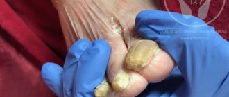
fibroma on the leg under the skin photo
Hardware removal of fibroids
Modern dermato-oncology has several effective non-invasive techniques for removing fibroids: laser removal, electrocoagulation, and radio wave surgery.
The peculiarity of the high-frequency radio wave surgery method is the possibility of non-contact excision with simultaneous tissue coagulation. The procedure is carried out using the Surgitron apparatus using local anesthetics. The radioknife not only removes fibroids, but also disinfects the wound tissue, after which a crust forms on it, preventing infection.
Another reason to choose this method is the possibility of completely removing the tumor and taking a full section of fibroma for histological examination. After removing the fibroma in this way, the wound heals quickly without leaving scars.
Treatment of fibroids is the field of activity of experienced dermato-oncologists. Under no circumstances should you try self-medication advice on yourself. Tying with a thread or, what sounds even worse, cutting the fibroma with scissors means injury and infection of the tumor, which can provoke malignant degeneration of the fibroma.
About diagnosing and treatment methods for fibrosarcoma abroad
Diagnosis of such tumors at the Meir Hospital in Israel involves pathomorphological studies of the tumor tissue area, histological examination of the tumor to determine the level of its malignancy. An accurate treatment plan is drawn up based on a set of data.
Diagnosis of fibrous tissue oncology is carried out using a number of methods, in particular:
- X-rays - using this technique, specialists determine the size of tumors, their area of location, and the level of tissue damage.
- CT - the procedure helps in conducting in-depth studies, and this allows for the development of metastasis to begin adequate treatment more quickly.
- MRI - diagnoses the localization of the process, the extent of tumor growth in blood vessels, soft tissues, nerve fibers, endings, as well as in the bone marrow.
- PET-CT allows you to evaluate the biological activity of formations. The procedure is carried out using expensive equipment, which is available only in leading foreign clinics.
- Biopsy is a microscopic examination of tissue to detect a tumor. A biopsy consists of a tissue incision or puncture.
The key approach used in the treatment of fibrosarcomas is its resection, along with the normal tissue surrounding the tumor. If the formation is located in the bones of the limbs, after its elimination, reconstructive surgery is used to restore the ability to move. If the tumor is located inside flat bones, the removed section of bone replaces the implant.
Absolute amputation of limbs in fibrosarcoma, as in the treatment of sarcoma in Israel, is practically not resorted to. It may be needed in situations where it is impossible to carry out a full-fledged reconstruction and when there is a high chance of infection due to malignancy of the soft tissues.
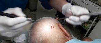
Removal of dermatofibroma
Treatment of the tumor involves its surgical excision. It is carried out by radio wave, laser or using a scalpel.
The procedure is performed under local anesthesia. Surgical removal of dermatofibroma is carried out on an outpatient basis without hospitalization of the patient.
After anesthesia, the tumor is removed with a scalpel, then stitches and a sterile bandage are applied to the wound. The rehabilitation period is 10-14 days. At this time, the wound should be carefully treated with antiseptics to eliminate the risk of infection. The sutures are removed 7-10 days after the removal procedure.
After excision, the tissue is sent to the laboratory for histological examination.
Possible complications
Possible complications after surgical removal of dermatofibroma:
- infection;
- development of bleeding;
- scars at the site of excision of the formation.
To avoid the development of any complications, trust the procedure to experienced specialists.
Treatment at the Podology Clinic
If you want to get rid of dermatofibroma, contact a specialized medical center. At the Podology Clinic in Moscow you will appreciate the following advantages:
- services are provided by experienced, qualified surgeons with extensive experience;
- use of high-tech equipment;
- attentive attitude to each patient;
- comfortable conditions in the Clinic.
The Podology Clinic uses safe and highly effective techniques to completely remove the tumor and avoid the formation of a scar. Trust your health to specialists!