Before removing any age spots, you need to consult a doctor and get a competent opinion about what kind of formation it is, whether it can be removed and how exactly. It is best to consult a dermatologist. Different types of nevi are suitable for different excision methods.
Benign nevi themselves are not dangerous as long as they maintain a clear shape, do not change color and remain flat. Some of them may begin to grow over time. Some skin lesions are not actually moles, but warts or seborrheic keratosis.
Under no circumstances should you remove a mole yourself at home, even if you are absolutely sure that it is benign.
A dermatologist can remove nevi that are dangerous from a health point of view only surgically so that a histological examination of the tissue can be carried out. For this there must be special indications:
- the appearance of itching in a mole;
- increase in size;
- change of form;
- color change (pallor, discoloration, streaks, redness);
- bleeding
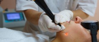
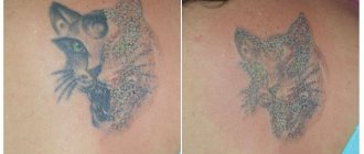
Laser mole removal
This innovative method is considered the most painless and fastest healing. Thanks to local anesthesia, the patient does not feel anything. The excision process takes place in stages: a directed laser beam removes the mole layer by layer.
However, after the procedure, a hole remains, which does not always completely disappear during healing. The beam evaporates the entire tissue of the mole, so it is not suitable for cases where an oncological study of the formation is necessary.
Pigment spots on the face require special attention. Since a healed scar is extremely important here, the specialist also needs to check whether keloid scars remain on the skin.
After removing a mole, the patient needs to protect the area affected by the laser from direct sunlight; this area on the face can be covered with a plaster. When the rehabilitation period is over, the protection can be removed.
Removing moles using the radio wave method
Removal of moles by this method is usually performed using a radio wave installation of the Surgitron type. The method is painless and effective; with not very large areas of treatment, it leaves almost no marks on the skin.
High-frequency radio wave vibrations are transmitted through tissue and cause minimal damage to the surrounding skin surface, less than a surgical scalpel. After radio wave treatment, a small wound remains on the body. After some time, it heals, leaving a small, gradually disappearing spot. The healing process takes from 3 days to 2 weeks, depending on the size of the treatment area.
The radiosurgical method is used to eliminate deep formations on the skin:
- warts,
- keratosis,
- fibroids,
- moles,
- facial capillaries or spider veins,
- papillomas,
- scars after acne.
Removal of moles using electrocoagulation
During the electrocoagulation procedure, tissue is removed using electric current for therapeutic purposes. This method allows you to visually control the depth of removal.
An electric current causes thermal damage to the mole and the area around it, which then forms a dry crust. During final healing, a noticeable mark may remain, so this method is not recommended for removing formations on the face.
But using this method, you can remove a mole in 1 session; moreover, the excised tissue is preserved and can be sent for histological examination. Electrocoagulation is carried out using direct and alternating current devices designed for this purpose. Different electrode shapes are used for different situations.
Contraindications
Excision of skin lesions using radio waves is a safe and fast method. But there may be limitations here. You cannot select it in the following cases:
tendency to turn into a cancerous tumor;- exacerbation of chronic diseases (including skin diseases);
- the presence of infection or herpes rashes;
- menstruation, pregnancy, feeding;
- high intraocular pressure (glaucoma);
- epilepsy;
- diabetes.
Mole removal with nitrogen
Cryodestruction is the instant freezing of a piece of tissue with liquid nitrogen. The substance, located at very low temperatures (from -100 to -180 ° C), causes freezing, destruction and rejection of tissue with a rapid recovery period.
The main advantage of cryodestruction is that dead tissue is not removed after destruction, but serves as a kind of bandage that protects the wound from infections. The wound heals painlessly. First, a crust forms, under which new, healthy tissue begins to grow.
However, the wound does not need to be treated with anything additional during the entire recovery period. This method is least suitable for the face, and the wound healing time here is much longer than with laser or radio wave removal.
Initially, after the scab falls off, the nitrogen-frozen area of skin will be lighter than the rest of the skin. But later everything will return to normal and traces of cryodestruction will become invisible.
However, the doctor is not in all cases able to control the depth of nitrogen exposure. Based on this, there is a possibility of frostbite of the tissues around the area, which can lead to the appearance of a scar. It is also possible that the mole will not be completely removed and the procedure will have to be repeated.
Treatment of cervical erosion using laser and radioknife
Cervical erosion is the area where the squamous epithelium has been replaced by cylindrical epithelium. During a gynecological examination, it looks like a bright red spot in the area of the uterine pharynx.
The affected area is easily injured and inflamed. A woman complains of bloody “spotting” after sexual intercourse or physical activity, profuse leucorrhoea with bloody streaks. Sometimes there are no complaints, and erosion is revealed only when examining the cervix on a mirror. Without treatment, the disease can develop into dysplasia and cancer.
Previously, electrocoagulation, which left scars on the neck, and cryotherapy (exposure to nitrogen), which did not make it possible to regulate the depth of cauterization and traumatic tissue, were used to remove erosion.
Now they have been replaced by new methods - radioknife and laser, which make it possible to treat erosion without damaging healthy tissue. The method does not leave scars, therefore it is suitable for treating erosion even in nulliparous women. Since the cervix in most women does not have nerve endings, anesthesia is not required to treat erosion. But, if the patient experiences pain, she will be injected into the cervix with 1% lidocaine solution with the addition of adrenaline.
A laser and electric knife allow conization of the cervix - removal of its cone-shaped fragment from the outer surface of the mucosa and cervical canal. The procedure is indicated for dysplasia (precancer) or large advanced erosion. Conization is more traumatic than conventional erosion removal and is therefore performed under local anesthesia with lidocaine.
On a gynecological chair, under the control of colposcopy, the patient’s tissue transformation zone is removed to a depth of 5-7 mm. Damaged vessels are “sealed.” Fragments of the obtained tissues are sent to the laboratory for analysis. Conization takes 10-15 minutes.
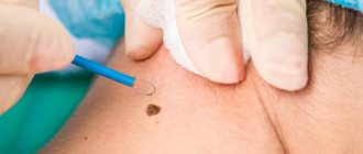
Surgical removal of a mole
Excision of the formation with a scalpel is considered the most painful. If the mole is on the face, this method is not advisable.
After surgery, the temperature may increase.
Surgical removal of moles requires not only the surgeon's qualifications, but also special care for the excision site after surgery. During the rehabilitation process, the patient will have to apply gauze bandages with an antibiotic or other healing ointment, changing them regularly (2 times a day). During the entire healing period, it is prohibited to swim or wet the removal site.
Consequences of mole removal
The consequences of removing moles on the body can be very different. Depending on the individual characteristics of the body, the chosen method and the equipment used, patients may quickly forget about the removed spot and not even recognize the remaining traces or suffer from complications that arise.
Removing skin lesions at home is absolutely unacceptable, even if the patient purchases special equipment for this. The possible consequences can only be minimized by a doctor in a specialized clinic.
You should immediately contact a specialist if after the procedure you notice the following symptoms:
- the appearance of suppuration;
- temperature increase;
- excessive secretion of lymph;
- strong pain;
- bleeding;
- negative changes in mental state.
Consequences of removing a mole on the face
There are many blood and lymphatic vessels on the face, and the skin on it is delicate and thin. Therefore, the removal of any formations on the face must be performed very carefully and professionally, using the most gentle methods possible.
The consequences of removing a pigment spot on the face may depend on:
- from the professionalism of the doctor;
- on the shape and size of the formation;
- on the selected removal method;
- on the individual health characteristics of the patient;
- from the patient’s thorough compliance with the recommendations of the rehabilitation period.
Proper care of the excision site largely determines how aesthetically pleasing the skin in this area will look.
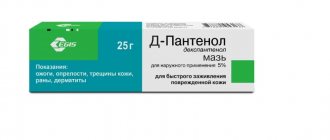
The damaged area of skin must be treated with antiseptics. The crust that has formed on the surface of the wound cannot be peeled off, otherwise the wound will take a long time to heal, and an unsightly scar will form in its place. Sooner or later the crust will fall off on its own.
During the healing period, it is forbidden to take a bath, go to the sauna, swim in ponds, apply cosmetics to damaged tissues, or stay in the sun for a long time.
After the procedure, the doctor will definitely tell you about the mandatory rules that you need to remember and follow to avoid complications.
Consequences of removing moles with liquid nitrogen
The procedure for removing nevi using liquid nitrogen does not provide any guarantees, although in general the results are positive. The fact is that when treating fabrics with a substance at such a low temperature, it is impossible to accurately calculate the depth of freezing penetration. This often leads to incomplete removal of the formation, so sometimes additional procedures may be required.
The healing process after cryodestruction is long, and after it there are burn marks - scars. Tissue restoration sometimes takes several months.
During the process of cryodestruction of moles, healthy nearby tissues may also accidentally be damaged. This type of damage usually looks like a burn and takes longer to heal than usual.
Consequences after laser mole removal
Laser removal of formations is currently considered the safest, does not require additional time for rehabilitation, and after the procedure there are no scar changes on the skin.
A laser removal session lasts several minutes, there is no cutting of tissue, and there is no risk of blood poisoning or bleeding.
Complications after laser mole removal are extremely rare. The only consequence is a thin, dry crust that disappears on its own after 7-10 days. After the skin is restored, almost no trace remains on it.
Consequences of mole removal by electrocoagulation
In most cases, electrocoagulation leaves a fairly noticeable scar, so the procedure is not recommended for the face and open parts of the body. However, there is no risk of bleeding.
- there is no point in arguing!
I believe that a professional phlebologist must be fluent in both methods and offer the patient the most optimal innovative treatment in each specific case. Leading European specialists successfully use both methods, since they both comply with the best government standards adopted in European countries.
Master class performed by Semenov A.Yu. on laser coagulation of veins for phlebologists from Ufa
In some city phlebological centers in Moscow, laser coagulation is still performed using end-mounted light guides on old hemoglobin (wavelength 980 nm) laser units. Most good European clinics have long used high-energy lasers (wavelength 1470 nm) and radial light guides in their work, which are not inferior in their characteristics to the radiofrequency procedure. In the best clinics in Europe and Russia, in addition to radial light guides, they have almost completely switched to a new generation of endovenous lasers with a wavelength of 1940 nm.
The latest laser generator with a wavelength of 1940 nm in our clinic
The principle of operation of the laser installation is to transmit laser radiation through fiberglass, which has a damaging effect on the vein wall, obliterating the lumen of the vessel, essentially gluing it together. Subsequently, the varicose vein undergoes lysis and, after a few months, completely resolves without a trace.
Radial light guide with wavelength 1940 nm
The operation at the Moscow City Phlebological Center takes place under constant modern ultrasound control. Therefore, complications with innovative procedures are practically nonsense.