Doctors Cost
Price list Doctors clinic
A mole, or nevus, is a benign skin formation that consists of cells filled with melanin pigment. This gives them their characteristic dark shade. Almost every person over 8–10 years old has moles. They appear in large numbers during puberty, which is explained by hormonal surges.
Pregnant women also often develop new moles, which is associated with endocrine changes. However, it is not at all necessary to resort to their removal during pregnancy if they do not bother you. It is necessary to show skin growths to a doctor; usually only observation is required.
Surgical removal of a mole is resorted to not only for cosmetic reasons. There are certain medical indications for removal.
When deletion is indicated
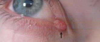
Epidermal nevi are benign formations and are harmless to health. However, any nevus carries a potential threat; there is a possibility of degeneration into a malignant tumor - melanoma. This is one of the most dangerous types of cancer. Therefore, it is important to monitor existing moles and consult a doctor if there is the slightest change.
You should visit a dermatologist as soon as possible in the following cases:
- the mole has increased in size, changed color, shape, outline;
- pain, itching, bleeding, burning, peeling, ulceration appeared, the surface became lumpy and uneven;
- the nevus was injured.
There is a high risk of degeneration for nevi located in open areas of the body. This is due to intense exposure to ultraviolet rays during the hot season. Also subject to removal are nevi that are constantly injured by elements of clothing - the handle of a bag, a collar, elastic bands of underwear, etc. Those formations that are located on the hairy areas of the body are also at risk - there is a possibility of tissue damage during shaving, waxing or sugaring.
General characteristics of formations
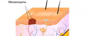
A mole is a pigment formation of one of the shades of the brown palette. The base contains melanin and the melanocyte cells that produce it. Melanins are high molecular weight pigments. They are characterized by a heterogeneous structure and complex chemical composition. Pigments are found not only in moles, but also in all skin, hair, iris, inner ear and even some parts of the brain.
Content:
- General characteristics of formations
- Indications/contraindications for the procedure
- What you need to know about melanoma?
- How to properly prepare for mole removal
- Technique of the operation
- Possible complications and side effects
- Features of rehabilitation
It is important not to confuse nevi and birthmarks. Moles do not form in the prenatal period, but as a person grows older and are closely related to his lifestyle. The cause of the formation of pigment spots can be a hereditary factor, exposure to ultraviolet radiation, or hormonal changes.
Each mole is a separate formation with a unique life cycle. At first it is a flat spot, which can increase and rise slightly above the skin as it grows and develops. The size and location of the formation depends on the level of melanocytes. If they are in the epidermis (top layer of skin), the mole will be flat. The deeper the melanocyte is immersed in the dermis, the higher the formation is located on the surface of the human body.
Convex nevi, which rise slightly above the skin, should not frighten their owners. This condition has no negative consequences and is considered a variant of the norm. The main thing is that the mole is miniature, round and uniform (in terms of color and structure) throughout its life cycle.
Can a mole disappear on its own and how safe is it? Yes, the formation can disappear without the influence of mechanical factors. This happens gradually and can alert a person. First, a white outline is formed around the formation, which resembles an orbit. This contour begins to increase and gradually covers the surface of the entire formation. The nevus disappears, and a small white spot remains in its place. Most often this occurs after severe sunburn. The disappearance of moles can also be a harbinger of the rare disease vitiligo.
If you notice changes in a mole (increase/decrease, uneven edges, change in shade or structure), be sure to consult a specialist.
Selecting a removal method
So, an oncologist or dermato-oncologist recommended removing the mole, or you decided to get rid of the formation. Consult with a specialist which removal method is best for your situation.
In many cases, histological examination of a removed mole is a mandatory procedure. It is important to evaluate the structure of the nevus tissue and make sure that the formation is truly benign and that nothing threatens your health. In this case, traditional mole removal with a scalpel is one of the most suitable methods. Some modern instruments do not leave a trace of tissue, so it will not be possible to study them in the laboratory.
If a large mole is located on the face or open areas of the body, discuss with a specialist the prospects of using a laser or radio wave removal method. These methods have fewer cosmetic consequences - they do not leave noticeable scars, so it is often advisable to resort to them. Remember that health is more important - if the doctor insists on conventional surgical removal, you should listen, because the prevention and early diagnosis of cancer depends on this.
Indications for removal of moles and papillomas
- Suspicion of a malignant disease
During the examination, the doctor can identify pathological structures in the formation, and therefore urgent surgical removal is necessary.
- Education is subject to frequent trauma
The inconvenient location of the mole, which in turn is often subject to friction and trauma, causes discomfort and bleeding of the formation in the patient.
In this case, there is a need for removal.
- Aesthetics
Some formations carry a cosmetic defect for the patient, and therefore require an removal procedure. After examination, the doctor selects an individual tactic for removing the formation, with maximum cosmetic effect.
Preparing for the intervention
No preparation is required for surgery: you can eat and drink water without restrictions, and you do not need to take medications. However, it is important for women to plan the day of surgery taking into account the menstrual cycle; it is better to do this after menstruation. The recommendation applies to any surgical intervention. Also, in cases where the nevus is located on open areas of the body, it is better to remove it from October to April. This will eliminate the need to hide the wound and protect it from ultraviolet rays.
If you are taking medications that affect blood clotting, tell your surgeon. You should not stop taking medications on your own—check with your doctor.
What you need to know about melanoma?
Every mole on the human body can degenerate into a malignant melanoma. What it is? Melanoma is one of the types of malignant tumors. It also contains melanocyte cells (producing melanin), but in melanoma the cells are more aggressive.
They constantly divide, displace healthy cells, enter the vascular bed and spread throughout the body. As soon as the first aggressive cell approaches the brain or, for example, the lungs, it settles inside the organ and begins to rapidly divide. This process is called metastasis.
Melanomas are extremely dangerous because they quickly metastasize and divide at an incredible rate. The main thing is to detect the pathology in time. Surgical removal of melanoma will prevent the process from spreading throughout the body and guarantee a complete cure. Failure to see a doctor in a timely manner or complete refusal of treatment can result in death.
Features of surgical removal
Surgical removal of nevi has several advantages:
- the mole is removed forever - there is no risk of reappearance;
- the operation requires only one visit to the surgeon;
- minimum contraindications;
- possibility of histological study of tissues.
However, there are also disadvantages - the need for reliable local anesthesia, the likelihood of a mark/scar forming at the site of the removed mole, and a relatively longer recovery period.
Removal steps:
- Pain relief with local anesthetics.
- Excision of nevus within normal tissue.
- Stitches (if the mole is small, this may not be necessary).
- Application of a special bandage.
Sutures are removed on average on the 5th–7th day. The overall healing period ranges from 7 days to 21 days depending on the health condition. The surgeon will give recommendations on how to care for the postoperative area at home. You can go home immediately after the intervention. As a rule, only the use of antiseptic solutions is required. You can also come to the clinic for dressing changes.
The doctor sends the material obtained as a result of excision to the laboratory for histological examination. A laboratory assistant examines it under a microscope for any atypical structure. You will receive the results within a few days, and if suspicious cells are detected, the specialist will definitely refer you to a dermato-oncologist.
As a measure to prevent skin cancer, your doctor may recommend giving up tanning beds and reducing the time you spend in direct sunlight. It is necessary to use sunscreen with a suitable protection factor, do not drink alcohol in excess and do not smoke. Patients with a large number of moles may be advised to visit a dermatologist for preventive purposes once a year.
Possible complications after surgical excision of nevi
After surgical removal of a mole, the following side effects may occur:
- Itching, pain, discomfort at the surgical site;
- Allergic reaction to a drug for local anesthesia;
- Prolonged bleeding;
- The appearance of keloid scars;
- Swelling in the area of intervention.
An increase in body temperature may indicate the presence of an inflammatory process, so in this case you should consult a doctor.
Surgical removal of moles is one of the effective methods of getting rid of nevi, which helps prevent the possible development of cancer pathologies. Our clinic employs qualified and experienced doctors who will help you efficiently and quickly get rid of not only nevi, but also other neoplasms.
Attention!
This article is posted for informational purposes only and under no circumstances constitutes scientific material or medical advice and should not serve as a substitute for an in-person consultation with a professional physician.
For diagnostics, diagnosis and treatment, contact qualified doctors! Number of reads: 4933 Date of publication: 08/06/2018
Dermatologists - search service and appointment with dermatologists in Moscow
Who is contraindicated for
Surgical removal of a nevus is contraindicated in the presence of the following diseases:
- exacerbation of chronic inflammatory diseases;
- infectious disease - viral, fungal, bacterial;
- fever of unknown origin, increased body temperature and other symptoms of intoxication.
Also, surgery may be postponed during pregnancy or breastfeeding - in each case, the doctor makes a decision taking into account the symptoms and condition of the formation.
Classification of moles:
1. Moles of epidermal origin (borderline, mixed, intradermal).
2. Moles of dermal origin (the most common representative is the blue nevus).
3. Combined moles.
4. High-risk moles (congenital and dysplastic nevi).
The most common type of moles are intradermal nevi - in appearance they are dome-shaped or papillomatous formations, from light brown to black in color, in some cases they can be reddish or whitish.
You can ask any questions you may have, sign up for a tumor removal procedure, or have a free consultation with a specialist by phone:
Sign up
Moles on the back - before surgery. Dermatoscopy:
Doctor: Grishko R.V.
Progress of the laser removal procedure
A laser removal tool is a beam of light that destroys tumor cells and evaporates them from the skin. Laser can remove a mole without bleeding. The procedure is performed under local anesthesia.
Removal lasts on average 10–15 minutes. Then a protective crust appears at the site of formation, which will disappear in a few days. There are no scars left at the site of laser treatment. After the procedure, it is necessary to follow medical recommendations: temporarily avoid sunbathing and contact with water, avoid injuring the skin area, treat, if necessary, with products prescribed by a doctor.
Our specialists
- Kiani Ali
Candidate of Medical Sciences, laser medicine specialist, dermatocosmetologist.
Sign up
- Stepanova Inna Igorevna
Candidate of Medical Sciences, maxillofacial surgeon, specialist in laser medicine.
Sign up
- Fedotova Marina Andreevna
Surgeon, dermatocosmetologist, laser medicine specialist
Sign up
- Popovkin Pavel Sergeevich
Surgeon, oncologist, laser medicine specialist.
Sign up
Advantages of laser “surgery”:
- Safety. There is no risk of blood poisoning - there will simply be no blood in the place where the “operation” is performed.
- No large scars. Small burns will not last long after you remove small nevi. This is especially important in the case of the face, where it is extremely important to avoid any marks.
- Versatility. Using a laser, you can destroy both large and tiny nevus. It doesn’t matter what size and shape the mole has - with a laser device, the cosmetologist will remove even a flat pigment spot.
- Low price and short term. Removing a nevus does not require special preparation. As a rule, in Moscow the “operation” is carried out immediately after the application - at a very affordable price.
Doctor's comment
Every person has pigment spots on their body. The risk of injury or change in the appearance of moles is an indication for their removal. Today there are different ways to remove such formations: destruction using laser, high temperatures, chemicals, as well as surgical excision of the nevus. To choose the method that is most suitable for you, you need to make an appointment with a doctor. Treatment tactics are prescribed taking into account the size of the formation and the presence of signs of malignancy, but for this you should undergo a painless examination. However, if you have moles, especially if they are located in the area of contact with parts of clothing, it is recommended to regularly undergo a dermatological examination; with the help of a simple and painless examination, you can promptly detect signs of malignancy of the nevus. In this case, timely treatment will avoid unwanted consequences. Thanks to the clinic’s equipment with modern equipment, we have the opportunity not only to carry out radical treatment, but also to achieve excellent cosmetic results.
Cryodestruction
Removing moles and warts using liquid nitrogen does not require pain relief. As a result of cold exposure, nerve fibers freeze and sensitivity is switched off. Liquid nitrogen is charged into a mini-cylinder with removable nozzles. The surgeon selects a tip 1 mm larger than the diameter of the tumor and applies it to the mole. The mole is frozen and rejected on its own. The disadvantage of this procedure is that it is difficult to calculate the depth of nitrogen penetration. If the exposure is too strong, healthy tissues are frozen, and if the exposure is weak, the nevus is partially removed. Damage to healthy tissue provokes cosmetic defects and increases the rehabilitation period.