Human skin
- a kind of indicator that reflects the state of all its organs and systems, protects against the penetration of infections, mechanical and chemical damage.
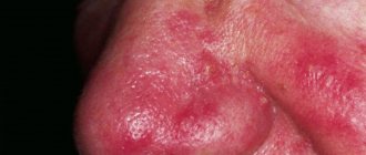
Skin diseases indicate
any disruption in the functioning of internal organs, an unhealthy lifestyle and bad habits, poor nutrition and failure to comply with personal hygiene rules by the patient. The variety of skin diseases is amazing. Many of them are difficult to treat and have a chronic, relapsing nature. Such ailments include eczema, psoriasis, neurodermatitis and dermatoses.
Plague of Galen
Medieval depiction of the Roman physician Claudius Galen
The Plague of Galen (an epidemic of the Antonine Plague) was brought to the Roman Empire by troops returning from campaigns in the Middle East. Beginning in 165 AD. BC, it raged throughout Asia Minor and much of Europe for 15 years, claiming the lives of two Roman emperors - Lucius Verus, who died in 169, and his co-ruler Marcus Aurelius Antoninus, who ruled alone until his death in 180 . Nine years later, the disease broke out again, as noted by the ancient Roman historian Dio Cassius, and began to claim up to 2 thousand lives per day in Rome alone.
Under Emperor Vera, the Roman army marched east when the Parthians attacked Armenia. The Roman defense of the eastern lands was difficult, since most of the soldiers fell, struck by disease. The consequences of the epidemic in the rest of the empire looked even worse. Many towns and villages, both in Italy and in the provinces, were completely depopulated. The epidemic spread as far north as the Rhine, affecting Germanic and Gallic peoples beyond the borders of the empire.
The great physician Galen left a description of the epidemic, among the symptoms are fever, diarrhea and sore throat, as well as a skin rash, sometimes dry and sometimes pustular, appearing on the ninth day of the disease. The scant information left by Galen does not allow us to accurately determine the nature of this disease, but many scientists diagnosed it as smallpox. According to various estimates, it is believed that the Plague of Galen killed about 5 million people.
Plague of Justinian
The Plague of Justinian, a pandemic of bubonic plague that swept through Asia Minor, Africa and Europe, struck Constantinople, the capital of the Eastern Roman (Byzantine) Empire, in the late spring and summer of 542 AD. e. After the plague began in Egypt, merchant ships and troops carried it throughout the Western world, allowing the disease to break out repeatedly over the next 50 years.
According to Procopius, the Greek historian and court insider who is our main source, the epidemic began near Ethiopia. Although ancient traditions claimed that diseases came from Africa, there may be some truth to Procopius's story. The plague bacillus appears to have originated in both central Africa and India, with the latter also being the likely home of a species of black rat that carried the plague. Ships sailing the Indian Ocean and Red Sea on their way to Egypt could have brought the rat and the bacillus together in a deadly combination.
People were terrified, many attributed the disease to the touch of a supernatural creature that appeared in a dream or in reality. To prevent demons from sneaking into their homes, people bolted their doors against all visitors, including family and friends. The mild fever that was the first symptom of the plague, however, did not cause alarm, and many people continued to lead their lives as usual until they developed bubonic tumors over the next few days.
Decades of war, famine and natural disasters in Mediterranean lands may have helped the plague reap a rich harvest; In Constantinople alone, about 300 thousand people died during the first year. Even Emperor Justinian suffered from it. The death and devastation caused by the plague prevented him from recapturing the western provinces and restoring the Roman Empire to its former size.
Diagnosis and treatment of skin diseases
A dermatologist diagnoses and treats skin diseases. In dermatology there are narrower specializations: dermatovenerologist, dermatocosmetologist and trichologist. If symptoms of skin diseases appear, you can first contact a therapist; he will tell you which specialist to contact in a particular case. To make an accurate diagnosis, a number of laboratory tests are often required, these may include: blood, urine and stool tests, serological culture, scraping, smear, biopsy.
In Volgograd, Volzhsky and Mikhailovka with symptoms of skin diseases, you can contact the DIALINE clinics. Experienced dermatologists using the latest diagnostic methods await you. To make an appointment, use your personal account on our website, where you can make an appointment at any of our clinics at a time convenient for you.
Black Death
The Black Death (black plague, bubonic plague) was a widespread epidemic that devastated Asia and Europe in the mid-14th century. This acute infectious disease, which in its earliest stages and in some places appears to have been predominantly of the pneumonic type (which helps to explain its rapid and frightful spread), is marked by swelling of the lymph nodes or buboes. It was also called the "Black Death" because of the black spots caused by subcutaneous hemorrhages that appeared on the skin of affected people near the time of death. Blood poisoning was rapidly fatal, with victims usually dying within two to four days. Bubonic plague was caused by the bacterium Bacillus pasteurella pestis
(
Yersinia pestis
), transmitted to humans by fleas from infected rats. Pneumonic plague, which occurs as a complication of the bubonic type and as an invasion of the lungs by bacteria, spread from person to person. In addition to black spots on the skin, the plague manifested itself in tumors in the groin or armpit and bleeding from the lungs; it was also characterized by very high fevers, delirium and prostration in its victims.
Originating in Central Asia, the disease killed an estimated 25 million Chinese, Indians and other Asians 15 years before it reached Constantinople in 1347. From there it quickly spread to Genoa, Naples, Venice, Marseille and other Mediterranean ports; ships carrying crusaders returning from the Middle East became a key factor in this regard. By the end of 1347, the plague had struck Dalmatia and the islands of Cyprus and Sicily. Thousands of people in southern France, Spain and Italy died from the black plague before it reached Paris in June 1348 and London a few months later. Raging in England and Ireland, the mysterious disease spread to the Netherlands, Germany, Norway, Sweden, Denmark and Russia, and by 1350, all of Europe (including Iceland and Greenland, according to some sources) was in the grip of the plague.
Pieter Bruegel the Elder "The Triumph of Death" (1562)
Historical estimates of mortality directly from the plague range from 1/4 to 3/4 of the populations of Europe and Asia; at least 25 million Europeans died between 1347 and 1351. They said that half of London (about 100 thousand people) died; 2/3 of Oxford University students died, 4/5 of the population of Marseille. The Pope in Avignon, where half the population had died, sanctified the Rhone River so that corpses could be thrown into it for Christian burial. More than a third of Italy's population died.
At the time, medical and lay authorities throughout Europe sought to provide rational explanations for a virulent plague that was clearly contagious. They published many treatises, trying to explain to the public the causes and symptoms of the disease and find methods of treatment.
The plague was attributed to anything and everything: tainted air and water, hot and humid south winds, the proximity of swamps, lack of cleansing sunlight, excrement and other impurities, putrid decomposition of dead bodies, excessive indulgence in food (especially fruits), God's wrath, punishment for sins and conjunction of stars and planets. Religious fanatics claimed that the terrible plague was brought about by human sins; they wandered from place to place, publicly flagellating themselves. In some places, everything was blamed on cripples, aristocrats and Jews, who were accused of poisoning public wells and were either driven away or tortured and burned. Panic reigned everywhere; men and women knew no other way to stop death except to run away from it.
The devastation caused by the Black Death helped to stop the Hundred Years' War for a time: both sides (the English and the French) signed a truce, which was extended three times (1347–1351); military operations resumed only in 1355.
For many historians, the Black Death marks the end of the Middle Ages and the beginning of the Modern Age. It gave impetus to the reorganization of society, landownership relations between the owner/farmer and the tenant/worker based on rent, which established a different balance between capital and labor.
Epidermolysis bullosa
Congenital epidermolysis bullosa belongs to a group of hereditary genetic skin diseases caused by mutations in a number of genes responsible for the synthesis of structural skin proteins. Currently, in Russia, on average, 3.64 cases per 1 million population are registered, and the forecast for the annual number of patients is 14–34 cases per 1.7 million newborns. The population frequency of this genodermatosis is 1:50000–1:300000.
The type of inheritance of epidermolysis bullosa can be either dominant or recessive. Epidermolysis is characterized by the formation of blisters and erosions on the skin and mucous membranes due to minor mechanical friction. This occurs due to disruption of intercellular connections in the epidermis and in the dermal-epidermal junction.
The clinical picture of this genetic skin disease includes blisters, erosions and milia on the skin, lesions of the nail plates (onychodystrophy), alopecia and aplasia of the skin, as well as palmoplantar keratoderma and macular pigmentation.
It is worth noting that BE nevi appear in place of large blisters, and erosions and milia resolve into atrophy, scars and fusion.
Treatment for VBE includes:
- atraumatic dressings;
- replacement therapy (transfusion of albumin, blood, plasma, immunoglobulins);
- correction of anemia (prescribe iron supplements);
- antibiotic therapy (if necessary);
- prevention and treatment of complications (esophageal stricture, osteoporosis, osteopenia, monitoring of neoplasms);
- correction of nutritional status (exclusion of rough foods, increase in protein products in the diet, use of specialized formulas for infants).
In addition to treatment, it is necessary to take into account the basic principles of caring for a child with epidermolysis bullosa. It is as follows:
- prevention of trauma to the skin - this is necessary to avoid the appearance of new blisters and erosions;
- careful care of erosions using modern atraumatic dressings made from special types of fabric;
- maintaining adaptive nutrition to improve epithelization of erosions;
- psycho-emotional support for both children and their parents.
An important point in care is the selection of dressing material. The main types of classical dressings include:
- hydrocolloid dressings;
- sponge dressings;
- calcium alginate dressings;
- dressings with alginate and collagen;
- cellulose-based dressings;
- dressings with activated carbon and silver impregnation.
Research into the treatment of epidermolysis bullosa is currently ongoing, and there are several promising methods that are still experimental. These include cell, gene and protein replacement therapies.
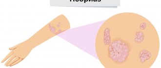
Ichthyoses
Ichthyoses are a group of hereditary genetic skin diseases characterized by a generalized disorder of keratinization of the skin, such as hyperkeratosis.
The estimated frequency of occurrence is 0.15–0.2%, that is, 1:300 thousand people. In 77.5% of cases, these are the first children in the family, born to young parents (22–25 years old). The incidence ratio by sex, female:m = 1.55:1.0.
In the international classification of diseases, ichthyosis is classified into:
- simple ichthyosis;
- X-linked ichthyosis;
- lamellar ichthyosis;
- congenital bullous ichthyoform erythroderma;
- Fetal ichthyosis, better known as “Harlequin fetus.”
Each type of ichthyosis has its own specific clinical picture, but there are also common manifestations, such as erythroderma, hyperkeratosis, peeling, joint contractures, nail dystrophy and unpleasant odor from the patient.
The basis of care for this genetic skin disease in the first year of a child’s life is the use of baths with oil additives, and after three years, baths with oil and baking soda are used alternately.
Also an important component of care is the mechanical removal of pronounced areas of keratosis. After 10–20 minutes of softening the keratoses in the bath, you should use silk rags or mitts to gently remove the elements. On average, full body care for severe ichthyosis takes 60–90 minutes per day.
Based on modern research, to relieve itching and reduce skin xerosis, it is recommended to use 10% paraffin and 15% glycerol, as well as creams containing 10% urea. As an alternative, local retinoids (0.05% retinoic ointment) are used, and in case of secondary infection, external antiseptics are used.
Severe forms of ichthyosis are referred for inpatient treatment, where systemic glucocorticosteroids, retinoids, intravenous immunoglobulins are prescribed and detoxification therapy is carried out.
Neurofibromatosis
Neurofibromatosis type I (Recklinghausen disease) is the most common of all autosomal dominant diseases, with an incidence of 1:2500–1:30,000. Males suffer from this genodermatosis more often than women.
The diagnosis is made based on the clinical picture and the presence of at least two of the following criteria:
- Cafe-au-lait spots on the skin. If there are 6 or more such spots up to 0.5 cm in size, in combination with other criteria, this genodermatosis can be assumed.
- Two or more neurofibromas of any type. Neurofibromas are soft nodules on the skin that vary in size and color (from flesh-colored to brown).
- Freckling in intertriginous areas. The presence of small hyperpigmented spots in areas such as the armpits, groin area, and under the mammary glands in women.
- Optic nerve glioma is a tumor of the glial cells of the optic nerve.
- Liche nodules are brown pigmented spots on the iris. The presence of two or more nodules is a diagnostic criterion for NF-I.
- Changes in bones (dysplasia of the sphenoid bone, thinning of long bones with pseudarthrosis, etc.).
- Presence of neurofibromatosis in first-degree relatives.
It is worth noting that the total number of spots does not correlate with the severity of NF, and Lisch nodules do not affect visual function.
Patients with this genetic skin disease are seen by a number of specialists - geneticist, ophthalmologist, dermatologist and others, but, unfortunately, treatment for neurofibromatosis still remains impossible.
Sometimes complete resection of elements is performed surgically or partial resection using a CO2 laser. In some cases, chemotherapy or radiation therapy is prescribed.
The patient’s management tactics are chosen by a council of doctors based on the form, symptoms and severity of neurofibromatosis.
Many genodermatoses look scary, and the very phrase “genetic skin disease” scares parents. But it is important to understand that with a timely diagnosis, timely therapy and proper care, the prognosis for many genodermatoses is favorable.
"Spaniard"
The 1917–1919 influenza pandemic killed more people than all the armies in the First World War. The total worldwide death toll was estimated at more than 21 million lives, and at least 200 (perhaps as many as 500) million people suffered from the “Spanish flu,” as the French and then others began to call this mysterious flu. Although the epidemic ranks with the Plague of Justinian and the Black Death as one of the most catastrophic disease outbreaks in history, it caused far less panic and unrest than other epidemics of the past. Perhaps people's feelings were too dulled by the First World War, battle losses and deaths.
When and where exactly the Spanish flu began remains unclear; however, it was so named because Spain (especially Madrid) was the first point of infection (about 8 million Spaniards fell ill in 1917–1919).
While presenting with the usual flu symptoms (headache, fever, chills, bone and muscle pain), the Spanish flu also caused complications such as severe pneumonia (with purple lips and ears and a pale face), purulent bronchitis, mastoid abscess and heart problems . Called by some “three-day fever,” it developed first calmly, with a cold, and then with a high fever, so that some diagnosed it as pneumonia.
Spanish Flu Hospital
After the signing of the armistice (November 11, 1918), which ended the war, the terrible disease seemed to subside, and after a year it no longer posed a threat. Among the most affected and devastated countries were China, India (where about 12.5 million people died), Persia (Iran), South Africa, Great Britain, France, Spain, Germany, Mexico, Canada, the United States (about 550 thousand Americans died ) and Australia.
In 2005, after nearly a decade of research, scientists from the Centers for Disease Control and Prevention (CDC) Mount Sinai School of Medicine and the Armed Services Institute concluded that the 1917–1919 pandemic was caused by an avian virus that spread to humans some fairly simple mutations. The scientists also focused on the H1N1 virus gene (which they recreated). Virologists fear the H1N1 strain could reappear, perhaps in a form as virulent as it was in 1918. Many pandemics originate in Asia, especially China, where large numbers of ducks, pigs and other virus-producing animals live in close proximity to humans.
Malaria epidemics
Malaria, along with tuberculosis and AIDS, is one of the world's leading killers. It is also one of the oldest known infectious diseases. However, because malaria does not leave marks on bones, it cannot be detected from remains.
The earliest information about malaria epidemics appears in the works of ancient Greek and Roman historians, who report that in the process of draining the swamps in the northern part of the Apennine Peninsula, many workers fell ill and began to die en masse. The disease was accompanied by fever, chills, and an increase in the size of the spleen and liver. Infectious diseases are transmitted from person to person after being bitten by female Anopheles
(“malaria mosquitoes”). Epidemics of malaria broke out in many parts of Europe during the Middle Ages and into modern times, with France being particularly hard hit. The first chronicled evidence of fever caused by malaria outside Europe was found in China. They date back to approximately 2700 BC. e., during the reign of the Xia dynasty.
How malaria came to the Americas and the Caribbean is unknown, but it may have arrived with Christopher Columbus in the late 15th century. At that time, the infection was widespread in Europe and Africa, and shortly after the arrival of Europeans in the New World, there were reports of malaria spreading throughout the Caribbean. Not all areas of the New World provided suitable habitat or climate for the mosquito vector, but by the 19th century the infection was widespread in the Mississippi Valley, California's Central Valley, and the coastal lowlands of northern South America.
At the beginning of the 21st century, the incidence rate was 350–500 million cases per year, of which 1.3–3 million resulted in death. According to the latest estimates of the World Health Organization (WHO), there are from 124 to 283 million cases of infection with malarial plasmodia per year and from 367 to 755 thousand deaths from the disease. The majority of cases of infection (almost 90%) occur in sub-Saharan Africa, and the vast majority of infections occur in children under the age of five.
As of 2022, the effectiveness of the current Plasmodium malaria vaccine is considered to be quite low (31–56%). Therefore, malaria epidemics continue to be a key issue on the WHO agenda.
Symptoms and their manifestation
Oral diseases can be diagnosed independently at home. It is only necessary to notice changes that have characteristic signs of a pathological process:
- The appearance of itching, burning or pain;
- Swelling;
- Formation of ulcers and pustules;
- Bleeding gums;
- Damage to tooth enamel;
- The appearance of an unpleasant odor;
- Weakness and sickness.
Cholera pandemic
Cholera is an acute intestinal anthroponotic infection transmitted through water, caused by bacteria of the species Vibrio comma
. The bacilli often do not survive in the gastric juices, but when this happens, they multiply rapidly in the digestive tract and cause radical dehydration from which the victim can die within hours. The horror of cholera lay in its symptoms, which included incessant diarrhea and vomiting, severe muscle cramps and prostration. Worst of all, the sudden loss of fluid in the body causes the facial features and soft tissues of the body to dry out, and the discoloration of the skin due to broken capillaries turns the shriveled victim black and blue, causing fear and horror.
Caricature of a cholera patient experimenting with medications. Around 1832
The history of cholera in the Western world is inseparable from the problems of 19th-century urbanization and public water supply. The growing population outpaced the ability of city authorities to develop adequate sewerage systems and provide clean water in sufficient quantities. Unclean water carries the cholera bacillus and causes most cases, but since the bacteria are passed into human feces, tiny particles of which can then be carried into food by unwashed hands or by flies and cockroaches, proximity to raw sewage and poor personal hygiene further contribute to the spread of the disease . Families were crowded into filthy tenements, people lived in coal-mining areas where unsanitary conditions were particularly bad, and laundresses and nurses who tended to the dirty bed linens of the sick were at particular risk.
The disease first appeared in the Sundarbans Forest of the Bay of Bengal in the Ganges Delta, where the bacterium Vibrio cholera
, probably mutated over thousands of years. This organism is found naturally in the environment in some coastal and brackish waters, where shellfish sometimes carry the infection.
However, it was not until the early 1800s, as the British opened new trade routes to India and sent troops across the subcontinent, that cholera began to spread beyond its home territory, first through India and then across the world in a series of huge pandemics. In August 1817, the British government received reports of a "malignant disorder" in the Sundarbans that was killing 20 to 30 people a day. Over the next few weeks, 10 thousand people died. From there, the disease spread throughout the country and then east and west to Nepal, Afghanistan, Iran, Iraq, Oman, Thailand, Burma, China and Japan.
This pandemic had barely subsided when the second began in 1826. Once again the source was the Ganges delta, and again the disease spread rapidly, returning to old habitats but also traveling further afield to the United States, Europe and Egypt. In Cairo and Alexandria alone, 33 thousand deaths were recorded per day.
By 1831, cholera was already in Moscow, devastating the great trading city of Astrakhan. Having reached St. Petersburg, she crossed the border between Europe and Asia, heading to Poland, Bulgaria, Latvia and Germany. The British watched its progress with alarm when, in the autumn of 1831, the disease crossed the North Sea from the Baltic coast of Germany and broke out at Sunderland Wharf. Over the next 70 years, pandemics spread rapidly across the globe, affecting countries on every continent and killing countless millions of people.
The third pandemic occurred in the 1850s and coincided with the Crimean War. In Russia alone, the number of victims exceeded 1 million people. This epidemic was the deadliest in the 19th century.
Today, endemic foci are present in Africa, South America, India and Southeast Asia.
Prevention of dental diseases
Some people are not even aware of what dental diseases with inflammatory processes and pathologies are provoked in other vital organs. For those who are not in the know, we inform you: these are gastritis, ulcers, tonsillitis, thrombosis, heart attack and stroke! And the pathogenic bacteria are to blame for everything, which, multiplying in the oral cavity, enter the throat, stomach, blood vessels and even the heart.
To prevent dental diseases, follow simple rules that will help significantly improve your oral health and overall well-being.
- Stop smoking.
A bad habit provokes vasoconstriction and deterioration of blood supply to the mucous membrane, which results in a lack of nutrients necessary for teeth and gums. - Eat a balanced diet.
The abundance of flour and sweet foods in the diet contributes to an excessive increase in microorganisms and inevitably leads to caries and its complications. Take care of strengthening your enamel by taking vitamin complexes. Then the disease when “teeth crumble”, which occurs due to a lack of calcium in the body, will not be scary for you. - Brush your teeth 2 times a day.
Regular hygiene is one of the most effective ways to keep your teeth safe and sound. In addition to the traditional use of brushes and pastes, you should not neglect dental floss. Flosses effectively remove food debris between the teeth, preventing the development of many diseases. - Get preventive examinations once every six months.
It is easier to cure any disease if it is detected at the beginning of its development. Some dental diseases in dentistry have symptoms that cannot be identified on their own. For example, caries at the junction of teeth is visible only on an x-ray.
A person is not able to influence factors that cause various types of dental diseases, such as poor ecology, heredity, and stress. However, with a responsible attitude towards hygiene and prevention, the risk of dental diseases can be significantly reduced or, at least, their treatment can be facilitated.
Typhoid
Angel of death poisoning the water. Image from the time of the typhoid epidemic, circa 1912
Typhoid fever and paratyphoid fever, also known as enteric fever, are believed to have a long history. A description of the disease can be found in the writings of the Greek physician Hippocrates, who lived in the 5th century BC. e.
An epidemic in Jamestown, Virginia, in the 17th century killed 6,500 of the 7,500 colonists. During the American Civil War (1861–1855), typhus is believed to have killed about 30,000 Confederate soldiers and 35,000 Union soldiers. In the Spanish-American War of 1898, it affected one-fifth of the U.S. Army, and six times as many people died from the disease as from wounds. In Russia, typhus raged especially actively during the civil war (1918–1922).
Typhoid fever and paratyphoid fever are similar diseases caused by different subspecies of the bacterium Salmonella enterica
, but paratyphoid fever tends to occur in a milder form, there is a lower mortality rate. Enteric fever, like cholera, is closely associated with poor sanitation, which prevails in slums, refugee camps and areas affected by natural disasters, where infrastructure such as sewage and water supplies has been destroyed.
Poster of the RSFSR, 1921
Typhoid fever remains a concern. According to experts, today from 11 to 20 million people a year fall ill with this disease and from 128 to 161 thousand die. Although improved living conditions and the use of antibiotics have sharply reduced morbidity and mortality in developed countries, typhoid fever remains a public health problem in parts of Africa, the Americas, Southeast Asia, and the Western Pacific. In these regions, anyone without access to clean drinking water and decent sanitation is at risk, with children being among the most vulnerable.
HIV AIDS
The HIV/AIDS pandemic is caused by the human immunodeficiency virus (HIV), which (in almost all cases) eventually destroys the immune system, leading to the fatal infections that characterize acquired immunodeficiency syndrome (AIDS). In the late 1970s, doctors first documented people with unexplained severe immunosuppression, but a clear syndrome was not identified until June 1981. Initially discovered in Africa, the Western Hemisphere, Australia and New Zealand, the epidemic reached almost every region of the globe by the mid-1990s; At the end of the decade and at the beginning of the 21st century, the rate of spread of HIV infection increased sharply in Eastern Europe, Central and Southeast Asia. Delays, omissions and inconsistencies in case reporting (not to mention the frequency of misdiagnosed symptoms) have made tracking the epidemic difficult. More than 32 million people have died from AIDS since the HIV/AIDS pandemic began in 1982, and in 2018 there were an estimated 37.9 million people living with HIV worldwide.
HIV is deadly because it attacks the cells that coordinate almost all phases of the immune response. Shortly after contracting the virus, a person may develop a short-term feverish illness similar to the flu or mononucleosis. After “recovery,” no other signs of infection may appear, sometimes for 10 or 15 years. However, during this asymptomatic period, the virus is active and can be transmitted to other people. By invading immune cells called CD4 lymphocytes, HIV causes them to become viral factories, sending more HIV to infect other cells in the blood as well as several tissues in the body. At least in the early stages, the immune system copes with the problem of HIV by creating immune cells daily to replace those destroyed by the virus. But HIV almost always wins. Even in the absence of symptoms, it constantly makes copies of itself, and is prone to errors in the process; With such a high mutation rate, HIV changes to become capable of resisting any weapon in the immune system's arsenal, including drugs. At some point, the continuous onslaught of HIV can lead to various non-fatal symptoms such as fever, fatigue, diarrhea, night sweats, swollen lymph nodes, recurrent yeast infections and forgetfulness.
The virus can be transmitted through direct contact of damaged or intact mucous membranes or damaged skin of a healthy person with the biological fluids of an infected person: blood, pre-seminal fluid (released throughout sexual intercourse), sperm, vaginal secretions, breast milk.
Transmission of the virus is more likely to occur through the use of contaminated needles and syringes (especially by injection drug users), as well as through blood transfusions (if medical personnel fail to comply with established blood donation screening procedures). Also, transmission of the virus can occur between mother and child during pregnancy and childbirth (infection through the mother’s blood).
At the moment, there is no vaccine against HIV, although news appears quite often that more and more new drugs are being successfully tested.
In January 2022, Chinese authorities confirmed the birth in Shenzhen of the world's first genetically modified people, who had the CCR5 gene responsible for interacting with HIV edited, and also began an investigation into biologist He Jiankui. It is assumed that genetically modified children will not be able to get HIV, but the experiment itself remains questionable.
Classification of dental diseases
Pathologies of teeth eruption and formation
Early detection of dental anomalies makes it easier to carry out subsequent bite correction. Irregularities in the sequence of eruption or asymmetry of opposing teeth should be a reason to contact an orthodontist. Diagnostic accuracy is ensured through x-ray examination.
Edentia
Characterized by partial or complete absence of teeth. The disease can be hereditary or develop throughout life. Sometimes the rudiments of teeth are completely absent from birth.
Supernumerary teeth (hyperdontia)
There are too many teeth in the mouth, and the extra ones can be located both in the dentition, disturbing the arrangement of the remaining teeth, and in atypical places - on the palate or on the front surface of the gums. The anomaly occurs in 5% of the population.
Impacted teeth
The teeth remain in the bone tissue of the jaw and do not erupt completely. In most cases, this is a disease of the molars - eights.
Dystopian teeth
The teeth are located in the wrong places in the jaw, at an angle, and only partially erupt. Pathology refers to diseases of wisdom teeth.
Macrodentia/Microdentia
An increase (or decrease) in the size of teeth compared to the standard norm.
Tooth enamel diseases
There are many chemical processes going on in the mouth that wear down the enamel. Under the influence of food containing dyes or acids, as well as cleaning products, the normal mineralization of teeth is disrupted. Pathologies of the endocrine system and work in hazardous industries are additional unfavorable factors.
Fluorosis
Damage caused by an excess of fluoride in consumed water or food. Fluorosis is commonly referred to as yellow tooth disease.
Enamel hypoplasia
A disease associated with impaired formation of the enamel structure. In most cases, it develops in the first nine months of a child's life.
Wedge-shaped defect
Formation of a “protrusion” at the neck of the tooth on the front side. A disease of the front teeth that can lead to chipping of the entire crown part.
Dental disease caries and its complications
Dental caries occurs in places where microbial plaque lingers and accumulates. The likelihood of developing the disease depends on the individual resistance of both dental tissues and the body as a whole. Resistance to caries is determined by the structure and composition of the enamel, the degree of its mineralization, as well as the characteristics of the dental system - the structure of the facial skeleton, jaws, bite and location of the teeth.
Caries
A pathology that develops in the hard tissues of the tooth, accompanied by demineralization of the enamel and the formation of a cavity. If left untreated, it provokes the development of inflammation in the pulp and periodontal tissues.
Pulpitis
Inflammation of the dental nerve. Characterized by acute pain radiating to the temples. May lead to tooth loss or exacerbation of periodontitis.
Periodontitis
The inflammatory process covers the apex of the tooth root. Without treatment, there is a risk of developing a cyst or abscess. The main manifestations are swelling of the gums, increased temperature.
Periostitis (flux)
Inflammation of the periosteum due to untreated periodontitis. Accompanied by swelling of the gums and severe pain.
Granuloma
Proliferation of inflammatory tissue in the area of the tooth root. Symptoms are redness and swelling of the gums, tooth pain. Ignoring the disease leads to the appearance of a cyst on the root of the tooth, fistula and diseases of the roots of the teeth.
Tooth cyst
A formation in the form of a capsule with pus at the apex of the tooth root, resulting from the penetration of infection into the root canals. The disease is asymptomatic, in some cases there is pain when chewing. The cyst is usually noticed on x-rays.
If you find signs of dental disease presented in the table, you must go to a dental clinic as soon as possible and consult a doctor. The specialist will examine the oral cavity, prescribe diagnostic procedures and draw up an optimal treatment plan for dental diseases to restore the health and beauty of your smile.
Ebola fever
The first officially recorded case of the Ebola virus disease is the story of the Flemish nun, Sister Beata. She died in a clinic in Kinshasa, in what is now the Democratic Republic of the Congo (DRC), in September 1976, suffering from terrible symptoms, including heavy bleeding. A few days later, the nun who brought Sister Beata to the hospital fell ill with the same symptoms. She was also admitted to the clinic, and she also died. She was followed by a young nurse caring for the nuns.
As Sister Beata lay dying, samples of her blood were sent to a research laboratory in Antwerp.
When the team examined the tissue samples under an electron microscope, they saw previously unknown worm-like structures that looked nothing like yellow fever but bore some resemblance to another deadly hemorrhagic disease called Marburg virus, which is indigenous to Africa. Marburg was only identified nine years ago in Germany, when pharmaceutical workers fell ill after working with monkeys imported from Uganda. Seven of the 25 people directly infected by the monkeys died, and six more became ill after contact with the victims. By then, the mysterious Yambuku disease outbreak had raged for three weeks, killing at least two hundred people. Three weeks after Sister Beata's death, the Centers for Disease Control and Prevention (CDC) announced the discovery of a deadly new virus, later named Ebola after the river near the Yambuku mission.
Since then, five strains of the Ebola virus have been identified. Four of them cause disease in humans: Ebola virus (Zaire ebolavirus), the Yambuku strain, the deadliest; Sudanese virus (Sudanese ebolavirus); Thai-Forest virus (Tai-Forest ebolavirus); Bundibug virus (Bundibug ebolavirus). Fifth, Reston virus ( Reston ebolavirus
), caused disease in monkeys and pigs, but not in humans.
Since 1976, the disease has regularly broken out in Africa, especially in the DRC and Uganda. Because it often occurs in isolated, sparsely populated areas, it is believed that the disease sometimes goes unreported.
From 1989 to 1994, four isolated cases of infection with the Reston strain of virus were reported in laboratories in developed countries, including the United States and Italy. However, all of these incidents involved laboratory monkeys.
Then, in March 2014, everything changed. Ebola struck West Africa, first in Guinea and then in Liberia and Sierra Leone. Over the next two years, the virus spread throughout the world, first affecting Mali, Nigeria and Senegal, and then from Africa to Italy, Spain, the UK and the US.
In the summer of 2014, panic gripped the world and Ebola dominated the news agenda for months and was compared to the great epidemics of the Middle Ages. From 2014 to 2016, 28,616 people worldwide contracted the disease and 11,310 of them died, with the vast majority of cases occurring in West Africa.
In 2016, the WHO declared the 2014 pandemic over and Ebola, at least for now, was no longer a threat. However, in the summer of 2022, Ebola resurfaced in a remote part of the DRC, infecting eight people, four of whom died. Despite the WHO statement, the question of when such a devastating epidemic as Ebola will end remains highly debatable.