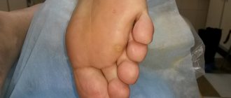
Plantar warts are benign epithelial tumors that are localized on the skin of the foot. They appear as hard skin growths that are off-white or yellowish in color. The location on the foot leads to frequent trauma to the formations when walking, and the appearance of severe pain.
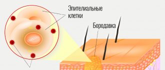
The cause of plantar warts is the human papillomavirus (HPV), which leads to proliferation of the epidermis and its local growth. The virus attacks the skin during direct contact, penetrating through abrasions, abrasions, and injured calluses. Favorable conditions for HPV infection are created in a humid and warm environment, when the skin is steamed and is more susceptible to the penetration of foreign microorganisms. Therefore, most often people become infected in public baths, swimming pools, and saunas.
In addition, the following factors increase the risk of infection:
- hyperhidrosis of the skin of the feet;
- dry skin of the feet;
- presence of skin lesions in the foot area;
- diseases that lead to impaired trophism of tissues of the lower extremities (diabetes, angiopathy);
- deformation of the feet (flat feet, deforming arthrosis). These conditions change the anatomy of the foot, which leads to chronic microtrauma of the skin and creates conditions for the penetration of the virus.
However, infection does not always mean the appearance of a wart on the skin. After all, many people come into contact with the virus, but only some of them have problems. The fact is that normally the activity of the virus is suppressed by the body’s defense mechanisms, in particular by the immune system. But if there is a malfunction in its operation or the presence of other factors, viral proliferation is triggered, due to which the cells of the dermis begin to actively divide, and a wart is formed.
The effectiveness of laser spine removal and the likelihood of recurrence
Benign formations caused by HPV strains bring discomfort and pain when walking. Laser removal will help cope with the problem. Benefits of the procedure:
- One session is enough to eliminate the growth.
- Ability to remove several warts at a time.
- Lack of blood due to cauterization of blood vessels.
- A chance to control the depth and area of laser exposure.
- Minimal impact on healthy skin around the spine.
- Low likelihood of complications after removal.
- Regeneration speed.
- Painless due to local anesthesia.
- Applicable to any age group.
- Does not leave scarring changes in the epidermis.
Laser removal of spines is considered a reliable method. The disadvantage is that there is no guarantee of relapses. If virus-infected cells persist, the disease can reappear. If the operation is performed correctly, the risk of the disease returning is minimal.
Symptoms of plantar warts
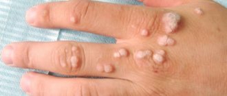
Plantar warts have the appearance of hard, rough papules (seals) with a violation of the papillary pattern. Their size ranges from a few millimeters to 1-3 cm. The formations can be level with the surface of the skin, or rise above it by 1-2 mm. As for the color, it may not be changed, but it may vary from light pink to brown.
At the initial stages, the surface of the wart is smooth, but as the epidermis grows, it becomes bumpy or rough. Education tends to promote deep inner growth. In this case, there may be a crater-shaped depression in its center. Also, black or dark brown dots are often observed on the surface of the plantar wart - these are thrombosed capillaries.
Typically, plantar warts are single formations. However, with high virus activity and due to constant pressure while walking, warts can spread through the mechanism of autoinoculation, infecting the surrounding skin. Thus, they are combined into mosaic clusters. As a rule, such warts are located on the foot in areas of greatest pressure.
Step-by-step procedure for removing formation
The algorithm is simple:
- The soles are examined, the area and depth of the spine are determined.
- Anesthetize the skin area and treat it with antiseptics - Chlorhexidine, Betadine.
- The disease is outlined with a defocused laser, and the formation is burned from the periphery to the center.
- They gradually move deeper until the root of the spine is eliminated.
- Clean the bottom of the wound, the edges, remove layers of keratinized epithelium around.
- They are repeated with an antiseptic and covered with an aseptic gauze bandage.
You can remove a spine with a laser in 15-20 minutes. A carbon dioxide device is often used. The radiation from it is not perceived by the human eye. For ease of targeting, a helium-neon mixture is applied to the desired area. The flow energy is absorbed by water inside and outside the cells. Rapid heating to 394 °C causes the liquid to evaporate, destroying the cells. The burn coagulates the vessels, which prevents the opening of capillary bleeding.
If the growths are located on both soles, they are operated on alternately.
The laser provides a bactericidal effect. Thanks to layer-by-layer removal of pathologically altered cells, it is possible to control the depth of spine removal. This avoids damage to healthy tissue and scarring. Old growths with deep roots pose a danger: bleeding may occur. Surface formations are easier to remove.
Is it painful to remove a spine with a laser?
Need advice from an experienced doctor?
Get a doctor's consultation online. Ask your question right now.
Ask a free question
The effect of the device on the skin is traumatic: the tissue is heated. The use of local anesthetics eliminates discomfort. Novocaine and lidocaine are used for pain relief. They are administered with syringes with a small diameter needle.
The anesthetic is used intradermally. They prick around the spine and inject to the depth of burning. After 5-10 minutes, sensitivity is checked. If there is no discomfort, removal begins. If pain occurs during the laser procedure, additional administration of the drug is allowed.
Types of calluses
In accordance with the international medical classification, all calluses (clavus) known to science are divided into 9 types:
- Hard – Clavus Durus.
- Soft – Cl. Mollis.
- With a thorn – Cl. Spina.
- Vascular – Cl. Vascularis.
- Neurovascular – Cl. Eurovascularis.
- Papillary – Cl. Papillaris.
- Neurofibrous – Cl. Neurofibrosus.
- Subungual – Cl. Subunguales.
- Millet – Cl. Miliaris.
Hard (dry) callus (CD)
Hard or, as it is also called, dry callus, is most often encountered in medical practice. This is a rounded area of dead skin rising above the healthy surface, under which there is a translucent glassy cone. A typical location is the back of the phalanges, middle and end joints. Due to the high hardness of the root, a dry callus on the toe causes pain when pressed from above. In most cases it does not cause problems when removed.
Soft (CM)
The soft callus between the toes (its inherent location) constantly softens due to increased humidity and always maintains a flat shape.
Callus with thorn (CS)
It looks like a clearly demarcated growth with a white edge and a transparent core in the form of a rod up to 4 mm long. In medical terminology it is called “core keratoma”, popularly – “chicken jelly”. Most often it forms on the little finger. Its elimination requires a certain amount of time and high professionalism of the podiatrist.
Vascular (CV)
A characteristic feature is vascularization (the formation of new blood vessels under the stratum corneum). Such formations occur in places of excessive stress. After removing the callus, black or red dots (tips of capillaries) remain on the surface.
Neurovascular (CEv)
The formation of a highly sensitive skin growth, penetrated by blood vessels and nerve endings, is a consequence of significant changes in the dermis. Often its apex is located directly under the layer of keratinized cells, and the “body” extends deep inside. Due to severe pain, removing calluses on the foot, in the area of the articular joints and on the tips of the fingers is associated with significant difficulties.
Papillary callus (CP)
At first glance, it is almost identical to solid. However, in this case, enlarged dermal papillae and clearer whitish contours are observed. Specialists who remove calluses on the feet note that the nuclei of papillary skin growths seem to “stick together” with the dermis.
Neurofibrotic (CNf)
A typical location is the sole (mainly the metatarsal part). Most often, it affects fairly large areas in areas of greatest stress. Removing rough skin with the dermal papillae that give it its characteristic roundness is much easier than eliminating core calluses.
Subungual (CSb)
It is located under the free edge of the nail plate, causes severe pain and often leads to hemorrhages in the nail bed area. To remove such a formation, preliminary excision of the part of the nail covering it is required.
Prosyannaya (CM)
It is a consequence of metabolic disorders and disruption of the keratinization process (cell death and deposition of keratin protein). The “favorite” places for localization of pinpoint keratinization are the low-rubbing inner surfaces of the feet.
Corns
Corns are compactions on the sole, presented in the form of large areas of rough skin that form in the area of greatest stress (usually on the ball of the foot and at the base of the toes). They have a yellowish or grayish color, are quite hard to the touch and are less sensitive than surrounding tissues. In some cases, they may have a rod (root) that has grown several millimeters deep into the body.
Removing calluses and corns
Skin calluses do not pose a threat to human health and life. However, they not only spoil the appearance, but can also cause serious inconvenience when walking. In such situations, the best solution is to contact a podiatrist, who, taking into account the condition of the foot, will prescribe safe and effective treatment for corns and local growths.
The Podology Clinic uses 3 main correction methods:
- conservative (wearing individual insoles and orthoses made taking into account the patient’s weight, height, structural features of the foot and load level);
- mechanical removal (using manual or professional hardware tools);
- laser treatment with the innovative PinPointe FootLaser device.
When choosing tools for treatment, the nature of the skin growth must be clarified. Thus, the removal of dry calluses, due to the absence of nerve endings and blood vessels, is a bloodless and painless procedure. However, even minor damage to nearby capillaries can cause bleeding. Therefore, when performing an operation, the boundary between the epidermis and dermis should be clearly defined, and this requires professional experience.
Callus removal at the Podology Clinic is carried out in several stages. First, using milling or cutting tools, the callus core and pathological hyperkeratosis are carefully removed without damaging nearby tissues, and then medications are applied. During the work, for more delicate treatment, keratolytics can be used to soften the stratum corneum. The elimination of skin compactions or the removal of dry calluses with a core on the toes is completed by careful grinding of the skin and restoration of the skin pattern.
The final stage is the use of special podiatric agents with anti-inflammatory components (Akileine, Suda, Peclavus, Gehwol). In some cases, local photodisinfection of wounds with the PACT system is necessary. Additionally, unloading bandages can be applied.
Treatment of calluses and corns with micropulse laser PinPointe FootLaser
At the Podology Clinic, all types of calluses and corns are removed.
Treatment of this pathology can be significantly accelerated using the PinPointe FootLaser laser system. This treatment is part of a comprehensive treatment. The device generates beams of rays that heat tissue to 79-80℃. The procedure has a physiotherapeutic effect in the form of: improving microcirculation, accelerating regeneration, and establishing keratinization processes. The wave penetrating into the tissue has a length of 6-7 mm. The beam does not cause any injury to the skin or soft tissues. The procedure does not cause pain and is performed without the use of local anesthesia for 5-10 minutes. And the main advantage of laser treatment of calluses is that there is no need for bandages, treatment of the injured skin area and additional visits to the doctor!
Modern methods of podological correction make it possible to achieve a positive effect even in the most problematic and advanced situations. You can see this for yourself by looking at photographs of the work of our podologists and reading real reviews left by patients of the clinic.
If you are concerned about the condition of your feet, contact the Podology Clinic, doctors will carefully and professionally solve any podological problems and restore comfort and beauty to your feet!
Wound care after the procedure and when the skin has recovered
After removal, a wound remains that requires care. If you follow the rules, complications and re-development of the spine do not occur.
| Treatment with antiseptics | The skin is cleansed twice a day with Chlorhexidine and peroxide, and the wound is covered with a sterile gauze bandage. In summer, feet often sweat; antiseptic measures are used more often after laser exposure. Do not allow surfaces to overheat. |
| Avoid infection | They provide rest to the lower limb on which the spine was located and reduce the load on the feet. Prolonged friction in the area of removal of a benign element contributes to the penetration of infection and the development of inflammation. It is better not to wear tight shoes after the laser procedure. Typical locations of the spine are the heels, toes, and feet. Parts of the leg have to be supported when walking. Jogging and long walks should be avoided until the wound heals. You cannot visit bathhouses, swimming pools, beaches, or ponds in the summer after laser removal of a tumor. |
| Refusal of cosmetics and procedures | The use of creams and peeling in the area of the burned spine should be limited until the skin is restored. |
| Stimulation of cell regeneration after clearing the spine | As a result of laser manipulation, a scab is formed in the area of the remote disease. They use products that help restore the epidermis: Zinc ointment, Salicylic ointment, Solcoseryl, Methyluracil, Levomekol, Panthenol. |
| Removal of crust is prohibited | Self-removal of the crust can cause capillary bleeding, slower regeneration, and scar formation. May lead to the appearance of hyperpigmented areas of the skin, increasing the risk of infection. A scab is a biological dressing that protects tissues from secondary infection by the spine after the laser. |
The reason for the development of complications is improper care of the wound surface. Undesirable consequences after removal include the formation of a scar, age spots, and suppuration.
Inflammation causes pain, redness, swelling, and a local increase in temperature. If you suspect the development of a purulent process after laser surgery, consult a doctor.
Recovery is 4-6 weeks. The crust at the location of the spine falls off on its own. Care and general condition of the body affects the time of complete recovery.
In what cases is the method not recommended?
Laser removal is not recommended for:
| Oncology | If during the preliminary examination there are doubts about the spine. |
| Diabetes mellitus | When the disease occurs, the processes of microcirculation and tissue regeneration are disrupted. This slows down the recovery of the laser wound after removal. There is a high probability of the formation of a trophic ulcer and secondary infection. |
| Pregnancy | Laser removal methods for pregnant women are not recommended. Carrying a child is not considered an absolute contraindication. If the spine bothers you, the procedure can be performed. If it doesn't interfere, there's no need to rush. After childbirth, self-healing without removal is possible due to increased immunity. |
| Inflammatory processes | An inflammatory reaction on the feet is considered a contraindication for laser surgery. It is necessary to eliminate the boil, then the spine. |
| Acute infections or exacerbation of a chronic disease | Intervention and the process of surgical removal are undesirable for the body. The disease will take away the strength to fight the infection. Wound healing is an energy-intensive process. |
You can get rid of painful symptoms and discomfort when walking by removing the spines that interfere with movements. A small cosmetic defect is eliminated using a laser. The procedure has advantages - it is safe for health, does not cause bleeding, discomfort, or relapses.
The article has been reviewed by the site editors
Reasons for appearance
Corns appear as a result of hyperkeratosis. The stratum corneum of the epidermis thickens due to constant friction or pressure. Later, a rod appears under the layer of dead skin cells. It is this that causes discomfort and pain. Removing calluses at home does not give results, because it is impossible to get rid of the hard core on your own. A callus looks like a grayish or yellow stratum corneum with a depression in the center.
Is it dangerous?
Corns are harmless, but they still need to be removed. There are several reasons to get rid of the growth:
- it causes pain and interferes with walking;
- over time the growth becomes larger;
- the formation can crack, bleed, and become inflamed.