Probable Causes
Keratolysis is formed due to penetration of infection into the skin. One of the causative agents of pathology is spherical cocci. They are found in the human body and do not pose a health hazard.
But if the microbes disappear into the skin of the feet, then a pathological process starts, as a result of which the surface layers of the skin dissolve. Cocci accumulate in the dimples of the skin, which prevents the immune system from restoring the layer. This is how holes are formed on the soles of the feet.
Another culprit of the pathology is pseudomonas. When exposed to this microbe, the disease occurs differently. The pathogen constantly needs a flow of oxygen, so it moves to other places and contributes to the formation of new holes on the skin of the legs. As a result, small pits connect, forming one large one.
The immune system tries to eliminate the pathological focus, microbes find new places. This indicates the progression of the leg disease.
Haglund's disease
Haglund's disease is a pathological bone growth in the area of the heel tubercle, leading to the development of pain and inflammatory changes in the soft tissues in the heel area. Like many other foot deformities, it is more common among women who like to show off their Louboutins. The heel of a high-heeled shoe forms a pressure zone along the posterior-outer surface of the heel tubercle and leads to compression of the soft tissues and synovial bursa located between the Achilles tendon and the heel bone.
But this disease often develops in men who wear classic boots with a hard back. In fact, Haglund's disease was first described by Patrick Haglund back in 1927 using the example of men playing golf, since traditional golf shoes are made of heavily tanned leather. Any shoes with a hard back, small or tight shoes, especially coupled with heavy physical activity, can lead to the development of Haglund's disease. In addition to hard shoes, high arches of the foot, varus deviation of the calcaneus, and shortening of the Achilles tendon play an important role in the formation of Haglund's disease.
As is often the case in the medical community, the term “Haglund's disease,” although widespread, is not correct. Currently, it is customary to distinguish between Deformity and Haglund Syndrome. Haglund's deformity refers to chronically enlarged posterosuperior and lateral calcaneal areas with periodic exacerbations of pain, and Haglund's syndrome refers to pain caused by inflammation in the retrocalcaneal bursa, Achilles tendon and superficial Achilles tendon bursa, which may not be accompanied by bone growth.
Haglund's syndrome is one of the common causes of pain in the Achilles tendon and heel area. The diagnosis cannot always be made based on the clinical picture, since many other diseases of this localization have similar symptoms, and the bone deformation may not be very pronounced. Thus, isolated retrocalcaneal bursitis, insertional tendinitis of the Achilles tendon, systemic diseases such as Reiter's syndrome and rheumatoid arthritis may be accompanied by the same symptoms. Accordingly, treatment in these cases will require completely different.
- “bump” on the back of the heel.
- pain in the area where the Achilles tendon attaches to the heel tubercle.
-swelling in the lower third of the Achilles tendon and its insertion site.
-redness in the lower third of the Achilles tendon and its insertion.
-all of the above symptoms can be observed on one or both feet.
A high arch, or high arch (pes cavus), may be one of the causes of Haglund's disease. Since the Achilles tendon is attached to the posterior surface of the heel tubercle, a change in the horizontal axis of the heel bone leads to tension and excessive injury during walking.
Due to constant trauma to the heel bone by the Achilles tendon, bone overgrowth can develop, and the retroachilles bursa becomes inflamed.
A second cause of Haglund's disease may be a rigid, tight, shortened Achilles tendon. This can be caused by both anatomical features and tendonitis or tendinosis of the Achilles tendon itself.
Another cause of the development of Haglund's disease is the varus position of the calcaneus. Normally, the human foot is characterized by a slight physiological valgus, that is, the heel tubercle deviates slightly outward from the longitudinal axis of the tibia.
With a varus position of the calcaneus, the outer part of the calcaneal tuber comes into conflict with the Achilles tendon, which is strongly stretched and rubs against it, which ultimately leads to the formation of a bone osteophyte in this area.
Diagnosis of Haglund's disease, in addition to the clinical picture, includes radiography, ultrasound and, in difficult cases, MRI.
Radiography for Haglund's disease. X-rays can reveal characteristic bone growth along the postero-outer surface of the heel tubercle, the disappearance of the Keger triangle due to retroachilles bursitis (clearance behind the Achilles tendon), thickening of the shadow of the Achilles tendon over 9 mm 2 cm above the edge of the heel tubercle due to tendinitis, enlargement the Chauveaux-Liet angle is less than 12°.
Ultrasound (sonography) examination of the Achilles region can reveal signs of retroachilles bursitis, insertional tendinitis of the Achilles tendon and Haglund's deformity itself.
MRI is an auxiliary method used in difficult cases. Allows you to visualize thickening and signal changes in the thickness of the Achilles tendon, retrocalcaneal and retroachilles bursitis, bone marrow edema in the area of the calcaneal tubercle. Allows you to differentiate Haglund's disease from advanced cases of insertional tendinopathy of the Achilles tendon and retroachilles bursitis.
Conservative treatment is aimed at relieving acute inflammation in the retroachilles synovial bursa and preventing its future trauma by wearing orthopedic shoes. To relieve pain and relieve inflammation, long-acting corticosteroids mixed with naropin or marcaine can be administered into the area of the retroachilles bursa. This manipulation can be performed under ultrasound guidance, but in most cases it does not present any difficulties given the subcutaneous location of the Achilles tendon. GCS should not be injected directly into the tendon as this will cause degeneration of its fibers and may subsequently lead to its rupture. After relieving acute pain, it is recommended to wear shoes with 5 cm heels with soft backs or without them at all.
Nonsteroidal anti-inflammatory drugs and local cryotherapy also help reduce pain in acute illness. If all of the above measures are ineffective, short-term plaster immobilization can be used.
Surgical treatment of Haglund's disease in most cases comes down to removal of the part of the calcaneal tuber that conflicts with the Achilles tendon, removal of scar tissue in the area of the retroachilles bursa, synovectomy of the distal portion of the Achilles tendon, and in case of pronounced changes in the tendon itself, removal of degenerative tissue and, if necessary, its plastic surgery.
To perform the operation, medial, lateral paraachillary, transachillary or endoscopic minimally invasive access can be used. When performing an open intervention, resection of the deformity is carried out using an oscillatory saw and Luer cutters. In this case, it is easier to control the completeness of the resection both directly visually and by palpation. However, a 3-4 cm incision looks less cosmetic and recovery time ranges from 6 to 12 weeks.
For open surgical treatment of Haglund's disease, a transahillary or paraachillary approach is used. In clinical studies, there was no significant difference in functional results depending on the approach used.
When performing endoscopic calcaneoplasty, pinpoint incisions are made on the skin on both sides of the Achilles tendon through which a camera and instrument are inserted. The diameter of the trocars is 4.5 mm, the diameter of the chamber and instrument is 3.5 mm. This provides excellent cosmetic results.
First, an arthroscope is installed on the lateral side, then, under visual control, a regular needle is inserted into the retroachilles bursa, after its positioning, a second incision is made along the needle on the medial side of the Achilles tendon. A 4.5 mm trocar is also inserted into the medial port, then the retroachillary bursa is removed using an abblator and shaver to improve visualization.
The periosteal layer covering the Haglund deformity is also treated with an ablator. To determine the location and extent of Achilles tendon impingement, the foot is placed in a position of maximum dorsiflexion.
Next, Haglund's deformity is removed using an arthroscopic drill.
Control of the completeness of deformation removal is performed radiographically.
The positive aspects of endoscopic intervention include excellent cosmetic results and faster rehabilitation. On the negative side, in some cases it is extremely difficult to assess the required extent of deformity resection based only on the endoscopic picture.
Surgical treatment for Haglund's disease is effective in 90% of cases.
Risk group
The following patients are most susceptible to developing holes in their feet:
- Military personnel.
- People who play sports professionally.
- Persons who, due to their professional activities, are forced to wear shoes for a long time.
Men suffer from pathology much more often than women: their feet sweat more often and are exposed to unfavorable conditions.
People at risk need to be especially careful about the health of the skin on their feet. If you experience frequent and profuse sweating of your feet, you should carefully monitor their hygiene.
Symptoms of the disease
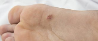
Manifestations of keratolysis primarily include the appearance of small holes on the soles of the feet. Outwardly, it resembles a fungal infection, so when diagnosing it is important to distinguish between these diseases.
Patients also develop the following symptoms:
- Itching of the skin of the extremities.
- Burning sensation.
- Whitish color of the affected areas of the dermis, which becomes more noticeable when the leg is wet.
- Thickening of the epidermis in diseased areas.
- Unpleasant foot odor.
Associated symptoms
Punctate keratolysis can be virtually asymptomatic, but sometimes the holes on the feet cause noticeable discomfort. Possible signs of the disease include:
- Damage to the skin of the feet, especially the heels. It is extremely rare that holes are found in the interdigital area.
- Hyperkeratosis is excessive thickening of the skin in the affected areas.
- Holes, with a diameter from 2 mm to 6 mm, similar to simple small holes. Sometimes they can merge and form erosions.
- Unpleasant foot odor.
- Unexpressed itching.
- Excessive sweating of the feet.
- Pain when pressing on the affected areas (not always detected).
Most people, when faced with holes in their feet, immediately suspect the development of fungal diseases. But the unjustified use of antimycotic (antifungal) agents can only worsen the situation.
How to identify the disease?
When small holes appear on the skin of your legs, you need to visit a dermatologist. It is better to do this immediately after identifying the holes. The doctor will first conduct a visual examination of the foot using a fluorescent lamp.
Then the doctor prescribes a scraping from the affected area of the skin to exclude the development of mycosis. A culture is also performed to detect the microbe and determine its type. This will help you choose an effective medicine to get rid of the disease.
Diagnostics
An experienced doctor can identify punctate keratolysis and distinguish it from other possible foot diseases. The specialist usually conducts:
- Visual examination of the patient.
- Anamnesis collection.
- Scraping the skin to exclude fungal infections.
- Sowing the collected material to isolate a specific pathogen. Such a study may take several days, but it allows you to select the most adequate and effective therapy.
A qualified dermatologist in an ordinary clinic can correctly diagnose punctate keratolysis. But less experienced specialists may find it difficult to make a diagnosis; in such a situation, the patient is advised to find a more competent doctor.
Popular means
An effective medication for treating holes in the skin of the legs is Erythromycin. It belongs to the antibacterial drugs from the macrolide group.
Available in different dosage forms - tablets, ointments, powders. This antibiotic helps destroy the infection.
In addition to Erythromycin, the following medications are prescribed for the treatment of ketarolysis of the feet:
- "Bactroban".
- "Clindamycin."
- Teymurov's pasta.
- Nitrofungin solution.
Which treatment methods to use is decided solely by the attending physician. Under no circumstances should you try to cope with the problem on your own. You can quickly get rid of keratolysis if you follow the doctor's recommendations.
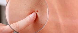
Skin on the feet with holes - what kind of disease is this?
Human feet are exposed to a lot of stress every day, but in addition, the skin on the feet is regularly attacked by various pathogenic organisms. Therefore, such areas of the body may suffer from calluses, warts and various fungal diseases. But sometimes people encounter a completely unexpected symptom - small holes on their feet. Doctors claim that the appearance of such a sign can be explained by punctate keratolysis. However, only a dermatologist can make an accurate diagnosis, because sometimes violations of the integrity of the epidermis on the feet are signs of fungal infections or the activity of HPV - the human papillomavirus.
Causes of punctate keratolysis
A disease such as punctate keratolysis has been known to doctors for about a hundred years. It is believed that it can develop in any person, but residents of the tropics or vacationers in warm countries, as well as some other categories of the population, are at risk. Punctate keratolysis is infectious in nature and usually develops due to continuous contact of the foot with tight shoes. Various pathogenic microorganisms can cause the development of the disease, but most often the culprit of the disease is:
- Pseudomonas or Pseudomonas aeruginosa. Such a pathogen is capable of dissolving the stratum corneum and forming defects in the skin. The stick needs a constant supply of oxygen, so after forming a crater in the skin, it captures new areas, causing the appearance of new erosions. In the future, the small pinpoint pits become one large one, and it can become inflamed. The human immune system tries to cope with this problem by stimulating the suppression of inflammation and tissue regeneration, but the rods migrate to other areas of the foot.
- Coccal microorganisms with a spherical shape. In general, these pathogens are not capable of causing harm to a person with full health and normal protective function of the epidermis on the feet. However, upon contact with sweaty skin, they are able to dissolve the uppermost layers of the epidermis and accumulate in the formed pits.