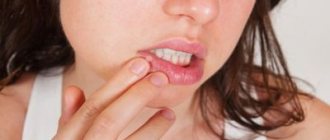
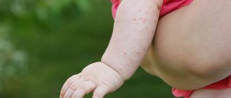
A newborn baby is a big responsibility for young parents. When visiting a pediatrician, parents usually ask a lot of questions. Especially if the child is the first. In my personal top list, feeding and colic take first place. Second - rashes!
In photographs on social networks, babies have clean, rosy cheeks and an angelic smile. And in life, redness, pimples, peeling and other unexpected things appear on the skin. Moms are not ready for this. They get scared and start looking for the reason. Neighbors advise you to follow a diet, grandmothers complain about “bad milk.” If you are unlucky, the doctor will scare you with allergies or prescribe tests, half of which will turn out to be unnecessary.
Let's try to figure out which skin problems in infants do not require treatment and are normal in infancy, and which do not. Let’s agree right away: any rash should be examined by a doctor in person. A diagnosis cannot be made from a photograph, because the doctor examines the elements of the rash, touches it by touch, and sometimes special diagnostic tests are needed. In doubtful cases, the pediatrician gives a referral to a dermatologist.
The information below is given so that young mothers stop being afraid of every pimple, understand conditions that do not require medical attention, and understand when to take their child to the pediatrician.
What does a newborn look like?
If the birth goes well, immediately after the birth of the newborn baby, the mother is placed on her stomach. His skin can be bright pink, or it can be pale, his hands and feet have a bluish tint. This scares young mothers if they have heard or read something about hypoxia and cyanosis before. But if the doctors in the delivery room are calm and do not provide resuscitation measures, then this is a variant of the norm. A healthy child can be like this in the first minutes of life. In addition, the newborn's body is covered with vernix - the baby looks as if it has been smeared with cream cheese. And this is also normal, although unattractive. Lubricant plays an important role in protecting the skin from germs, so the baby’s skin is only lightly blotted without being completely wiped.
5-10 minutes after birth, the newborn’s skin becomes bright pink, almost red. This is a normal condition - physiological erythema . The air pressure is less than the pressure of the fluid in which the baby was in utero. When the skin ceases to experience resistance, blood actively flows to it. The body gets used to the new form of life. Gradually, the blood vessels will narrow, and the skin will become its usual pink color by the end of the week in full-term babies and after 2-3 weeks in premature babies.
Helping mom: examining baby's skin
We all know the expression “skin like a baby.” However, many people imagine a newborn as a small lump with very soft and pink skin. But most often this is not the case. Let's figure out what skin rashes frighten parents in the first month of a baby's life and what they talk about.
At the reception, parents often hear complaints that the child has “some pimples” on the skin. And rashes are often associated with allergies.
The first month of life is a period of adaptation. At this time, the child’s skin is actively populated by microflora and gets used to the air environment. This means that its appearance also changes. Parents are often concerned about the following harmless conditions.
Simple erythema - red spots of small diameter. They can be located on any area of the skin. The cause is vasodilation. Most often noticeable on the second or third day after birth, they do not bother the baby. They go away on their own in a few days.
Erythema toxicum is also a red spot, but there is also a small “pimple” in the middle. In everything it is similar to its “namesake” - simple erythema, but it just begins due to the colonization of the skin by microflora.
Peeling. It can be large or small, reminiscent of peeling skin after sunbathing. You can notice it in the first days of life.
Milia are small white dots, often located on the nose. These are clogged hair follicles. There is no need to crush them, they will disappear on their own.
Neonatal acne, or infant acne , is one of the most interesting conditions. The rashes really look like pimples, sometimes even with a white head. You can find them on the face, shoulders and chest. One baby will have single pimples, while the other will be densely strewn with them. The time of occurrence is approximately the end of the first month of life.
It is acne that many parents tend to mistake for a manifestation of allergies. But the reason is completely different.
This is just a reaction of the sebaceous glands to hormones in breast milk. It is important that the general condition of the baby does not change in any way. Acne is the longest lasting condition of all of these. It can remain on the child’s skin from one to two weeks to a whole month. It goes away on its own and leaves no traces.
All of the above changes in the skin are physiological. They do not affect the general condition of the child in any way, do not require treatment and go away over time.
A number of other symptoms require active parental involvement. For example, prickly heat occurs due to the fact that the child is in very warm conditions for a long time. The ducts of the sweat glands in babies are normally closed. But the sweat glands themselves produce sweat when overheated. It is this that causes irritation inside the skin around the sweat glands. To cure it, create comfortable environmental conditions. It is important to maintain an air temperature at home of 20–22 °C and a humidity of 40–60%. And under no circumstances should you wrap the baby.
Diaper dermatitis, or diaper rash , is irritation of the skin under the diaper due to prolonged contact with physiological secretions. Frequent use of wet wipes may also be the cause. Irritation often appears as severe redness with clear boundaries. Treatment is based on correcting hygiene rules. They are simple, and if you follow them, diaper rash goes away in a few days:
- Reduce the time you use diapers and give air baths more often.
- Wash your child under running water only after defecation.
- Pat your skin dry after washing.
- Use fragrance-free wipes only when water is not available.
- Use protective creams under the diaper.
The skin of newborns is very delicate and easily injured. Therefore, she requires a very careful attitude towards herself. And remember: not all rashes are associated with allergies. But if rashes appear, it is better to show them to a doctor and not self-medicate.
Daniyar Zhuraev, pediatric neurologist.
Photo depositphotos.com The author's opinion may not coincide with the opinion of the editors.
Marbled leather
During the first month of life, a baby's skin sometimes looks marbled. A uniform mesh pattern of white, purple, and reddish spots is the reaction of skin vessels to cold. Therefore, this phenomenon is most often noticed during swaddling. If the marbling is symmetrical and disappears when warmed, then there is no cause for concern. In other cases, it is worth discussing this symptom with your pediatrician so as not to miss the early diagnosis of diseases of the heart, lungs or skin itself.
Peeling
On the second or third day after birth, the newborn’s skin begins to peel off. Peeling is observed in everyone, but to varying degrees of severity. Most severe if the baby is post-term. The reason is the same restructuring of the body to exist in the air. The skin loses moisture, and the top layer gradually peels off. Physiological peeling does not require treatment and goes away on its own within a few days.
If the peeling is profuse and leads to deep cracks, the skin is lubricated with special moisturizers - emollients. In case of pronounced peeling, it is necessary to exclude congenital skin diseases such as ichthyosis, hyperkeratosis and others.
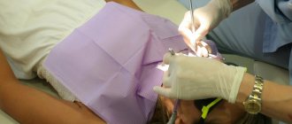
Treatment of milia
Milia do not go away on their own; it is almost impossible to get rid of them with the help of cosmetics. Under no circumstances should you squeeze out milia yourself, as this will damage the hair follicle and sebaceous gland. Such self-medication often leads to the subsequent formation of a larger acne or the addition of an infection, which can cause the formation of a rough scar.
Treatment of milia is carried out by a dermatologist and consists of opening and removing the cyst capsule with its contents. The choice of removal method depends on the location, number, size and depth of the milia. For single milia, mechanical removal is carried out (using a sterile needle and curettage) with preliminary and subsequent antiseptic treatment of the skin. Small wounds left after such a procedure heal on their own without leaving a trace.
For multiple whiteheads, modern methods are used - removal of milia with a laser, radio wave method or electrocoagulation. The crusts formed at the cauterization site disappear on their own within 10-14 days. Removing more than 10 elements at one time is not recommended to avoid significant trauma to the skin and disruption of the sebaceous glands.
Erythema toxicum
By the second or third day of life, 70% of full-term newborns develop a small red rash on the skin. Rarely, this condition occurs in premature babies. First, spots appear, and within an hour a yellow bump 1-2 mm in size forms in the center. Elements of the rash are located on the face, torso, arms and legs, and can merge with each other. The cause of erythema toxicum is unknown, but the rash goes away without treatment in 1-7 days and is not dangerous.
If the clinical picture is atypical or other symptoms are present, the doctor will prescribe additional examination to rule out other causes of the rash, such as infection.
Newborn acne
20% of newborns develop a rash similar to teenage acne from the third week of life. Small pustules surrounded by a red halo. They are located on the face, head, chest, shoulders, and less often on the stomach. The condition is called “acne,” although in the literature the term “cephalic pustulosis” (from the words “cephalo” - head and “pustule” - abscess) or “neonatal pustulosis” is more often used. The main two reasons are the influence of maternal hormones during breastfeeding and the colonization of the skin by microorganisms. Elements of the rash resolve without treatment after 1-3 months. This condition is not dangerous, does not cause discomfort to the child and leads to independent recovery, so there is no need to prescribe medications. The exception is severe cases when the rash is profuse, leads to secondary infection or heals with the formation of scars.
How to speed up the process of resorption of white spots in a child
Parents want to save their child from suffering, they try to help the newborn and eliminate the signs on the nose. Pediatricians do not recommend that adults interfere with the physiological processes associated with the excretion of excess amounts of hormones. The baby adapts to new environmental conditions, coping with emerging defects on his own.
Need advice from an experienced doctor?
Get a doctor's consultation online. Ask your question right now.
Ask a free question
White spots on the nose of a baby disappear until the age of two months; at the end of this period, the epidermis of the newborn is cleared. If the causes of milia are natural, the pimples will disappear without treatment. Excessive care can delay the process of restoring the condition of the integument, prolong the period of rashes, and aggravate the situation.
To make sure that the white formations on the baby’s nose are not dangerous and do not indicate a severe pathology, make an appointment with a pediatrician to determine the nature of the spots and prescribe treatment.
If a newborn has corn, it is important to avoid injuring the white growths on the nose: scratching problem areas can lead to infection. To get rid of formations, observe hygiene standards:
- trim the baby’s fingernails and toenails in a timely manner, preventing damage to blisters and spots;
- ensure the cleanliness of the child's epidermis.
To speed up the process of cleansing a newborn’s skin from white pimples located on the nose and cheeks, parents regularly perform the following manipulations:
- Wash a baby who has spots on his face with herbal compositions from string and chamomile. The dry substance is brewed with boiling water, filtered, and added to the water when bathing a newborn. The liquid relieves irritation, forms a protective layer on the baby’s skin, and helps get rid of problems on the baby’s nose.
- Wiping the areas affected by small white dots with a cotton pad soaked in breast milk. The procedure ensures the removal of elements without the risk of developing allergies or complications.
- Using water with the addition of potassium permanganate to relieve skin irritation in a newborn, get rid of white grass and spots. The solution has a light pinkish tint and helps remove growths. The concentration of the crystalline substance cannot be exceeded; there is a risk of aggravating the situation and increasing the area of the rash on the nose.
- Regularly wash the baby with boiled warm water, the procedure is performed 3 times a day.
By ensuring the cleanliness of the integument and adhering to hygiene standards, you can rid the newborn’s dermis of white nodules and dots.
Infant acne
The same rash in children after three months of age is called infant acne. It persists as persistent acne of newborns or reappears at 2-3 months. It differs from newborn acne only in the age of the children and the duration of the rash. Self-resolution occurs within 6-12 months. Infantile acne may be a risk factor for acne during adolescence.
Prickly heat
Develops in children from the second week of life when overheated. The rash has different manifestations: small red spots, nodules, pustules or blisters in the thickness of the skin. Miliaria can occur on any part of the body: in the folds, on the face, scalp, torso, arms and legs. The cause of the rash is blockage of the sweat glands.
There are several factors contributing to this:
- high room temperature;
- increased body temperature in a child;
- thick clothing, non-breathable fabric, for example, an oilcloth mattress cover;
- lying in one position for a long time.
Treatment consists of general care:
- daily bathing without soap and other chemicals;
- using lighter and looser clothing;
- maintaining the air temperature in the room at 22-24 degrees C;
- ventilation.
When these conditions are met, prickly heat disappears within a few hours. The recommendations listed above also apply to prevention.
For walks outside, the child is dressed like an adult plus one layer of clothing. No need to re-wrap. There is also no need to wear a cap in warm weather.
During the cold season, undress your child during a long stay in a store, shopping center or other premises. While awake, place your baby on his stomach and hold him vertically in your arms to diversify his movements.
The reason for the appearance of white spots on the face of a newborn
In the first year of life, a child develops light pimples on the nose due to an excess of hormones in the mother. The child's body received them along with nutrients during intrauterine development; the supply of elements after the birth of the baby occurs during feeding with milk.
The appearance of small white dots on the cheeks and nose of a newborn may be a consequence of blockage of the sebaceous glands. Milia are small pimples with secretions accumulated in them; people call them millet pimples.
The blockage of glands in a baby is caused by underdevelopment of vital systems: the newborn’s body is developing, it is not able to ensure the timely outflow of sebum. As you grow older, the white spots that form on the nose tend to go away on their own.
Diaper rash
A more serious skin reaction to overheating is diaper rash. Occurs in natural folds. It looks like redness, sometimes with weeping and erosion. With a long process, a bacterial or fungal infection may develop, then the surface of the affected skin is moist, bright red, with clear or purulent discharge. Prevention measures are the same as for prickly heat.
The skin in the folds is treated with powders. If normalizing the microclimate and caring for the newborn does not lead to a reduction in diaper rash, you should see a doctor. If complications occur, antibiotic therapy or topical steroid ointments may be needed.
Diaper dermatitis
The distinctive feature of this rash is that it appears only in the area under the diaper, other areas of the body remain clean. The reason is increased humidity and irritating feces.
The rash is found in the buttocks, thighs, lower abdomen in the form of individual elements or continuous areas of changed skin. The elements of the rash are varied: red spots, bumps, pustules or peeling.
To alleviate the condition, you need to change the diaper and wash the baby immediately after bowel movement. While changing clothes, you can arrange air baths, that is, leave the child to lie naked, you can cover it with a regular dry diaper. Lubricate the skin with a special diaper cream.
Change the diaper every 3 hours, even if it is only slightly full. More often when very full. If these measures are ineffective, you should consult a doctor. Local glucocorticoid agents may be needed.
Preventive measures
Almost all acne in babies is not dangerous. Their occurrence can be easily avoided if you follow a number of measures:
- watch your diet;
- if there is a reaction to the mixture, replace it;
- properly care for your baby and maintain hygiene;
- give the baby air baths;
- wash your baby’s underwear with specially designed powders.
If acne does not go away for a long time and bothers the newborn, doctors usually prescribe medications: Panthenol, Bepanten or Zinc ointment.
Seborrheic dermatitis
Appears in infants soon after birth and lasts up to a year. Yellow scales and peeling are most often located on the scalp and behind the ears, but can also be in other folds - axillary, elbow, inguinal. In everyday life they are called “milk crusts”. Because of its similarities to atopic dermatitis, seborrhea is sometimes considered a manifestation of allergies. The causes of this condition are not fully understood, but the relationship with allergies has not been identified.
Seborrhea goes away without treatment, in most cases in the first year of life. Unlike atopic dermatitis, the rash does not cause itching and does not cause discomfort to the child. If for the sake of aesthetics you want to get rid of seborrheic crusts, before bathing the baby’s head is lubricated with oil and then combed out with a soft comb.
In doubtful cases, when seborrhea is located on the body, it makes sense to consult a doctor to clarify the diagnosis.
Seborrheic dermatitis in children: how to help your child
Seborrheic dermatitis in children
Modern medicine has reached a qualitatively new level of knowledge in the last decade, which has made it possible to take a fresh look at the causes of many widespread diseases, including seborrheic dermatitis.
What is seborrhea or seborrheic dermatitis?
According to the definition, seborrheic dermatitis (SD) is a chronic recurrent skin disease associated with increased secretion of sebum, changes in its qualitative composition and characterized by localization in areas of accumulation of sebaceous glands - on the scalp, on the face, upper body, folds.
Causes. Pathogenesis.
The cause of seborrheic dermatitis is excessive colonization of the yeast fungus Malassezia spp., which belongs to the opportunistic microflora of the human body. There are many reasons why this microorganism begins to actively multiply, leading to the development of an inflammatory process on the skin. Among the most likely factors are emotional stress, dysfunction in the neuroendocrine and immune systems, the presence of foci of chronic infection, diseases of the gastrointestinal tract, excess body weight, imbalance of microelements, and the use of certain medications, such as psychotropic drugs. Additional factors contributing to the severity of the process will be a warm environment, increased secretion of the sebaceous glands, individual sensitivity (skin hyperreactivity, decreased immune status, disruption of the epidermal barrier).
It should be noted that Malassezia spp. are lipophilic microorganisms, and their life cycle depends on the quantity and quality of sebum, the production of which varies significantly at different age periods.
The intensity of Malassezia colonization changes with age and is directly related to the functional activity of the sebaceous glands, and therefore the manifestations of seborrheic dermatitis in children: it is most pronounced in newborns, infants, adolescents and young adults, and minimally in children from 2 to 9 years of age.
| Age period | Manifestations of seborrheic dermatitis in children |
| 0-2 years | Often |
| 2-3 years | rarely |
| 4-5 years | rarely |
| 5-7 years | rarely |
| 7-10 years | rarely |
| 11-12 years old | often |
Symptoms of seborrheic dermatitis in children
The first episode of seborrheic dermatitis may occur during the newborn period. This condition is defined as gneiss or cradle cap and occurs in more than half of newborns and infants. In a baby, multi-layered fatty scales or crusts of various shades of yellow appear on the scalp; the number of crusts and scales varies widely in each child, and can manifest as minimal damage in the parietal area or scalp, moving to the face.
The reason for the development of this condition in the first year of a child’s life is the active work of the sebaceous glands under the influence of the mother’s sex hormones; as a rule, the process resolves within 1-1.5 months. In case of minimal manifestations, drug treatment is not required. Just gentle care is enough.
The photograph shows a child aged 6 months, born from a second pregnancy. The mother suffers from atopic dermatitis. The boy has dry, yellowish crusts on his head in the parietal region that adhere tightly to the skin and are difficult to remove. Gentle care and topical therapy are recommended.
However, in 10-15% of children, the process may be more widespread. The rashes are represented by erythematous-squamous lesions, yellowish scales.
In the photograph, a child is 1 month old, born from the first pregnancy. The mother's obstetric and gynecological history was not burdened. Gentle care and external medications are recommended.
Complications
In very rare cases, the pathological process spreads far beyond the “classical” seborrheic zones, affects the folds of the skin, and weeping appears. The child's general condition suffers. In such a situation, hospitalization is required and external remedies alone will not be enough.
In the photo, the child is 1 month old, born at term from the first pregnancy, the mother is healthy.
In most children, seborrheic dermatitis subsides during the first year of a child's life. And at the age of 2 to 9 years it is extremely rare.
With the onset of puberty, the sebaceous glands and sweat glands are activated, the qualitative composition of sebum changes, in addition, during prepuberty and puberty, significant changes occur in the immune and nervous system, which also leads to a change in the balance between macro and microorganisms.
Diagnostics
Rashes with seborrheic dermatitis are located on the skin of the scalp, face, ears, upper body, mainly in the area of the sternum and shoulder blades, and in large folds.
The inflammatory skin process begins with redness and slight infiltration of the skin with the spread of flaky spots and round papules along the periphery of the lesions. Moreover, the severity of seborrheic dermatitis does not depend on the number of fungi.
Very often, seborrheic dermatitis in children can be combined with damage to the follicular apparatus of the skin, which is manifested by small papules on the shoulders, chest, and in the interscapular space.
It is extremely important to begin treatment for seborrheic dermatitis on time, since the symptoms of the disease carry a significant psychological burden and cause physical suffering.
The activity of the pathological process can have varying degrees of severity.
In most patients, this is a mild course, dandruff appears on the scalp - a moderate amount of flakes, which can be either dry or oily, redness of the skin is mild and, as a rule, is accompanied by intermittent itching of varying intensity.
Treatment of seborrheic dermatitis in children
For mild seborrheic dermatitis, a medicinal shampoo containing activated zinc pyrithione - Skin-cap - has proven itself very well.
Using shampoo allows you to:
- inhibit the activity of opportunistic microflora
- control inflammation, since zinc pyrithione exhibits its activity not only against the fungus of the genus Malassezia spp, but also against S/aureus
- reduce skin itching
- eliminate excess peeling
- allows you to achieve stable remission and maintain normal corneometric (oily, moisture) parameters of the skin, which, of course, improves the quality of life of patients.
It is very important to use Skin-cap shampoo correctly; it should be applied to damp scalp, left for 1-2 minutes and then rinsed with enough water.
In some patients, seborrheic dermatitis may have more severe symptoms. The skin is bright red, swollen, with a significant number of yellowish-gray scales that are difficult to separate from the skin and reveal wet erosions with sticky discharge. The itching in this condition is very intense. The process is localized at the edge of hair growth, on the occipital, parietal region.
Moderate seborrheic dermatitis requires the use of combination creams that contain a steroid (hormone) and an antifungal agent. If there is a risk of a bacterial infection, then three-component drugs are used. The choice of drug and the duration of its use is determined by the doctor.
With such a course of seborrheic dermatitis, it is critically important to rationally select second-line therapy (without hormones), and Skin-cap shampoo with antifungal activity will fully meet the task.
When the severity of the process subsides, you can control the process using Skin-cap cream. The cream has a very light texture, does not stick, does not leave marks on clothes and hats, and is ideal for containing the process. One of the dosage form options is Skin-cap aerosol. It is convenient because it is very easy to apply even to hard-to-reach areas. The course of treatment can range from 14 days to 1 month.
It should be noted that Malassezia spp. can act as a constant irritant, contributing to the development of an allergic reaction to fungi, which can seriously aggravate diseases such as atopic dermatitis and bronchial asthma.
Symptoms are especially pronounced in a child with an atopic history. Then we are talking about severe seborrheic dermatitis. This course will be characterized by erythema, small papulopustular elements covered with fatty, white-yellow scales. Areas of predisposition include the nasolabial folds and upper lip near the nostrils, eyebrows and root of the nose, pre- and retroauricular areas, the process can also spread to the sternum, less often the back. Sometimes the eyes are involved in the pathological process, which manifests itself in the form of seborrheic blepharitis.
In addition to the aesthetic disadvantage, chronic seborrheic dermatitis in children seriously disrupts the child’s daily life.
In the photo, a 17-year-old teenager suffers from bronchial asthma. After a stressful situation and a diet violation, an inflammatory process occurred on the scalp and neck within 3 days. Against the background of hyperemic, edematous skin there is pronounced peeling, weeping erosion, and intense itching.
In addition to systemic therapy, the patient was recommended to use Skin-cap aerosol. The convenience of this form is determined by the fact that the patient can apply the product without outside help to hard-to-reach areas.
Unfortunately, in adolescents suffering from atopy, seborrheic dermatitis is combined with folliculitis and/or lichen asbestos. This form of the disease is very difficult to manage and requires constant monitoring.
The pictures show a 15-year-old patient with atopic dermatitis since childhood. With the onset of puberty, itchy rashes began to appear on the head in the armpit, groin area, and on the skin of the chest.
The patient does not tolerate milk protein. In addition to drug therapy with systemic antifungal drugs and external combination agents, she was recommended Skin-cap shower gel for constant use.
Cream, shampoo, shower gel, Skin-cap aerosol help cope with the task of treating seborrheic dermatitis in patients of any age, there are no age restrictions for the shampoo, gel, aerosol formulation, from 1 year for the cream formulation. The vector of action of the drug is aimed at suppressing excessive colonization and activity of Malassezia spp. fungi, bacterial flora, and helps reduce the intensity of the inflammatory process in the skin. The excipients included in the Skin-cap have a moisturizing and reparative effect.
The possibility of using the Skin-cap line both as an independent product and as part of complex therapy for the treatment of seborrheic dermatitis of any severity allows you to expand the possibilities of long-term control over the disease.
Prognosis and prevention
When consulting a patient with seborrheic dermatitis, the pediatrician should inform that seborrheic dermatitis on the head, face, body is a chronic process, its relapses are associated with various trigger factors, such as stress, medication use, dietary errors, and many others. And adequate control over the disease is determined not only by drug therapy, but also by rational care of the child’s skin.
Letyaeva Olga Ivanovna
Professor, dermatologist
Bibliography
- Panyukova S.V., Piruzyan A.L., Korsunskaya I.M. Seborrheic dermatitis: how to help the patient. Consilium Medicum. 2020;22(7):46-48
- Fassakhov R. S. Zinc pyrithione in complex therapy of atopic dermatitis: pathogenetic basis and research results. Medical advice. 2017;20:171-176.
- Clinical dermatovenerology: manual. In 2 volumes. Volume 2. GEOTAR-Media / Ed. Yu. K. Skripkina, Yu. S. Butova. 2009. Chapter 16. Diseases of hair, sebaceous and sweat glands. pp. 469-475.
- Mokronosova M.A., Pyzh V.V., Kashaeva O.V., Reznikov O.V. Therapeutic effect of activated zinc pyrithione in patients with atopic dermatitis/eczema syndrome with sensitization to yeast-like fungi. Ros allergol journal 2005; 3: 83–87
- Skin microbiota from the point of view of fundamental medicine. Published in the journal: Effective pharmacotherapy. 2022. T. 16.
Atopic dermatitis
This is a chronic inflammatory skin disease. It occurs in 15% of children, of whom two thirds experience the first symptoms before one year of age, sometimes even in infancy. The rash appears on the face, torso, and extensor surfaces of the arms and legs. There may be redness, peeling, crusts, and when scratching, infection and the presence of pustules may occur. Elements of the rash itch and bother the child.
Only 30% of children have atopic dermatitis associated with food allergies. Therefore, a nursing mother does not always need a diet.
The cause of atopic dermatitis is increased skin permeability. For treatment, first of all, creams that restore barrier function are used. They are called emollients. There are quite a few brands that produce such products. The choice is based on personal preference. From a medical point of view, there is no advantage to any particular drug.
If you suspect atopic dermatitis, you need to see a doctor to clarify the diagnosis and receive recommendations for treatment. You can contact your pediatrician or dermatologist. If the diagnosis is confirmed, keep in mind: treatment will be long. It includes skin care and constant use of emollients; in more severe cases, hormonal ointments may be needed; for food allergies, a diet. It is important to understand that with strict implementation of the recommendations, stable long-term remission can be achieved.
Rashes in babies
Babies are generous with various rashes. According to the “good” tradition, most of them are considered allergic with all the consequences - a strict diet for a nursing mother, transfer to artificial feeding, prescription of medicinal mixtures, etc. In fact, true allergic rashes in infants are not that common. Allergic diseases affecting the skin in infants include: atopic dermatitis, acute urticaria and Quincke's edema. Acute urticaria is extremely rare in infants - it is an acute allergic reaction in the form of peculiar blistering-type rashes (like a nettle burn, hence the name), which suddenly appear on the skin and just as suddenly disappear without leaving any trace behind, usually do not exist on the skin for longer than a day and are accompanied by severe itching, which manifests itself in the child’s general anxiety. The most common causes are food proteins (for example, cow's milk), viral infections, insect bites and medications (for example, antibiotics). In severe cases, it may be accompanied by swelling and redness of the soft tissues of the face, neck, larynx, arms, legs, genitals or abdominal cavity - Quincke's edema, which requires immediate medical attention.
Let's figure out what is most often unfairly called an allergy:
Erythema toxicum of newborns is a transient, benign rash whose exact cause is unknown (possibly due to skin irritation by environmental factors).
Appears at birth or in the first 24–48 hours of life. Localization - face, torso, limbs, except palms and soles. Disappears on its own within 5–7 days, sometimes 3 weeks. Does not require treatment.
Newborn acne (infantile acne, neonatal pustulosis) is caused by stimulation of the baby's sebaceous glands by androgens.
The peak of rash occurs in the 3rd week of life. It is most often localized on the face, sometimes spreading to the scalp, less often to the collar area. Resolve spontaneously. The skin requires cleansing and moisturizing; in some cases, the use of medicated creams may be required.
Miliaria is a rash that occurs in poorly “ventilated” areas as a result of blocked sweat glands. Can occur at any age.
Localization - folds of skin, buttocks and back surface of the body, sometimes the face (after sleep). Depending on the depth of the lesion, there are crystalline prickly heat, prickly heat, deep prickly heat (superficial).
The duration of the rash ranges from several hours to several days.
Treatment - cool water baths, air baths, prevention of overheating. Calamine-containing lotions and corticosteroid and antibiotic creams may be used to treat some cases of prickly heat and miliaria.
Seborrheic dermatitis is a skin disorder that forms on sebum-rich areas of the skin. The exact cause is unknown (a certain role is played by the skin saprophyte - the Malassezia fungus, which grows well and multiplies in sebaceous secretions).
It can be focal or widespread, dermatitis with pityriasis-like scales, which can form a crust (“cap”, gneiss) on the scalp.
Favorite localization is the scalp, face, folds (!).
It can begin from the 1st - 2nd week of life or later, and resolves spontaneously over several weeks or months.
Treatment involves softening the crusts with oil or cream and then removing them, moisturizing the skin and, in some cases, applying antifungal and anti-inflammatory creams.
Simple contact dermatitis is a nonspecific damage to the skin due to prolonged or repeated exposure to a number of substances - saliva, fruit juices, foaming bath products, detergents (their residue on the walls of the bathtub), etc. In infants, saliva often causes dermatitis in the contact area with the pacifier and in the folds of the neck.
As a rule, elimination of the offending agent and short-term administration of anti-inflammatory creams quickly leads to recovery, but some children are so sensitive that it is almost impossible to identify the causative factor.
Diaper dermatitis (a prototype of contact dermatitis) is a skin lesion that occurs under the influence of physical (overheating), chemical, enzymatic (contact with sweat, urine and feces) and microbial factors. Localization - area of the diaper or diaper area.
Treatment is carried out using the abbreviation ABCDE (air, barrier, cleansing, diaper, education). Frequently changing diapers, washing the skin and drying it thoroughly helps. Dermatitis is effectively prevented by applying products to clean skin that completely cover it (vaseline, zinc paste). In persistent cases, medicated creams containing corticosteroids, antibiotics, or antifungals may be recommended.
And now a few words about AD:
Atopic dermatitis is a chronic allergic inflammation of the skin, genetically determined, associated with loss of the skin barrier and, therefore, accompanied by dryness, itching and various rashes. In a third (!) of cases it is combined with food allergies (the most common “culprits” are cow’s milk, chicken eggs, wheat, fish, soy, nuts).
Most often it starts no earlier than 3 months of life.
The most common localization up to 2–3 years is the face (cheeks, forehead, chin), convex parts of the limbs (extensor surfaces) and the torso; it never occurs in folds in infants (!).
Exacerbations are triggered by various factors - stress, dry air, sweat, food (histamine liberators), infections, contact with tobacco smoke, animal hair, rough fabric, detergent residues on clothes, etc.
It is treated with careful skin care and the use of anti-inflammatory creams.
There are also:
Pseudoallergic reactions are reactions that are externally similar to allergic ones (for example, various rashes), but are not such, due to the non-immune mechanism of their development.
The reason is the increased content of histamine (tyramine, serotonin) in foods, or the ability of foods to enhance the release of these substances in the body, or their increased absorption, due to pathology of the gastrointestinal tract (enzymopathies, inflammation in the intestinal wall, etc.). These products include chocolate, cocoa, strawberries, citrus fruits, honey, sauerkraut, marinades and spices, seafood, fish, caviar, pork, mushrooms, cheeses, nuts, smoked meats, preservatives, dyes and flavor enhancers.
Treatment includes dietary advice, skin care, and in some cases, antihistamines and anti-inflammatory creams.
The clinical manifestations of atopic dermatitis, simple contact dermatitis in highly sensitive children and the manifestation of pseudo-allergic reactions are very similar to each other, so the main task remains the creation of a “skin barrier” by constantly moisturizing the skin with emollients, stopping exacerbations with anti-inflammatory creams and excluding exacerbation factors.
And the last thing:
Skin infections - herpes virus, staphylococcal pemphigus, candidiasis also occur in infants, do not forget about them. You should consult a doctor immediately if the child is lethargic, has a fever, refuses the breast or bottle, skin rashes are accompanied by pus or are covered with purulent crusts, there are blisters or a group of blisters, erosion (violation of skin integrity), severe swelling and redness of the skin.
Author:
Eroshkina Maria Sergeevna pediatrician
Mastocytosis
A rare disease that can appear at an early age, sometimes even from birth. Outwardly, it resembles allergic urticaria, but spots and blisters last longer, and when the exacerbation subsides, pigmentation remains. A typical diagnostic test is the appearance of blisters on the skin due to mechanical irritation of areas of pigmentation.
Treatment for childhood forms of the disease is not required; recovery occurs spontaneously within several months, sometimes several years. If itching is present, antihistamines are used.
There are various causes of hypopigmented spots on a child's face, ranging from conditions that resolve on their own within a few weeks to conditions that can last a lifetime.
Causes of white spots on a child's face
White spots can be caused by the following skin conditions:
Milia
Milia are also called milk spots. These tiny white bumps usually appear on the face and rarely on the upper torso or limbs. Although milia can be seen at any age, it is common in newborns.
Causes and diagnosis of milia
Milia occurs due to entrapment of keratin (skin scales) beneath the surface of the skin. If there is no improvement after a couple of weeks, you should consult a doctor. Diagnosis is made by visual examination and no tests are required.
Prevention and treatment of milia
There is no cure for milia and it often goes away within a few weeks or months. There is no need to treat milia in children.
How can you reduce milia at home?
- Wash your child's face daily with mild soap and water.
- Pat your face dry after washing.
- Be careful not to scratch the milia to prevent skin damage and infection.
- Do not apply lotions or oils to your baby's face.
Note: Some may confuse baby pimples or Epstein pearls with milia. However, acne can cause red bumps and pustules on the face. Epstein's pearls are small, white-yellow cysts that often resemble milia, but they appear on the roof of the mouth and gums.
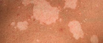
Pityriasis alba (lichen alba)
Pityriasis alba is a self-limiting skin condition that causes dry, thin, scaly and pale patches on the face. It is common in children and adolescents and is considered a type of eczema or dermatitis.
The name of this condition comes from the characteristic appearance of the skin.
Causes of white ptyriasis
The causes of white ptyriasis have not yet been clarified. However, atopic dermatitis and dry skin often coexist with it. Exposure to the sun can make it more visible by tanning the surrounding skin.
Suspected causes such as under- or over-bathing, low serum copper levels, ultraviolet radiation, or Malassezia yeast have not yet been proven to cause hypopigmentation.
Symptoms and signs of ptyriasis alba
One or more white spots, varying from 0.5 to 5 cm in diameter, are a characteristic sign of pityriasis alba. It may also cause mild itching in some children.
Diagnosis of pityriasis versicolor
The condition can be diagnosed through a physical examination by a doctor. A skin biopsy may show mild spongiotic (fluid accumulation between cells) dermatitis with decreased melanin pigment. A skin scraping may also be taken to rule out a yeast infection.
Treatment of white lichen
Treatment is not recommended in asymptomatic cases. Moisturizers are helpful for dry skin, and mild hydrocortisone (topical steroid) creams can help reduce itching and redness in the area.
Tacrolimus ointment, pimecrolimus cream, and calcineurin inhibitors have been shown to be effective in the treatment of pityriasis alba.
The appearance of the skin may gradually return to normal over several months or two to three years.
Prevention of white deprivation
Limited sun exposure may help reduce your risk of developing this disease.
Vitiligo
Vitiligo is a depigmentation of the skin due to the loss of melanocytes, which are the cells that produce the skin pigment called melanin. Vitiligo can affect both sun-exposed and unprotected areas of the body. Also, with the disease, depigmentation of the lips and gray hair are often observed.
Causes of vitiligo
The exact causes of vitiligo are unknown. The disease may be associated with dysfunction or loss of melanocytes (the cells that produce the pigment melanin), which give the skin its color. This may be due to genetic factors or an autoimmune condition in which the immune system attacks the melanocytes.
Vitiligo can occur at any age, but the onset of the disease is typical in children and adolescents.
Diagnosis of vitiligo
Vitiligo can be diagnosed through a physical examination. Rarely, a skin biopsy is performed to help confirm the diagnosis by the absence of melanocytes (pigment cells) on the skin. Thyroid disease and diabetes are checked as these may increase the risk of vitiligo in many people.
Treatment of vitiligo
Mild vitiligo may not require treatment: the spots will disappear over time. However, there are several treatments available to make your skin tone even.
Corticosteroid creams, photochemotherapy (PUVA), narrow band ultraviolet B (UVB) therapy, and depigmentation are a few treatment options. Using sunscreen can reduce tanning of the skin around vitiligo patches, and concealers can hide white spots on the skin.
Although vitiligo can be distressing to many people due to its appearance, it is not a medically dangerous condition. Vitiligo is not an infection, skin cancer, or a contagious disease.
Shingles
Shingles is a fungal skin infection that causes lighter or darker patches on the skin. The disease occurs at any age, but is most often seen in adolescents and young adults.
Causes of herpes zoster
Shingles is caused by a yeast that is usually present on the skin. Their excessive growth under the influence of environmental factors can lead to the appearance of spots on the skin. Malnutrition and excessive sweating (hyperhidrosis) can also lead to yeast growth.
Note: Shingles cannot be passed from one person to another (not contagious) since most people have Malassezia yeast on their skin. This condition is not caused by poor hygiene.
A weak immune system, oily and moist skin, or hot and humid climates may increase the risk of contracting shingles. Children taking corticosteroid medications may also be susceptible to this condition.
Signs and symptoms of shingles
Although white patches can appear on the face, they are often found on the chest, back, and forearms. The spots may be pink or light brown and have scales. Skin changes are usually limited to the outer layer of the skin and often do not cause any pain or itching.
Diagnosis of herpes zoster
A physical examination is sufficient to diagnose shingles. In rare cases, doctors may collect a skin scraping to confirm the diagnosis.
Treatment of herpes zoster
Shampoo containing selenium sulfide is the main treatment method. If the condition does not go away, then you can use antifungal or anti-dandruff shampoos. The skin may improve over a short period of time in many children. However, it may take several months to achieve an even skin tone. To prevent relapse of the disease, monthly use of shampoo is recommended.
Idiopathic guttate hypomelanosis
Idiopathic guttate hypomelanosis (IGH) is a skin condition with small, white, oval patches.
IGH is more common in older, fair-skinned people than in children and often goes undetected.
Causes of idiopathic guttate hypomelanosis
The exact cause of hypomelanosis is unknown. Although this has not yet been proven, it is believed that it may be related to sun exposure.
Diagnosis of idiopathic guttate hypomelanosis
A physical examination is sufficient to determine the condition. Rarely are biopsy samples taken. There is usually a decrease in the number of melanocytes in the affected areas, although they are not completely absent as in vitiligo.
Treatment of idiopathic guttate hypomelanosis
IGH spots are benign and do not require medical treatment. Most procedures are aimed at improving the cosmetic appearance of the skin.
Home remedies may include regular use of sunscreen and physical barriers to prevent sun exposure, as it can promote or accelerate hypomelanosis.
References:
- Milia; Healthychildren; The American Academy of Pediatrics
- Milia; MedlinePlus; The United States National Library of Medicine
- Pityriasis alba; DermNet NZ; The New Zealand Dermatological Society
- Donald N. Givler, et al.; Pityriasis Alba; The United States National Library of Medicine
- Pityriasis alba; C. S. Mott Children's Hospital; The University of Michigan 6. Pityriasis alba; The Australasian College of Dermatologists (ACD)
- Vitiligo; The mission of the National Institute of Arthritis and Musculoskeletal and Skin Diseases
- Vitiligo; National Health Service
- Diagnosing Vitiligo; NYU Langone Medical Center
- Vitiligo: Diagnosis and Treatment; The American Academy of Pediatrics
- Tinea Versicolor; Harvard Medical School
- Pityriasis Versicolor; National Health Service
- Falon Brown and Jonathan S. Crane; Idiopathic guttate hypomelanosis; The United States National Library of Medicine
- Idiopathic guttate hypomelanosis; DermNet NZ; The New Zealand Dermatological Society
Infantile hemangioma
Hemangioma is a benign tumor of endothelial cells (endothelium is the inner lining of blood vessels). It may be congenital or appear at 1-2 weeks of life. Superficial hemangiomas are bright red and protrude above the surface of the skin. Deep - blue-purple, sometimes warm to the touch. The element size ranges from a few millimeters to an entire anatomical area (hand, buttock, etc.). In the first 5-6 months, the hemangioma may increase in size and become brighter, then the growth ends and spontaneous regression begins.
Up to 90% of hemangiomas can resolve without treatment.
In this regard, surgical treatment is no longer used routinely. The child is observed by a pediatrician and a surgeon; if hemangiomas are located on the face and neck, in the genital area and near large vessels, conservative treatment is prescribed. The operation is indicated only if it is ineffective.
Are milia dangerous in a baby and when should you see a doctor?
If spots appear on the nose, cheeks, forehead, or eyes of a newborn, consult a doctor for diagnosis. The rash can be caused by physiological reasons or serious diseases.
White pimples caused by natural factors in the development of the body will go away on their own. After a nine-month stay in the mother’s womb, the newborn needs to adapt to living conditions; as he adapts, the problem of the appearance of dots will no longer be a concern.
White formations on the nose may indicate that the baby has health problems. Seek medical help when observing symptoms in a child:
- there is a negative reaction of the newborn to touching areas of skin with light spots, indicating discomfort;
- an increase in the size of elements on the nose, accumulation of pus inside (a sign of an infection);
- the presence of redness and swelling in the area where white painful spots are located, which is a sign of an inflammatory process in a newborn;
- rashes on the baby’s nose are accompanied by an increase in body temperature.
Symptoms are characteristic of many diseases that require serious treatment. The doctor makes a diagnosis and prescribes treatment for white dots formed on the nose, which will ensure a speedy recovery of the newborn. Attempts by parents to independently diagnose the disease and carry out treatment are unacceptable: there is a risk of harm to health.
Vascular abnormalities
Congenital anomalies of vascular structure are varied
- A simple nevus is a capillary anomaly. It appears as a pale pink spot or several small spots. The common name “stork bite” or “angel kiss” arose due to its location on the back of the neck and head, on the forehead, bridge of the nose and upper eyelids. The spots fade by the age of two, but can appear when crying or physical activity. No treatment required.
- Port wine stain or flaming nevus. A dark red spot that looks like a hemangioma. It may be one of the manifestations of pathological syndromes. Lasts for a long time. Treatment is not necessary, but can be done if the spot creates a cosmetic defect.
- Venous anomaly - blue-violet painful nodules under the skin. They can also masquerade as a hemangioma. Occur alone or in combination with other anomalies. The danger lies in the possibility of thrombosis of the altered veins. Treatment consists of wearing compression clothing and using aspirin to prevent blood clots. Surgical excision is possible.
- More rare lymphatic, arteriovenous and combined anomalies are found as part of various pathological syndromes.
Prohibited actions for milia on the nose of a baby
Parents should not interfere with the natural processes associated with the gradual cleansing of the baby’s skin from spots caused by physiological factors. It is recommended to limit yourself to bathing the newborn in a decoction of string or chamomile.
Doctors prohibit actions aimed at eliminating white spots on the baby’s nose and face:
- Squeezing out the points: infection gets into the wound, manipulation threatens to injure the delicate baby skin.
- Use of alcohol-containing products. Pediatricians prohibit smearing the sensitive skin of a newborn with lotions and tinctures that contain this component. The action threatens the baby with burns, which are more difficult to treat than milia on the nose.
- Use of medications, brilliant green, iodine in the fight against white specks and dots on a child’s face.
- The use of fatty cosmetic creams in the care of the dermis of a newborn. The remedies will not help get rid of the problem; they will cause blockage of the sebaceous glands on the nose. Their use threatens new acne, delaying the process of removing the millet.
- Abuse of powder while caring for a child: the product causes overdrying of surfaces and the formation of spots on the body.
- Self-prescribing medications to remove white formations can harm the baby’s health. The appearance of milia on the nose due to natural causes in a newborn does not require treatment with pharmaceutical drugs. If the rash is a sign of a serious illness, it is diagnosed by a pediatrician.
The white dots on the baby's nose will disappear when they reach two months of age if hygiene is maintained.