This article uses excerpts from the chapter of the same name in the Face Anatomy atlas. It is of particular importance for specialists involved in the correction of age-related changes and contouring using fillers. The title “Dangerous triangle of the face” was not chosen by chance; it is in this zone that there are prerequisites of an anatomical and functional nature that can interfere with medical manipulation and cause side effects, including severe ones. Let's look at the topography of this triangle and the reasons for its danger.
Triangle structure
If you go in order, says Tatyana Romanenko, moving from top to bottom along the arterial blood flow, then the first will be the facial artery (a. facialis). “It is a continuation of the external carotid artery (a. carotis externa). The facial artery enters the face, bending around the inner edge of the angle of the lower jaw at the level of the anterior edge of the masticatory muscle, after which it goes into the thickness of the muscles. On the face, it passes near the corner of the mouth, wing of the nose and anastomoses (connects) in the medial corner of the eye with the artery of the dorsum of the nose (a. dorsalis nasi), which is a branch of the ophthalmic artery (a. ophthalmica), belonging to the basin of the internal carotid artery (a. carotis interna),” notes Tatyana Romanenko.
The therapist also says that, having reached a place just below the corner of the mouth, the facial artery gives off a branch: the inferior labial artery (a. labialis inferior) and next to the corner of the mouth - the superior labial artery (a. labialis superior). They both hide in the thickness of the orbicularis oris muscle (m. orbicularis oris) and anastomose with the arteries of the same name on the opposite side. This creates a single anastomosis, consisting of four arteries of the lips (top and bottom), around the mouth.
Veins accompany it along the entire length of the facial artery and its branches, says the therapist. Top down:
- superior ophthalmic vein (v. ophthalmica superior).
The tributaries of the superior ophthalmic vein are:
- nasofrontal vein (v. nasofrontalis);
in the medial corner of the orbit it anastomoses with
- angular vein (v. angularis), which is the root of the facial vein;
- and the inferior ophthalmic vein (v. ophthalmica inferior); at the medial corner of the eye it flows into the angular vein.
From the medial outer corner of the orbit, the inferior ophthalmic vein goes deep into it, then divides into two trunks. One of them flows into the cavernous sinus (sinus cavernosus) or into the superior ophthalmic vein, the other passes through the inferior orbital fissure and flows into the cavernous sinus (sinus cavernosus), which lies at the base of the skull.
Marks of old age. Why does age-related pigmentation appear? More details
Causes of cyanosis
Whether constant cyanosis is characteristic of a particular disease or is transient in nature depends on the cause of its occurrence.
If after examination the cause of cyanosis of the skin cannot be determined, then it is called primary, or idiopathic.
Cyanosis may not have a pathological basis, but in order to protect the patient, it is necessary to undergo an examination and exclude organic pathology. Secondary cyanosis occurs as a result of some disease.
Central cyanosis most often occurs in respiratory failure, when there is high pressure in the pulmonary arteries, pathology of the bronchi, lungs, or if the patient has a congenital defect of the interventricular or interatrial septum. The most intense cyanosis will be in those areas where the skin is the thinnest; for example, cyanosis of the lips will be characterized by an almost purple color. A characteristic sign is also cyanosis of the nails.
Peripheral cyanosis is more often observed in patients with heart failure, as the speed of blood flow is significantly reduced. Areas of the body that are exposed to low temperatures have a bluish color.
Zonal risks
Such detail is extremely important to understand the potential danger of the nasolabial area. “The rich vascularization (good blood supply) of this area, anasomoses (connections) between the vessels sharply increase the risk of the spread of any infection. Hence the frightening name that this area of the face received for a reason, since the infectious process in a very short time can spread to the sinuses (sinuses), then the meninges can be involved in it, and meningitis develops,” emphasizes Tatyana Romanenko.
The therapist also notes that if the process progresses unfavorably, thrombosis may develop. “This is an extremely dangerous situation, very often leading to death. Therefore, the phrase “Don’t crush pimples!”, which everyone often heard in adolescence from their mothers and grandmothers, should be taken very seriously. Modern medicine has already learned to cope with acne, including those that form in the area of the nasolabial triangle, using effective and safe methods. The main thing is to see a doctor in time,” says Tatyana Romanenko.
Complications can also arise during cosmetic procedures, in particular contouring, says Tatyana Romanenko. “There are risks of compression or embolism of the inferior or superior labial arteries, which can lead to ischemia and/or necrosis of the tissue supplying this area,” the doctor emphasizes.
The therapist also says that one of the most formidable and dangerous complications is blindness as a result of embolism of the ophthalmic artery (a. ophthalmica). “Embolization of this artery can occur due to the introduction of an embolus in any part of the facial artery. If it gets into the bed of the facial artery and some of its branches, the embolus with blood flow can be delivered to the place of blood supply to the eyeball through the ophthalmic artery. Such cases are rare, but the existing risks cannot be underestimated. Remember that the face is our beauty and a high-danger zone, so all manipulations should be performed only by professionals, and cleanliness and proper care will help you avoid health problems,” notes Tatyana Romanenko.
Sew a young face. How is the popular thread lift applied? More details
Introduction
So, a dangerous triangle of the face. We can describe this zone in this way: the apex of the triangle is located in the glabella area, its legs enclose the nasolabial folds and reach the base, which is located under the lower lip [Fig. 1].
Rice. 1. Dangerous triangle of the face.
The area within this zone is often corrected with filler: just think about glabellar lines, nasal hump correction, nasolabial folds and lip remodeling. The anatomical feature, or originality of this zone, which makes it so “insidious”, lies in its blood supply and especially in the topography of the arteries.
Historical reference
To many, it seems far from them (or even a medical exaggeration) the situation when you can die due to damage in the area of the nasolabial triangle. In fact, history knows many examples when people died almost instantly. For example, the composer Scriabin . He was in England on tour when he developed a boil on his upper lip. Its appearance was accompanied by fever, headache and intoxication. Measures in the form of bandages with a special ointment seemed to have yielded results.
But almost a year later, when the composer had a small boil again, he simply tore it off with his hands. And after a while he felt bad. An ordinary pimple led to swelling, an increase in temperature to 40 degrees, and the development of infiltration. The composer developed a carbuncle. With such a pathology, fatal blood poisoning often developed. The composer's condition rapidly deteriorated, and pleurisy began. As a result, 7 days after he felt unwell, and a little less than two weeks after he accidentally popped a pimple on his face, Scriabin died.
There are many other examples of how intervention in such a controversial and sensitive area has caused serious health problems and deaths.
Therapeutic strategies to prevent and manage complications
It is obvious that it is necessary to use techniques that minimize the risk of developing these dangerous complications. The text of the atlas describes, area by area, all the precautions and manipulations that must be taken to reduce this risk: the use of a cannula, the depth of injection, the quantity and quality of filler injected, and so on. Pallor of the skin and patient complaints of sudden pain in the injection area are signs that blood flow has stopped in this area. We must be able to control this situation.
All measures are aimed at restoring blood flow: urgent dissolution of the filler (if hyaluronic acid was used), warm compresses, massage, etc. Then there are prescriptions that need to be followed at home: antibiotic therapy to prevent bacterial superinfection, antiplatelet agents, topical medications.
In the introduction to the atlas I placed the inscription
“Only non-practitioners do not make mistakes; only through practice does it become possible to reduce the risk of error.”
If all measures are carried out on time and correctly, the spread of the necrosis zone will be minimal, a large area of skin will be preserved and, therefore, the chance of restitutio ad integrum will be higher.
1.General information
It is not difficult to guess that there is no such diagnosis as “smoothness of the nasolabial fold” in any classification, just as there is no such disease. Of course, this is only a symptom, one of many phenomena objectively observed and recorded by a doctor - which, however, has great diagnostic significance in neurology and often serves as an informative sign of a pathological process.
Nasolabial folds are paired narrow grooves or wrinkles on the skin that run from the wings of the nose to the corresponding corners of the lips. It should be noted that close attention to these skin folds is shown by another medical specialty that has been rapidly and successfully developing in recent decades, namely aesthetic medicine. The fact is that many people - and not only women, as one might think - their own nasolabial folds can be perceived as too deep, unnatural, “aging” or even disfiguring, which is the reason for seeking help, say, to a plastic surgeon. However, such a problem, which can be subjectively very acute or even extremely valuable, at least does not pose a direct threat to health, while an asymmetric change in the shape or relief of the folds can be a very alarming sign in the neurological sense.
It is known that in a living person, in any case possessing a unique individuality in all aspects, the face is never completely symmetrical. Human facial expressions are so rich, varied and individual that this is reflected in the shape of wrinkles, habitual tension and relaxation of facial muscles, etc. However, a certain degree of asymmetry - the smoothness of one of the nasolabial folds with the same depth and length of the other - ceases to be a normal personal feature and becomes a pathology.
A must read! Help with treatment and hospitalization!
Benefits of contouring
Before the invention of fillers, the only way to get rid of nasolabial folds was through surgery, tightening the skin.
Contour plastic surgery has a number of undeniable advantages over surgical intervention:
- This is a non-traumatic, minimally invasive procedure. The gels are inserted through tiny punctures using thin needles or cannulas. Injection marks heal in a couple of days, small bruises and slight swelling disappear quickly;
- There is no need for special training. All that is required is to refrain from taking anticoagulants and prolonged exposure to the sun for two weeks before the procedure. During the day before your visit to the clinic, it is better not to abuse alcohol and salty foods to avoid swelling;
- Fillers are fully compatible with leather and other fabrics. Hyaluronic acid is a biodegradable substance, which means that over time, fillers naturally dissolve and are removed from the body without causing harm;
- Painless. Fillers usually contain anesthetics, and the puncture sites are additionally treated with a cream that reduces skin sensitivity;
- This is an accessible technique, the procedure is performed on an outpatient basis;
- Natural look. Correctly introduced gel is invisible under the skin, there are no unnecessary swellings, and facial expressions do not suffer;
- Instant effect. The folds are instantly smoothed out, and within 4-7 days not a trace remains of them;
- You can get rid of asymmetry. With malocclusion, nasolabial wrinkles may look different on different sides of the mouth. In this case, the asymmetry can be eliminated by adjusting the amount of fillers;
- No rehabilitation required. Restrictions after contouring are minimal: you need to avoid touching the treated area, protect your face from the sun, do not go to the bathhouse or sauna, and also refrain from playing sports and drinking alcohol. You can leave the clinic immediately after administering the drug; a doctor’s supervision is not required.
The choice between plastic surgery and contour plastic surgery is obvious: a gentle cosmetological technique allows you to quickly and painlessly rejuvenate your face, getting rid of unsightly wrinkles.
The nasolabial triangle is burning. The nasolabial triangle is not visualized.
Girls, who for a long time could not see the nasolabial triangle? My friend is very worried. They didn’t look at it at 18 weeks, nor at 24. The doctors are silent. They are silent about the pathology, but they don’t reassure either. Has anyone had this happen and everything is fine with their child?
- 155 replies
- 42 replies
Our choice
First signs of pregnancy. Polls.
Sofya Sokolova published an article in Pregnancy Symptoms, September 13
Are there reliable signs of pregnancy that indicate pregnancy already before the delay? We conducted polls on our forum. All common first symptoms of pregnancy.
Recommended
BabyPlan.ru, Friday at 21:54
Wobenzym increases the likelihood of conception
Irina Shirokova published an article in Medicines, herbs, dietary supplements, September 16
Wobenzym. This is not a dietary supplement, but an enzyme drug, which consists of plant and natural enzymes and is prescribed for the treatment of inflammatory processes, and also regulates the immune system.
Recommended
BabyPlan.ru, Friday at 21:54
Gynecological massage - fantastic effect?
Irina Shirokova published an article in Gynecology, September 19
Have you heard about such a medical procedure as gynecological massage? It is considered quite common to massage sore muscles of the head, legs, and back. It would seem quite logical to resort to massage for “female” pain, because muscle tissue is also present in the genitals.
- 57 replies
- 30 answers
Recommended
BabyPlan.ru, Friday at 21:54
AMH - anti-Mullerian hormone
Sofya Sokolova published an article in Analyzes and examinations, September 22
There is no biological clock. A woman's fertility (her ability to conceive) decreases with age. Anti-Mullerian hormone is considered an indicator of female fertility.
Recommended
BabyPlan.ru, Friday at 21:50
Douching. Everything you wanted to know...
Irina Shirokova published an article in Gynecology, September 23
Many women use douching for preventive and hygienic purposes, as well as as a way to protect themselves from unwanted pregnancy, but it is not as harmless as it seems.
How do fillers work?
Fillers are viscous gels of varying densities - most often based on stabilized hyaluronic acid. They are injected under the skin in the place where there is a lack of volume. The gel fills voids and lifts and smoothes the skin above them. Additionally, hyaluronic acid moisturizes the skin, attracting moisture and stimulating collagen production for greater elasticity of the dermis.
To correct nasolabial lips, the same fillers are usually used as for lip augmentation. In some cases, the use of denser fillers for the dermis or subdermal layer is indicated. Moderate amounts are used - usually 0.5 ml of filler on each side.
Do you always need to correct nasolabial folds? In some cases, the use of fillers is not advisable. For example, with the deformational type of aging, fillers can only aggravate swelling and make the face even heavier. In this case, it is better to use threads with notches for tissue lifting. Comparative study - Correction of the nasolabial fold by restoring lost cheek volume with fillers compared to thread lifting.
The method of introducing the gel is selected individually depending on the type of folds, type of aging, and individual characteristics. For example, if the appearance of creases is associated with a loss of volume in the zygomatic area, then fillers need to be injected not into the nasolabial triangle, but directly into the cheekbones: it is enough to build them up a little - to create “support points” lost with age in order to lift the tissue and eliminate folds.
If folds appear as a result of age-related changes such as loss of skin elasticity, then it is recommended not to limit yourself to the introduction of fillers. It is worth additional work to improve turgor. The lifting effect will be provided by procedures such as peelings, biorevitalization, plasma therapy, laser and photorejuvenation.
Thus, when you come to the doctor for an appointment with the desire to get rid of unsightly “nasolabial lips,” you should be prepared for the fact that the cosmetologist will offer a set of various procedures aimed at achieving the desired goal, and will not limit himself to injections into the problem area. The types of influence are selected individually each time.
Why does asymmetry of nasolabial folds appear?
Physiological reasons
The faces of most people are asymmetrical, which is explained by slight differences in the structure of the right and left halves and the formation of facial wrinkles. Asymmetry of the nasolabial folds is especially noticeable in the habit of smiling at one corner of the mouth, curling the mouth to express dissatisfaction, sleeping on one side, or chewing gum on one side of the mouth. Symmetry disorders progress with age, but in the absence of other causes they do not reach the level of a noticeable cosmetic defect.
Facial neuritis
The most common neurological cause of asymmetry of the nasolabial folds is considered to be facial neuritis (Bell's palsy), accompanied by unilateral weakness of the facial muscles. The pathology occurs primarily as a result of a cold or complicates the course of the following conditions:
- Otitis media
Symptoms develop against the background of shooting pain in the ear. - Parotitis.
The appearance of asymmetry is preceded by an enlargement of the salivary gland, changes in facial contours, and signs of general intoxication. - Herpetic infection.
The manifestation of neuritis is caused by a special form of herpes zoster - Hunt's syndrome, in which ear pain, skin rashes, hearing loss, and dizziness are observed. - Facial nerve injuries.
The nasolabial fold is smoothed out due to a violation of the integrity of the nerve trunk or its compression by scar tissue. - Melkersson-Rosenthal syndrome.
Occurs with periodic relapses. Complicated by neuritis in 2% of patients. Other manifestations include dense facial swelling and a folded tongue. - Alternating syndromes.
Facial paresis in Millard-Gübler syndrome is complemented by the opposite hemiparesis, in Gasperini syndrome – strabismus, hearing loss, and sensitivity disorders. Brissot-Sicard syndrome is characterized not by paresis, but by spasm of the facial muscles with a deepening of the nasolabial fold.
Neuritis is diagnosed with tumors of the brain and the area of the facial nerve, for example, neuroma of the internal auditory canal. In addition, Bell's palsy occurs against the background of neuroinfections, which include:
- Encephalitis.
A group of diseases of fungal, bacterial and viral nature with intoxication syndrome, general cerebral and focal symptoms. - Polio.
The lesion is caused by the polio virus and is observed in the stem form of the disease. - Brain abscess.
Limited accumulation of pus in the brain tissue, accompanied by focal symptoms and severe intoxication. - Neurosyphilis.
In the early stages, focal, cerebral, and general infectious manifestations are detected. Subsequently, mental disorders, progressive dementia, and stroke-like symptoms are detected. - NeuroAIDS.
Paresis is combined with aphasia, ataxia, mnestic disorders, and psychopathological manifestations. - Botulism.
Develops acutely after eating canned food. Paresis and paralysis are typical, respiratory and cardiac problems are possible.
In the initial stages of neuritis, asymmetry appears due to smoothing of the nasolabial fold on the affected side. In the absence of treatment or inadequate treatment, patients develop contracture of facial muscles. In this case, the nasolabial fold on the affected side, on the contrary, becomes more pronounced.
Asymmetry of nasolabial folds
Cerebrovascular disorders
Asymmetry of nasolabial folds is of great practical importance in the development of acute cerebrovascular accidents. This symptom is clearly visible and detected at an early stage. Along with other signs (slurred speech, weakness of the limbs, deviation of the tongue to the side), it allows you to quickly determine the nature of the problem and promptly deliver the patient to a medical facility. It is detected in the following variants of stroke:
- transient cerebrovascular accident;
- ischemic and hemorrhagic strokes;
- migraine stroke;
- subarachnoid hemorrhage.
The frequency of occurrence of changes in the configuration of nasolabial folds in different types of stroke varies. The symptom is quite typical, but not pathognomonic for this pathology; it occurs due to damage to the parts responsible for the functioning of the facial nerve. The absence of a sign is not a basis for excluding stroke.
Traumatic brain injuries
As in the previous case, asymmetry of the nasolabial folds develops as a result of disruption of the brain centers that regulate the activity of the facial nerve. Can be observed with the following traumatic brain injuries:
- brain contusion (mostly moderate and severe);
- acute compression of the brain;
- diffuse axonal damage;
- intracerebral, subdural, epidural hematomas.
The severity of asymmetry differs significantly. The pathology is most noticeable with acute compression and is combined with a “sailing cheek” and lagophthalmos.
Innervation disorders in children
The symptom often accompanies various forms of dysarthria in children. Gross changes in most cases are observed in cerebral palsy. Slight asymmetry of the nasolabial folds is found in children with erased dysarthria associated with inadequate innervation of the tongue, lips, and soft palate. Several cranial nerves are affected, the asymmetry is complemented by limited movements of the lower jaw and tongue, hypersalivation, and impoverished facial expressions.
Dental problems
Asymmetry of nasolabial folds can be congenital or acquired. Caused by the following reasons:
- Lack of teeth.
With a long-term absence of molars and premolars on one side, the contours of the face gradually change, the nasolabial fold deepens. Bilateral absence of teeth causes deepening of the folds on both sides, the severity of the asymmetry is determined by the location of the remaining dental units. - Crossbite.
There is a crossing of the dentition when the jaws are closed. The chin moves, the lip sinks, which entails a violation of the symmetry of the lower parts of the face. - Tumors of the salivary glands.
Distortion of the nasolabial folds can be determined by adenomas, lipomas, angiomas, neuromas, sarcomas, carcinomas, and is formed secondary to compression or germination of the facial nerve passing near the salivary gland. - TMJ diseases.
Restriction of movements in the temporomandibular joint due to arthrosis, ankylosis, and contractures causes lateral displacement of the lower jaw and distortion of the face. - Tumors of the jaws.
Asymmetry becomes one of the first symptoms of a neoplasm of the upper jaw when it is located in the projection of the nasolabial fold. - Jaw defects.
Developmental defects, post-traumatic deformities, defects after tuberculosis, osteomyelitis, and removed tumors of the upper jaw lead to receding cheeks and smoothing of the nasolabial fold. In patients with mandibular defects, asymmetry is formed due to displacement of the jaw when opening the mouth. - Injuries.
With fresh jaw fractures, asymmetry is provoked by swelling and displacement of fragments. In the long term, changes in the contours of the nasolabial folds are caused by improper fusion of bone fragments and excessive formation of callus.
Consequences of aesthetic procedures and operations
The lesion is often potentiated by the introduction of fillers based on calcium hydroxylapatite, polycaprolactone, and L-lactic acid polymer. The listed agents cause increased formation of fibrin fibers and proliferation of connective tissue. Some time after the procedure, uneven fibrosis or the formation of coarse fibrous strands causing asymmetry may be observed.
Collagen-based fillers quickly dissolve, which necessitates the need to inject an excess amount of the drug and possible overcorrection of the nasolabial folds. Sometimes, after the use of such fillers, granulomas and areas of compaction appear. The formation of granulomas is also observed after the injection of the patient’s own adipose tissue.
In some patients, asymmetry occurs after ligature lifting of the nasolabial folds and the use of various methods of surgical facelift. The reasons for the changes are insufficiently careful planning or violation of the intervention technique, the patient’s failure to comply with medical recommendations, and complications in the postoperative period.
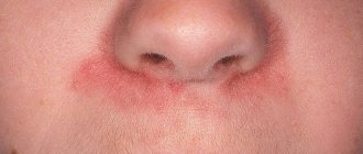
Causes of redness of the nasolabial triangle on the face. Treatment of an allergic reaction
What to do, how and with what to treat allergies on the face?
The decision must be made based on the speed at which symptoms appear. There are two options. Treatment methods are somewhat different.
Lightning-fast appearance In case of allergies, a patient's face swells, and red spots often appear. A doctor's consultation is required. If you have symptoms of angioedema, call an ambulance immediately. Before the doctor arrives, give the victim antihistamines. They will relieve swelling. Suprastin, Tavegil, Diazolin, Zyrtec, Cetrin are effective.
Pay attention to new generation drugs. They act just as quickly, but do not cause drowsiness. Write down the names: Lordestine, Fexofenadine, Norastemizole.
Don't you keep such products in your first aid kit? In vain! Be sure to buy one of these drugs. A simple precaution can save your or your children's life.
Hives are a milder form of an allergic reaction, accompanied by red spots on the face and a blotchy rash. Taking antihistamines is effective.
Slow-motion view Symptoms do not appear immediately; a rash appears on the face. Favorite locations are the cheeks, chin, and nasolabial triangle.
What to do and how to get rid of allergies on the face:
- find out the cause of the irritation. Remember what you ate and what medications you took. Perhaps you were on a picnic outside the city or trying out a new face cream;
- stop contact with the source that caused the allergy. Without complying with this condition, you risk getting a chronic form of the disease;
- Before visiting a doctor, gently wipe your face with a decoction of chamomile, calendula, string, and sage. Herbs have a calming, anti-inflammatory effect;
- make a compress with 1 tsp. boric acid dissolved in a glass of water. Apply wet gauze, re-wet it in the solution every 5 minutes, and hold it on your face again. Repeat 2-3 times;
- take an antihistamine tablet. The sooner you do this, the sooner the unpleasant signs will disappear;
- visit a dermatologist, allergist. Specialists will prescribe tests. The test is effective for identifying allergens;
- on the recommendation of a doctor, buy a suitable allergy medication;
- use chamomile cream for allergies. The product with natural ingredients moisturizes, disinfects, and soothes the skin;
- in severe cases, special allergy ointments are recommended. It is advisable not to use hormonal drugs, which have a number of side effects;
- If you have a food allergy, follow a diet. Eliminate foods that cause harsh skin reactions from the menu.
3. Symptoms and diagnosis
The smoothed nasolabial fold is visually perceived as less deep and prominent, with a simultaneous lowering of the associated angle of the mouth and facial muscles on the same side of the face. The severity of the symptom can be very different - from barely noticeable asymmetry to the complete absence of the fold (for example, with Millard-Gubler syndrome).
The localization of the symptom is also diagnostically important: in particular, with sensory cortical aphasia of Broca-Wernicke, the right nasolabial fold is always smoothed out.
The diagnosis, of course, is not the skin fold as such, but the neurological status of the patient. A detailed medical history is collected, a thorough neurological examination is carried out with the study of reflexes, skin sensitivity, muscle strength of the limbs, coordination of movements, and speech. Instrumental studies are prescribed as necessary; The most informative is tomographic methodology, but almost always the electrical activity of the brain is also studied using EEG; Dopplerography may be indicated as a way to measure major cerebrovascular vessels, etc.; in some cases, it is advisable to conduct a neuropsychological examination and/or other additional diagnostic methods.
About our clinic Chistye Prudy metro station Medintercom page!
