A rash is a change in the color and texture of the skin in the form of nodules, blisters, spots, and peeling. The skin of babies and older children is very sensitive to external environmental conditions and general health. Foot rashes in children are caused by allergies, infections, care errors, and autoimmune disorders. It may not bother you or may be accompanied by severe itching and pain. To combat it, it is important for parents and a doctor to find out the cause.
Allergic reactions
Allergic rashes on the feet of a child are caused by various factors. Depending on the type of irritant and the method of contact with it, they appear only in certain areas or throughout the body.
What causes allergies on the feet?
- Medicines. Hypersensitivity reactions are caused by both active and excipients in the drug. Most often it is provoked by antibiotics, sweet syrups, solutions, and lollipops. Within 2–3 hours after taking the product, a red rash, swelling, and itching appear on the child’s body and feet;
- Poisonous plants (poison oak, ivy, sumac). When contacting them on the child’s feet, blisters, a rash in the form of spots or pimples, and swelling form. In this case, you need to thoroughly wash your skin with soap and water and wash your clothes. To prevent future exposure to poisonous plants, teach your children what they look like. When walking, avoid places where they grow;
- Insect bites (bees, wasps, mosquitoes, ticks, fleas). In addition to itching, red rashes on the feet and legs of a child from insect bites, severe reactions are possible - anaphylactic shock and Quincke's edema. These conditions are life-threatening and require emergency care. To prevent bites, stay away from areas where insects congregate. Make sure your child does not run on the grass barefoot or in sandals with bare feet. In the summer, before going outside, apply protective creams or aerosols to exposed areas of your baby’s skin;
- Physical factors. Allergies can be caused by cold, heat, sun rays, pressure, compression. In this case, when freezing or overheating, the child develops a hives-like rash on the palms or soles, which turns pale when pressed. If you notice this reaction, take steps to prevent it from happening in the future;
- Cosmetics for skin care. If a cream or soap contains synthetic fragrances and dyes, then there is always a risk of allergies. Choose products with natural ingredients and no fragrances. Test the effect of a new product on your skin on a small area first.
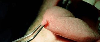
A tendency to allergic reactions can trigger eczema. This is a chronic disease with a hereditary predisposition. Very often it occurs in childhood. There are several types of eczema. One of them is dyshidrotic. Appears on the palms, soles, sides of the feet, and toes. The elements of the rash look like clusters of fluid-filled blisters up to 5 mm in size, accompanied by itching. After the bubbles open, erosions and crusts form.
If an allergy is detected, you need to be careful with irritants and, if possible, avoid contact with them. As first aid, give your child an age-appropriate antihistamine. Then be sure to contact your doctor to find out what to do next.
Infections
Infections that cause a rash on a child's feet include diseases caused by viruses, fungi, and parasites. They can affect only the feet or be systemic, spreading to the entire skin. The shape of the rash elements, their number, localization, nature, and speed of appearance are important diagnostic criteria.
Viral infections
With many viral diseases, in addition to a rash on the body and feet, the child has fever, weakness, loss of appetite, sore throat, and runny nose. Moreover, high fever is often the first symptom of infection, which appears several days before the rash. This development is typical for chickenpox, measles, rubella, roseola, and hand-foot-mouth disease (Coxsackie virus), which are easily transmitted by airborne droplets. These conditions resolve on their own; treatment is symptomatic.
The rash is different for each of them:
- Chickenpox. A rash in the form of blisters, which then burst, dry out, and become crusty. Appears first in the chest and back area. Quickly spreads to the face, head, including the scalp, abdomen, arms, legs. Causes severe itching;
- Measles. The rash appears on the third day from top to bottom. First, a brightly colored rash forms on the child’s face, neck, shoulders, then on the torso, arms, legs, and feet. The spots tend to merge with each other. The lower on the body they are, the rarer and less red they are. Pigmentation at the site of the spots lasts up to three weeks;
- Rubella. Pale pink spots appear immediately after the first symptoms and do not merge with each other. First - on the stomach and chest, then quickly spread to the whole body: arms, legs, face, back. And after a few days they disappear without a trace;
- Roseola (sudden exanthema). The rash appears as small, pink, flat spots or slightly raised bumps. Appears on days 3–5. First on the chest, back, then on the arms and legs. There are no other symptoms. The rash lasts up to 4 – 7 days. Roseola mainly affects children under 2 years of age;
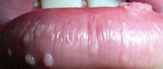
- Hand-foot-mouth disease. It is accompanied by ulcers in the mouth, a rash on the palms and soles of the child’s feet, and sometimes on the buttocks. The elements of the rash are painful, but not itchy. They look like flat red spots, sometimes with small bubbles. After 7–10 days they disappear without a trace.
For a photo of what a rash on a child’s feet and body looks like with the listed viral infections, see below:
Scabies
Scabies is an infectious disease caused by a microscopic mite. Infection occurs through clothing, bedding, towels, and washcloths. In children, parasites are transmitted through toys.
The disease is characterized by blisters, a rash elongated in a line, reminiscent of mite burrows (scabies), and itching that intensifies in the evening and at night. Typical places for a rash in a child are on the hands, feet, torso, lateral surfaces of the fingers, and in the spaces between the fingers.
If scabies is detected, the whole family should be treated. The doctor prescribes a special cream that destroys parasites. The product is applied not only to the rash, but also to the entire body, including the face, scalp, and ears. Antihistamine cream or hydrocortisone cream can help relieve itching. For effective treatment, you need to disinfect clothes, towels, and bed linen.
Fungal infection
Fungal skin infections are quite common. At risk, children who go to the pool wear unnatural or too hot shoes, which create a “greenhouse effect.” The elements of the rash are scaly pink spots of a round shape. With a fungal infection, the skin at the site of the infection becomes dry, begins to peel, and becomes covered with microcracks. Sometimes children experience itching and burning. To combat fungal infections, doctors prescribe local antifungal ointments, solutions, and creams.
Diseases that occur with pustular rashes on the palms and soles include palmoplantar psoriasis, palmoplantar pustulosis, acrodermatitis persistent pustular Allopo, and Andrews pustular bacteridus. There are disagreements among researchers in the interpretation of the concepts palmoplantar psoriasis and pustulosis. In some sources, palmoplantar pustulosis is classified as a separate nosological form, or is considered a type of palmoplantar pustular psoriasis [1]. There are localized pustular psoriasis of the Barber type (syn.: pustulosis of the palms and soles; chronic palmoplantar pustular psoriasis; persistent palmoplantar pustulosis; pustular psoriasis of the extremities). Many domestic scientists are of the opinion that various forms of pustular psoriasis are nosologically independent [2, 3]. At the same time, the question of whether non-infectious pustular dermatoses belong to pustular psoriasis continues to be discussed. Some domestic scientists [4] believe that in terms of clinical and morphological manifestations, palmoplantar pustulosis is closer to Andrews bacterid, and not to psoriasis. Some foreign authors [5] also consider palmoplantar pustulosis as a separate nosological entity. Data from genetic, immunological, and histological studies allow dermatologists to prove these statements. Perhaps this is due to the different meanings attached to the concept of “palmoplantar pustulosis”. In the generally accepted ICD-10, the form palmoplantar pustulosis (L40.3) is distinguished, but it is considered under the heading of psoriasis (L40), as well as acrodermatitis persistent Allopeau (L 40.2). With acrodermatitis persistent Allopeau, pustular rashes of a different localization may appear, sometimes similar to generalized pustular psoriasis, which also indicates that this disease belongs to psoriasis [5]. This issue is also important in terms of differential diagnosis, since errors in diagnosis and patient management tactics often occur. Damage to the palms and soles causes a lot of physical and emotional suffering for patients, disrupting performance, interpersonal relationships, and affecting the quality of life.
Localized pustular psoriasis (Barber type), a type of pustular psoriasis characterized by erythematous-squamous and pustular elements in the palms and soles [1], is widespread throughout the world, more common than the generalized variety of pustular psoriasis [6, 7], but significantly less common than psoriasis vulgaris. The disease occurs in the period from 20 to 60 years, mainly at the age of 40-50 years, very rarely after 60 years and only in 19% of cases before the age of 20; in women 3 times more often than in men [1], while psoriasis vulgaris more often affects men [5]. A family history of this form of psoriasis is noted only in 7-24% of cases; the connection with antigens of the HLA system has not been established [1]. Analysis of some genetic studies [5] allows us to assert the nosological independence of palmoplantar pustulosis and psoriasis, but other studies [8] do not allow us to identify clear differences between these pathologies, which coincide in many aspects. According to the Tyumen Regional Dermatovenerologic Dispensary for the period 2008-2012, limited palmoplantar psoriasis accounts for only 1.4% of the total number of registered cases of vulgar psoriasis and 2% of newly diagnosed cases of psoriasis (Fig. 1, 2)
.
Figure 1. Patient V., 33 years old, with a smoking history of 20 years. Clinical manifestations of palmoplantar psoriasis. Symmetrical lesions of the skin of the hands (a, b) and feet (c) with a typical plaque located on the skin in the lower leg area (d).
Figure 2. Patient K., 51 years old, with a smoking history of 35 years.
Clinical manifestations of palmoplantar psoriasis. Symmetrical lesions of the skin of the hands (a) and feet (b). The exact cause of localized pustular psoriasis is unknown. Provoking factors are foci of focal infection, primarily odontogenic and tonsillogenic [1], Helicobacter pylori
, as well as stress, anxiety, the use of certain medications, and liver dysfunction [2]. Sometimes the disease first appeared after long-term treatment with lithium drugs. Smoking and unfavorable environmental factors (high humidity and temperature) aggravate its course, and a more pronounced association has been identified between palmoplantar pustular psoriasis and smoking, especially in women [1, 8]. Smoking cessation was an important treatment intervention given the observed abnormal response to nicotine [5].
Localized pustular psoriasis occurs with or without psoriasis vulgaris rashes, most often in patients with a personal or family history of psoriasis, although often independently [1].
A possible mechanism for the formation of pustules in pustular psoriasis is considered to be a decrease in the activity of antileukoprotease in the skin as a result of a protease/antiprotease imbalance [5]. To date, extensive material has been accumulated on the significant role of infectious agents in the occurrence, spread and activation of the psoriatic process [6]. The contents of the pustules are sterile, although some authors, in particular G.Ya. Sharapova et al. (1983) noted the presence of Staphylococcus aureus in some patients. According to numerous studies [7], microorganisms that sensitize the patient’s body against psoriasis are often streptococci and Staphylococcus aureus, as well as foci of chronic infection. Staphylococcus aureus and streptococci secrete exotoxins that play the role of superagents capable of binding to proteins of the major histocompatibility complex of tissues on antigen-presenting cells, keratinocytes, T-lymphocytes, monocytes, which leads to the formation of cytokines that enhance the proliferation of keratinocytes [5].
Subjective symptoms boil down to burning, itching and a feeling of discomfort in the palms and soles; often these sensations precede the appearance of fresh elements. Cases when in the early stages the disease is represented by single plaques or is asymmetrical in nature are rare [1]. With severe severity of rashes, the quality of life of patients is significantly reduced due to pain, inability to stand, walk, or work with their hands [5].
Clinically, on a scaly erythematous background with sharp boundaries, there are pustules, which, unlike generalized pustular psoriasis, are located deep in the epidermis, which is due to the morphological structure of the skin of the palms and soles. Localized pustular psoriasis is manifested by sterile yellow pustules with a diameter of 2-5 mm, approximately the same size, clusters of which within a few hours appear symmetrically on the apparently healthy skin of the palms, mainly in the thenar area, less often - the hypothenar, even less often - the middle and distal part of the palm and soles, mainly in the area of the arch of the foot [1]. Subsequently, a halo of hyperemia appears around individual elements. New rashes are associated with places of favorite localization and rarely spread to the back of the hand and foot, wrists, and fingers. The lesion is usually symmetrical, but a unilateral arrangement of elements may be observed. Sometimes the pustules spread to the dorsal surface of the fingers, toes, or the inside of the wrist. The skin of the terminal phalanges of the fingers is usually not affected. The disease lasts for years, with off-season exacerbations followed by remissions [9]. Episodes of new pustular eruptions occur over varying periods of time and are strictly limited to their preferred locations. Occasionally, lesions of ordinary psoriasis appear, but more often the rash remains localized [10].
Subsequently, the color of the pustules changes to yellow-brown and dark brown. Without opening, the pustules dry out to form brown crusts, which resolve within 8-10 days. The presence of pustules at different stages of evolution (evolutionary polymorphism) gives the lesion a multi-colored color. Later, the pathological process manifests itself as sharply defined erythematous-squamous plaques, within which and along the periphery there are multiple yellow and brown pustules, some of which merge to form “purulent lakes” [1]. Remission begins with a decrease in the number of new pustules, although the skin of the palms and soles may remain erythematous, erythematous-squamous, or hyperkeratotic, clinically resembling eczema. Remissions continue for weeks or even months [11]. Thus, unlike other forms of psoriasis, the course of pustular psoriasis of the palms and soles is chronic. According to W. Enfors and L. Molin [1], at the time of examination 10 years after the diagnosis of pustular psoriasis of the palms and soles, its manifestations were absent in only 28% of patients.
Localized pustular psoriasis is often associated with hyper- and hypothyroidism, diabetes mellitus, arthropathy; it is often diagnosed in patients with SAPHO syndrome (synovitis, acne, pustulosis, hyperostosis, osteitis), namely with chronic recurrent multifocal osteomyelitis, lesions included in this syndrome sternoclavicular and sternocostal joints, pustular arthroosteitis, peripheral arthritis, pseudo-infectious arthritis involving the sacroiliac joints in the pathological process [1, 5].
In the differential diagnosis, fungal, scabies, eczematous dermatitis, and Andrews pustular bacterium are excluded.
The diagnosis of pustular psoriasis of the palms and soles is established on the basis of the characteristic clinical picture and course of the disease, and in difficult cases should be confirmed histologically.
Andrew's pustular bacterium
(syn.: Lever's pustulosis of the palms and soles) is a type of pustulosis of the palms and soles, which some authors classify as localized pustular psoriasis [1, 5]. A.A. Kalamkarian et al. [12] deny the existence of this disease as a nosological form. This variant of pustulosis was isolated by G. Andrews in 1934 and is characterized by the formation of small pustules with sterile contents on the unchanged skin of the palms and soles.
The etiology and pathogenesis are not clear, but since the occurrence of the disease is facilitated by foci of chronic infection in the body, especially chronic tonsillitis [1], chronic recurrent osteomyelitis, osteoarthritis [7], an infectious-allergic origin of the process is assumed, or more precisely, a hypersensitivity reaction to streptococcal antigens.
Clinically, the disease manifests itself as multiple blisters and pustules on the palms and soles. The rashes, as a rule, are symmetrical, starting from the center, gradually covering the entire surface of the palms and soles, including the lateral parts, but the fingers are not involved in the pathological process. The blisters quickly turn into pustules, which grow rapidly. They are deeply embedded, located on apparently healthy skin or partially surrounded by a narrow rim of erythema [6]. When merging, the diameter of the elements sometimes reaches 0.5-1.0 cm. Patients are concerned about itching and soreness [1].
The course is recurrent. The process lasts 2-3 weeks, possibly 1-2 months. Remission occurs after the elimination of the factor provoking the disease, the infectious focus. Relapses are accompanied by more intense itching and pain. The process may be protracted, but atrophic changes do not occur [9].
Acrodermatitis pustularis persistent Allopo
(syn.: persistent Crocker dermatitis, persistent Setton dermatitis) is a dermatosis of unknown origin, characterized by pustular non-bacterial rashes localized in the acral zones (fingers and toes) [1]. This is a rare disease of pustular, sterile eruptions on the fingers or toes that progress slowly in a proximal direction. Subsequently, prolonged pustulization causes destruction of the nail and atrophy of the distal phalanx [5].
In 1888, H. Crocer described recurrent bullous and pustular rashes on the hands and feet, and later F. Hallopeau (1890-1897) and R. Sutton (1911) gave a detailed description. Some authors classify it as a localized form of pustular psoriasis, while others, based on the similarity of the histological picture, classify it as a localized form of Gebra's impetigo herpetiformis, Dühring's dermatitis, and enteropathic acrodermatitis. There is also an opinion about it as an independent disease [1, 10, 12]. The disease develops at any age, more often in men; provoking factors can be trauma, pyoderma, and zinc deficiency [6].
The clinical picture is characterized by lesions of the pustular, vesicular or erythematous-squamous nature of the terminal phalanges of the fingers, less often the feet, and a gradual transition to adjacent areas of the hands and feet without proximal spread. The lesion can be one-sided for a long time [7]. Initially, small pustules appear, leaving a shiny surface against an erythematous background on which new pustules develop [5]. In some cases, secondary atrophic changes in the skin are observed. Pathognomonic lesions of the nails, usually one finger, leading to involvement of the nail bed in the pathological process, to onycholysis, onychomadesis. Features of clinical manifestations mainly depend on the intensity of exudation processes. If they are insignificant, erythematous-squamous changes are found in the lesions with increased redness along the periphery, layering of dry shiny scales, and multiple superficial cracks. If pustular rashes dominate the clinical picture, the disease is more severe [6].
There are seven clinical forms of the disease [1].
In purulent form
At first, the process resembles paronychia: hyperemia and swelling of the nail folds develop, from under which pus is released when pressed. The affected areas of the fingers are red, swollen, covered with multiple pustules, merging into “purulent lakes” of a wide variety of shapes [6]. Pustules appear on the skin of the entire nail phalanx (club-shaped-thickened), turning into erosions, crusts, and scales. After opening the pustules, small erosions are formed, covered with scaly crusts; after removing the layers, new pustular elements become visible through the reddened skin. The rash is accompanied by a burning sensation and pain. Flexion and especially extension movements are limited due to severe pain, the fingers are in a half-bent position. Pustules are sterile. Radiographs sometimes reveal atrophic changes in the bones of the corresponding phalanges, osteitis, osteoporosis, and possible mutilations. At the site of regression of pustules, the skin is slightly atrophic, shiny, and has a reddish tint. The nail plates are dystrophically changed and, as a rule, are rejected.
Vesicular
(bullous) form is characterized by a similar clinical picture, but instead of pustules, vesicles or, less commonly, blisters appear; mild itching and soreness are a concern [1].
With erythematous-squamous
(abortive) form, the clinic is limited to the appearance of hyperemia, peeling, cracks.
Phlyctenulous
this variety is more malignant: conflicts spread to the palms, soles, and sometimes to the ears; the process can lead to atrophy and mutilation of the terminal phalanges.
With vegetative
form, miliary pustules are grouped into plaques with peripheral growth and a tendency to resolve in the center; The formation of semi-soft vegetation along the periphery is characteristic.
Generalized form of Аudry
distinguishes a malignant course with the spread of the process to the entire skin, primarily to the hands, groin areas, genitals, elbows and thighs. There is clinical similarity to Hebra's impetigo herpetiformis. Damage to the nails and periungual tissues is often observed. In this case, the clinical process is characterized by onycholysis, often with complete loss of the nail plate.
In Allopeau's acrodermatitis, damage to the mucous membranes (as in generalized pustular psoriasis) is characterized by migrating ring-shaped erythematous elements located in the tongue area, covered with swollen white scales (annulus migrans). With the long-term existence of the process, signs of atrophy of the skin and muscles of the fingers appear, mutating changes due to trophic disorders. The prognosis for life is favorable, but the course is long, often relapsing, resistant to therapy. Spontaneous improvement is rare, and episodes of acute pustulization appear for no apparent reason [5].
Thus, in the presence of pustular rashes on the skin of the palms and soles, it is necessary to have knowledge of differential diagnosis in order to develop management tactics for this category of patients (see table)
.
Limited palmoplantar pustulosis can be understood in the meaning of palmoplantar psoriasis, since there are no significant differences in the categories of patients and the clinical picture that make it possible to distinguish this pathology into a separate nosological group. The presence of typical psoriatic elements of a different localization in patients with palmoplantar pustulosis also confirms this. Therapeutic measures in both cases correspond to the general principles of treatment of psoriasis.
Care
Rashes often appear in children due to improper care. It is not dangerous to health and quickly disappears on its own when errors are corrected.
In newborns and babies up to 3 months, a rash on the face, arms, and torso is a normal physiological phenomenon caused by hormonal changes and the body’s adaptation to new conditions. This condition is called neonatal acne. It looks like small red pimples all over the body.
If the baby's skin has been wet for a long time or overheated, then sometimes a small pinpoint rash appears on the child's feet. A common cause is shoes that are too hot. Therefore, if possible, choose breathable shoes for your baby made from natural materials with good thermoregulation. For treatment, baths with decoctions of string, chamomile or oak bark help.
Coxsackie virus: calm, only calm
The news of an outbreak of infection with this virus shook up the ranks of vacationers in hot countries and their compatriots spending the summer at Russian resorts. Citizens frightened by the “epidemic” are hastily looking for a panacea, and those who have a vacation ahead of them, in a panic, decide to completely abandon the tours to the coast purchased in advance. And if you cool down and calmly figure out what kind of virus this is with the mysterious name Coxsackie and what disease does it cause?
Who is Coxsackie?
These enteroviruses were discovered by chance in 1949, when American scientist Gilbert Dalldroff was testing the feces of polio patients to create a cure or vaccine. The attempt to create a cure for polio failed, but Dalldroff went down in history as the discoverer of enteroviruses, to which he gave the name Coxsackie - after the small town on the Hudson River, from where the scientist received the first samples of material for research.
Despite the rather exotic sounding name, Coxsackie viruses do not represent anything extraordinary. These are common enteroviruses that thrive in the human gastrointestinal tract. They are distributed throughout the world, their activity depends on the season and climate.