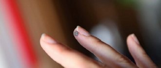
How to remove a callus from a finger?
The situation with the appearance of calluses on the hands is familiar to every person. The symptom may be caused by the specifics of work or overexertion on the eve of the event. It’s easy to get rid of negative manifestations.
For this, there is an arsenal of folk methods and pharmaceutical preparations. It is necessary to first understand the reasons for the formation. By excluding them from your life, you will be able to prevent the appearance of calluses in the future.
Symptomatic means problematic
The growths are visible to the naked eye. The reason for their occurrence can be judged by their appearance.
Hard and dense lumps on the joints, covered with smooth skin, usually form in men over 40 years of age and indicate gout. They are usually called gouty tophi. They grow during periods of exacerbation of the disease. The patient becomes weaker and complains of malaise. The temperature in the area of the tophi rises, and the pain sometimes becomes unbearable.
If the skin on the lump is rough and red, and the growth itself is round and elastic, it is a hygroma. It usually forms in women 20-30 years old and can be single-chamber or multi-chamber. Hygromas differ from other types of growths in that their size gradually increases, and pain is felt when pressed.
Formations on the interphalangeal joints that appear after 60 years are called Heberden's nodes. They are equally likely to be found on any phalanx and are signs of osteoarthritis. This disease leads to stiffness of the hands and deformation of the joints.
Nodules up to 2 cm in diameter, which form in 20% of patients with rheumatoid arthritis, are rheumatoid nodules. They do not cause pain, but the course of the disease is accompanied by other unpleasant symptoms: lethargy, fatigue, fever, and sometimes loss of body weight.
Types and causes of callus formation
The formation of callus occurs for the following reasons:
- Senile age.
- Radiation sickness.
- Obesity.
- Pregnancy.
- Syphilis.
- Osteoporosis.
- Amyloidosis.
- Exhaustion.
- Hypoproteinemia.
- Tuberculosis.
- Endocrine disorders.
- Avitaminosis.
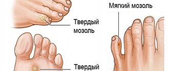
Among the forms of calluses are:
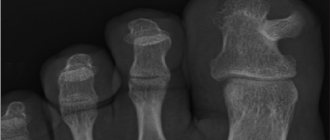
- Periosteal or external. Grow after a bone fracture, rapid healing is possible.
- Intermedial. Appear in the space between fragments of broken bones. The zone is filled with a network of vessels. There is a high probability of eliminating the callus.
- Endosteal or internal. Occurs in bone marrow cells.
- Paraosseous. Unfavorable form. The callus grows from soft connective tissue and healing is slow. Forms a large protrusion.
Diagnostics is everything
To identify the root cause of the disease and select an effective course of treatment, the doctor gives the patient appointments for ultrasound, X-ray, MRI, and urine testing. The study of uric acid salts will determine the presence of gout. An x-ray will reliably demonstrate the presence of osteoarthritis or rheumatoid arthritis. However, the results of a visual examination and verbal complaints from the patient are often sufficient to make a diagnosis.
Hallux valgus
In fact, it’s not even a growth, but a protrusion of the big toe, or a violation of the anatomy of the foot. In other words, this is an acquired skeletal deformity. For what reasons does this disease appear? It could be:
- Tight, uncomfortable shoes. When the foot remains in an uncomfortable position for a long time, the bones begin to shift so that they take on shapes similar in shape to a pair of shoes.
- Increased load. Most often we are talking about athletes. The risk group includes people who professionally lift weights (bodybuilding, weightlifting) or who have prolonged activity on their feet (dancers, football players). Increased load on the forefoot is also present in those women who prefer to walk in high heels. A pair of shoes leads to such consequences if the heel is over 5 cm.
To correct the situation, you first need to eliminate the source, the cause of this condition. Of course, few people will decide to give up professional training, but you can temporarily limit the load. If it's all about the shoes, then you need to replace them with anatomically correct ones. By the way, it is absolutely not necessary to buy shoes in specialized stores, because they cost a lot of money there.
It is enough to purchase special correctors. They can be made of plastic or silicone and are inserted into shoes from the inside. Such correctors fix the finger in the correct position and evenly distribute the load. They should be worn for a long time, several months.
Over time, the arch of the foot will acquire its previous correct shape. Most likely, the patient will be prescribed a course of anti-inflammatory or painkillers, since hallux valgus is usually combined with other diseases (bursitis, arthrosis, etc.).
How to treat a lump on your thumb that hurts
Unfortunately, this problem is usually caused by chronic diseases and cannot be completely cured. Only hygroma can be removed once and for all. All other types of growths will remain for life - but exacerbations can be avoided if stable remission is achieved.
Rheumatoid arthritis is treated with corticosteroids or non-steroidal anti-inflammatory drugs. These same non-steroidal anti-inflammatory drugs, as well as colchicine, are effective against gout.
For osteoarthritis, chondroprotectors, finger therapeutic exercises and physiotherapy sessions are prescribed: multi-channel electrical stimulation and laser therapy. With multichannel electrical stimulation, muscles are stimulated with short pulses of electricity to relieve tension and improve blood flow. During the laser therapy procedure, a laser beam is applied to the joint growth to relieve inflammation and activate blood flow within the tissues.
Osteoarthritis is also treated with physiotherapy: ultrasound (reduces tissue volume), shock wave therapy (activates blood flow, eliminates swelling and pain by exposure to acoustic impulses), laser therapy.
Surgery may be required if the cartilage and surfaces of the joints are severely damaged in the later stages of arthritis or arthrosis, and the fingers become swollen, stiff and painful. In especially severe cases, endoprosthetics is prescribed, when a worn-out joint is replaced with an artificial one. The operation can be performed in the presence of a malignant tumor. The oncologist prepares an individual therapy program for each patient.
Traditional medicine is powerless when there is a lump and pain on the finger joint. Not a single homeopathic remedy contains components that would dissolve the lump. Therefore, relying on chance in the case of such a disease is extremely undesirable: all hopes for a miracle can ultimately lead to serious consequences.
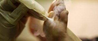
Removal of callus with liquid nitrogen (cryodestruction)
Cryodestruction is an innovative, highly effective method of removing calluses on the foot, heel or toes using liquid nitrogen.
During the procedure, the keratinized area of the skin is frozen, after which the destruction of the callus is observed.
It has been noted that the procedure is associated with an extremely low rate of callus re-development.
Many dermatologists note that no other method for removing core calluses gives results as favorable as cryodestruction.
Removing callus with liquid nitrogen takes about 10-20 minutes.
It is performed under local anesthesia, so the procedure is completely painless and safe.
Side effects are extremely rare (no more than 3% of cases).
Include callus recurrence, pain, discomfort, bleeding, infection, scarring.
The first few days after the procedure, the formation of a cold bubble is observed, which under no circumstances should be pierced.
Within 10-14 days, the healing process occurs; a crust forms in place of the bubble, which falls off on its own.
The process is not accompanied by the formation of scars.
The procedure is usually well tolerated, with minor discomfort possible.
Some disadvantages of cryotherapy:
- If the rod is extremely deep, cryodestruction is not always able to completely rid the patient of the callus.
- If you do not properly care for the skin after the procedure, there is a risk of an infectious process.
- Freezing the callus with liquid nitrogen is not used when large areas of the epidermis are affected. Cryodestruction used on large calluses can lead to a number of complications. For this reason, in advanced cases, they resort to other therapeutic methods.
A few words about prevention
The best prevention for growths is a healthy lifestyle: a balanced diet, regular consumption of fresh vegetables and fruits, giving up bad habits, sweets and fried foods. Wear protective gloves before handling chemicals. Avoid stress and lack of sleep.
If you are a schoolchild or student, you have a lump on your finger and it hurts - you probably write too much and hold the pen incorrectly. In this case, the growth is an ordinary callus that will go away over time. There is no need to put a band-aid on it or try to get rid of it by other methods. It's better to buy a soft-bodied pen, try to hold it a little differently and not squeeze too much with your fingers.
Treatment of callus with medications
It is immediately worth noting that drug treatment of calluses has the least therapeutic effectiveness.
This phenomenon is associated with the inability of drugs to penetrate deep into tissues, eliminating the root of the formation.
The products used for core calluses often contain keratolytic components and can be effective at the beginning of callus development.
As a rule, products based on salicylic acid are used.
Salicylic acid is a keratoplastic and keratolytic agent.
In addition, it has an antibacterial and antifungal effect.
Salicylic acid works by softening keratin, a natural protein that is part of the skin's structure and produced by the body.
The drug has a softening effect on the stratum corneum of the dermis, which facilitates its removal.
Salicylic acid is often used in combination with other substances.
Allows for a higher therapeutic effect.
When using salicylic acid preparations, the simultaneous use of the following drugs is not recommended:
- Alcohol-containing medications.
- Any other local medications that contain dibenzoyl peroxide or retinoid.
- Soapy substances that have a drying effect.
- Cosmetics that have an exfoliating effect.
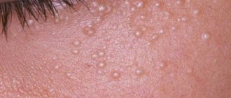
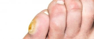
Hygroma
Hygroma is a lump that often appears on the phalanges of the toes, especially the big and little fingers. Inside the finger, with this benign formation, a capsule is formed with liquid contents inside (fibro-mucosal or fibro-serous fluid). Most often, the growth does not stand out much above the rest of the finger. It is stable, that is, it does not move, since it is closely connected to the tendon apparatus.
Such small hygromas may not cause problems to a person, and therefore many simply do not notice them. As the size increases, painful sensations appear, because the formation affects neighboring areas with nerve endings. If such symptoms are ignored, a person may experience limited mobility of the toe joints.
The most effective method of treating hygroma is puncturing it, that is, puncturing the area where the fluid is localized. This is usually combined with physiotherapeutic methods that restore blood microcirculation and improve local immune processes. In some cases, when a capsule with liquid has just formed, it may be reabsorbed on its own when the load on the foot is reduced.
Dermatofibroma
This formation, unlike the previous one, has a solid structure. It often appears on the top of the fingers, on the back side. To diagnose it yourself, just try to collect a patch of skin in this place. If a dermatofibroma is present, the skin will pull inward into a fold.
Normally, this area of skin has a darker reddish tint. If the dermatofibroma grows to a large size, then in the center you can clearly see a tubercle, more solid in structure than the entire formation.
Causes of dermatofibroma:
- insect bite;
- untimely opened boils;
- penetration of foreign objects into the thickness of the skin.
Its main danger is that it has the ability to degenerate into dermatofibrosarcoma, and this is an oncological diagnosis with all that it implies. And, of course, it causes a lot of inconvenience to a person, preventing him from leading a normal active life. It is almost impossible to treat dermatofibrosarcoma at home.
The thing is that this formation lies deep under the thickness of the skin layers. It is most reliable to remove it using the traditional surgical method.
Classification
Depending on the nature of the course of the disease, primary gout is distinguished - an independent disease that occurs against the background of exposure to provoking factors, and a secondary form of the disease, which develops against the background of concomitant pathology. Based on the prevalence of the pathological process, gout is divided into the following types:
- Monoarticular arthritis. Urates accumulate in the area of one joint. Characteristic of the onset of the disease. The most common site of monoarticular arthritis is the big toe joint;
- Polyarticular arthritis. Inflammation affects more than 2 joints. Polyarticular arthritis is characteristic of chronic gout. Localized in the joints of the elbows, wrists, legs, feet and hands.
Make an appointment