Exostosis of the finger is a rare and not serious disease, but it can significantly reduce the quality of life of patients. Neoplasms of this kind are rarely found on the hands; more often they form in the feet, especially on the big toe. By exostosis we mean an osteochondral growth on the surface of the phalanx bone in the form of a linear, spherical or comb-shaped formation. It can form on any part of the bone, including under the nail plate. In the latter case, they speak of the presence of subungual exostosis.
Reasons for development
Exostoses of the fingers and toes may represent an osteochondroma, i.e., a benign tumor, or be a consequence of injury, chronic inflammation, or even prolonged wearing of tight shoes. The latter factors more often provoke the formation of exostoses in adults.
Osteochondromas are mainly typical for children and adolescents under 20 years of age. They can be single or solitary, as well as multiple. Solitary osteochondromas of the fingers and toes are a rare occurrence. More often, in the presence of exostoses with such localization, similar lesions of other skeletal bones are found, in particular the femur, tibia, humerus, spine, clavicle, etc.
The reasons for the formation of osteochondromas have not yet been fully established. It is believed that isolated neoplasms of this kind are a consequence of displacement of the epiphyseal plate. This may be due to disorders of embryonic development, radiation therapy at an early age, or exposure to other types of ionizing radiation. Epiphyseal plates are areas of bone growth that are made up of cartilage and are located directly under the “head” of the bone. Due to the fact that its cells are in the process of constant mitotic division, the child’s bones lengthen as they grow older. Subsequently, the cartilage cells located furthest from the epiphyseal plate ossify and form bone tissue.
If, due to the action of one factor or another, a fragment of the epiphyseal plate moves to the side, it continues to synthesize new cells, which also gradually ossify. This is how exostosis forms in children. Initially, it is represented only by cartilaginous tissue, but over the years it becomes dense and hard, but the cartilaginous cap remains. It usually grows in proportion to the rate of bone growth, and therefore is usually detected during puberty, when a sharp growth spurt occurs.
Multiple exostosis disease is considered to be a hereditary disease. Massive skeletal damage by exostoses is usually detected in early childhood and requires dynamic monitoring, since with it the likelihood of malignancy of neoplasms increases. Single exostoses become malignant in less than 1% of cases.
A few words about prevention
The best prevention for growths is a healthy lifestyle: a balanced diet, regular consumption of fresh vegetables and fruits, giving up bad habits, sweets and fried foods. Wear protective gloves before handling chemicals. Avoid stress and lack of sleep.
If you are a schoolchild or student, you have a lump on your finger and it hurts - you probably write too much and hold the pen incorrectly. In this case, the growth is an ordinary callus that will go away over time. There is no need to put a band-aid on it or try to get rid of it by other methods. It's better to buy a soft-bodied pen, try to hold it a little differently and not squeeze too much with your fingers.
Symptoms
In some cases, exostosis of the toe or hand is asymptomatic. If it forms on the side of the finger, it can cause manifestations of soft tissue hyperkeratosis. But since their volume is relatively small, a full-fledged callus is not formed. If you remove areas of thickened skin, the discomfort does not go away, and the tissues soon become keratinized again.
When exostosis reaches a large size, it injures soft tissues and provokes inflammatory processes in the joints. This leads to discomfort or even pain, especially when wearing tight shoes. It can also protrude beyond the physiological boundaries of the finger. When palpated, exostosis is a dense bony protrusion with a smooth or rough surface. This further aggravates the discomfort.
With active growth of the tumor, the phalanx may become deformed, as well as neighboring fingers. This already leads to the development of an aesthetic defect.
At a certain location, exostosis can compress the neurovascular bundle. The consequence of this is local swelling of the finger, a feeling of numbness or goosebumps.
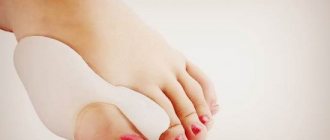
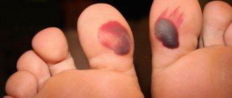
The most common condition is exostosis of the big toe. This may be accompanied by valgus deformation of this finger, which is manifested by its deviation from the normal axis towards neighboring fingers. As a result, 2-3 toes may also become deformed, acquiring a hammer-like shape.
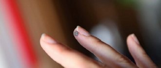
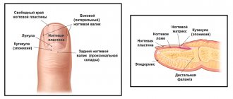
With subungual exostosis, there is a protrusion at the end of the phalanx of the finger, which looks like a thickened subungual ridge. In this case it is observed:
- pain when pressing on the nail, fingertip, or during physical activity;
- impaired nail growth, up to ingrowth or peeling;
- redness of soft tissues;
- callus formation.
Why do warts appear?
The formation is caused by the human papillomavirus (HPV) of various types (currently there are more than 100 varieties). Infection occurs through contact (from person to person) and household contact (through common objects, contaminated surfaces - swimming pools, saunas, gyms). Cases of self-infection are common.
The virus that enters the body through microtrauma actively multiplies in the surface layers of the skin. The latent period lasts 1-6 months, then typical nodules appear.
In practice, most often we encounter flat and vulgar warts. Vulgar ones are predominantly located on the dorsum of the hands. These are multiple painless dense rounded gray papules with an uneven, keratinized surface of a flesh-colored or yellow-brown color. Flat warts are located on the back of the hands, forearms, as well as the face and mucous membranes. Clinically, they are represented by small multiple papules the color of normal skin. On the surface of both, black dots can be seen - these are thrombosed capillaries.
Diagnostics
The appearance of signs of exostosis requires contacting an orthopedist-traumatologist. At the appointment, the doctor carefully examines the finger, palpates the growth and finds out the nature of the symptoms. If a dense bone formation is detected, an x-ray is indicated.
With its help, you can not only diagnose exostosis of the toe, but also evaluate its location and size. The images also provide data on the degree of deformation of the distal phalanges and allow you to plan the most effective course of treatment. In rare cases, CT and MRI are additionally prescribed.
What does it look like?
Warts are small, dry, flesh-colored lumps. They have a rough surface and are sometimes painful. On the hands, this can lead to a pronounced cosmetic defect.
When warts appear on the feet, severe pain appears, making it difficult to walk. Another type of wart, genital warts, develops on the genitals. This can make intimate relationships difficult. It is important to note that in women, condylomas of this localization can be caused by strains of the virus that increase the risk of cervical cancer.
Such situations require immediate treatment.
Treatment of exostoses of fingers and toes
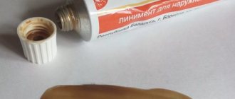
To relieve pain and inflammation, patients are prescribed drug therapy. It is selected individually depending on the complexity of the situation and the nature of the patient’s chronic diseases. Most often, NSAIDs are prescribed in the form of ointments, gels, creams or oral forms. But their use does not lead to the resorption of the osteochondral growth, but only helps to eliminate the symptoms.
The only effective way to treat exostosis of the phalanx of the finger is surgery. It is shown when:
- large amounts of exostosis;
- finger deformities;
- persistent pain syndrome;
- the development of complications or the appearance of signs of malignancy.
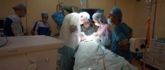
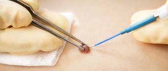
The operation is not technically difficult and can even be performed under local anesthesia. They mainly resort to the method of marginal resection of the tumor. It involves making a transverse incision in the projection of the bone growth. Its magnitude depends on the size of the formation, but is usually on the order of several millimeters. The soft tissue is carefully separated from the bone to obtain a clear view of the exostosis and accurately determine its boundaries.
After this, using a surgical chisel or other instrument, the growth is carefully removed within healthy tissue. It is important for the surgeon to completely remove the entire tumor along with its cartilaginous cap, since otherwise there is a high risk of relapse. The surgical wound is actively washed with saline to wash out the smallest bone particles, and only then is it sutured and covered with a sterile bandage.
If a patient is diagnosed with deformity of the phalanges of the fingers, a corrective osteotomy is indicated. The operation involves not only removing the osteochondral exostosis, but also cutting the bone, followed by juxtaposition of the resulting fragments in such a position that the phalanx acquires an anatomically correct shape. The bone is fixed with special metal systems in a given position, after which the wound is sutured and covered with a sterile bandage.
Features during operations
The mechanism of development of Taylor's pathology or tailor's disease largely mirrors the pathogenesis of hallux valgus in Hallux Valgus. this allows surgeons to use identical treatment and surgical techniques.
The purpose of surgical intervention is to restore the biomechanical connections in the forefoot, improve the condition of the ligamentous apparatus, and eliminate the aesthetic defect. It is necessary to remove the valgus deformity of the fifth metatarsal bone and return the parabola of the little finger to its natural position.
Rehabilitation
The duration and complexity of the recovery period are determined by the type of surgery performed. After marginal resection, discharge can be carried out on the day of surgery, but patients are advised to limit physical activity for 2 days. Drug therapy is also prescribed to reduce the risk of developing infectious complications and eliminate pain. After 2 days, a dressing is required, the stitches are removed after 7-10 days.
When performing a corrective osteotomy, recovery is more complex and lengthy. It involves immobilization of the operated finger, which is necessary for the healing of an artificial fracture.
Thus, exostoses of the fingers and toes are a rare phenomenon, but can significantly reduce the level of physical activity, cause cosmetic defects, pain, and generally worsen the quality of life. The solution to the problem is only possible through surgery. In this case, the operation is usually simple and does not require complex recovery. The main thing is to contact an orthopedic traumatologist as soon as possible after signs of exostosis appear, before its active growth provokes deformation of the fingers.
How to treat a lump on your thumb that hurts
Unfortunately, this problem is usually caused by chronic diseases and cannot be completely cured. Only hygroma can be removed once and for all. All other types of growths will remain for life - but exacerbations can be avoided if stable remission is achieved.
Rheumatoid arthritis is treated with corticosteroids or non-steroidal anti-inflammatory drugs. These same non-steroidal anti-inflammatory drugs, as well as colchicine, are effective against gout.
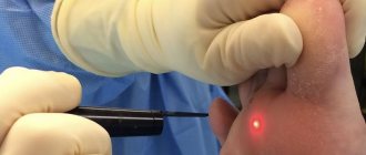
For osteoarthritis, chondroprotectors, finger therapeutic exercises and physiotherapy sessions are prescribed: multi-channel electrical stimulation and laser therapy. With multichannel electrical stimulation, muscles are stimulated with short pulses of electricity to relieve tension and improve blood flow. During the laser therapy procedure, a laser beam is applied to the joint growth to relieve inflammation and activate blood flow within the tissues.
Osteoarthritis is also treated with physiotherapy: ultrasound (reduces tissue volume), shock wave therapy (activates blood flow, eliminates swelling and pain by exposure to acoustic impulses), laser therapy.
Surgery may be required if the cartilage and surfaces of the joints are severely damaged in the later stages of arthritis or arthrosis, and the fingers become swollen, stiff and painful. In especially severe cases, endoprosthetics is prescribed, when a worn-out joint is replaced with an artificial one. The operation can be performed in the presence of a malignant tumor. The oncologist prepares an individual therapy program for each patient.
Traditional medicine is powerless when there is a lump and pain on the finger joint. Not a single homeopathic remedy contains components that would dissolve the lump. Therefore, relying on chance in the case of such a disease is extremely undesirable: all hopes for a miracle can ultimately lead to serious consequences.