| "Before and after" | Prices | Reviews | Doctors | Sign up |
Milia are dense formations in the superficial skin layer (epidermal cysts), in the form of white or yellowish nodules.
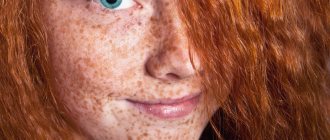
Milia rise above the surface of the skin as small white pimples from 0.5 to 2 mm. They are also called millet cysts, miliary cysts. Most often, milia are multiple and are located on the eyelids and under the eyes, around the eyes. They also occur in other areas of the face, less often on the body.
How to get rid of milia?
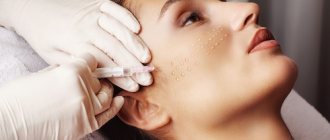
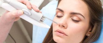
Today, the “gold standard” is CO-2 laser removal of milia
Benefits of laser milia removal:
- no direct contact with skin
- bloodless aseptic method
- high-precision impact
- does not affect healthy tissue
- effectiveness of the method
- excellent cosmetic result
At the Gradient clinic, milia removal is performed by experienced dermato-oncologist surgeons. The doctor will carefully examine the formations, make sure of their origin, carefully remove them and prescribe subsequent skin care at the sites of removal.
Causes
Milia occur in patients with a thick form of oily seborrhea, characterized by hyperfunction of the sebaceous glands and changes in the chemical composition of sebum and creating a favorable background for the development of acne.
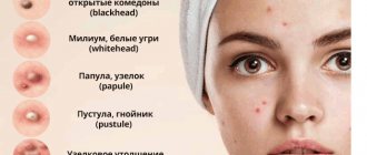
There are closed comedones - whiteheads (milia) and open comedones - blackheads (blackheads). Thick seborrhea is also characterized by the appearance of deep cysts of the sebaceous glands - atheromas and acne vulgaris. Milia have the appearance of small rounded dense nodules of a white-yellowish hue, 0.5-3 mm in size with clear boundaries, protruding above the surface of the skin and resembling millet grains (popular name - “millet”). Milium is characterized by an uninflamed head, since the whitehead does not have a natural outlet and contact with the environment. Milia do not change in size for a long time. When microorganisms enter the milium, inflammation may develop with the formation of an abscess. Milia (milia) can often be found on the wings of the nose in recently born babies; they are considered borderline conditions in newborns and soon disappear spontaneously without any treatment.
Milia are recognized based on an examination of the skin by a dermatologist-cosmetologist based on characteristic external signs.
Problems of the elderly. Eyelid diseases: diagnosis and treatment
| Figure 1. Entropion of the upper eyelid with cicatricial pemphigus |
What are the most effective methods to control the development of blepharitis? When to operate on patients with entropion and ectropion? What signs of eyelid tumors indicate their malignancy?
The main function of the eyelids is to protect the cornea from external influences. The eyelids also maintain a constant environment for the eye, including protecting the tear film necessary for the functioning of the cornea.
For adequate functioning of the eyelids, it is necessary to have eyelashes of a certain length and shape, normal tone of the eyelid muscles, and intact blink reflexes. If at least one of these components is impaired, the ability of the eyelids to perform their functions is also impaired, which leads to serious visual impairment and, possibly, blindness. The most common diseases of the eyelids in old age are caused by involutional changes associated with aging, the development of tumors (both benign and malignant) and systemic diseases.
- Blepharitis
Blepharitis, inflammation of the eyelids, is a very common disease that can only be controlled, but not cured. Patients usually complain of irritation, burning, and a sensation of a foreign body in the eyes. Symptoms are severe in the morning and decrease during the day. Clinically, blepharitis is distinguished between anterior (involving the edges of the eyelids) and posterior (mainly affecting the meibomian glands), although a combination of both is usually observed.
| Figure 2. With entropion, eyelashes scratch the cornea |
Anterior blepharitis requires eyelid care and topical antibiotics. Eyelashes are cleaned with diluted baby shampoo or soda (a quarter spoon of soda per cup of cool boiled water; only a freshly prepared solution is used). Eyelid toileting should be done regularly. Antibiotics are applied to the edge of the eyelid for a short time, immediately after the hygiene procedure.
Posterior blepharitis is corrected with hot compresses and eyelid massage (to facilitate the outflow of stagnant meibomian gland secretions).
The course of systemic antibiotics, such as tetracycline, doxycycline and erythromycin, for meibomitis ranges from 6 weeks to 6 months. Their mechanism of action is unknown; they are thought to stabilize lipid secretion.
- Entropion
This is a violation of the position of the eyelid, in which the edge of the eyelid is turned inward towards the eyeball. Recognizing the condition is important because rolled up eyelids and eyelashes can damage the cornea, leading to ulceration and secondary infection.
Table 1. Diseases of the eyelids in old age
| Bacterial infections, inflammation | |
| Barley | Infection of Zeiss glands |
| Chalazion | Meibomian gland infection/inflammation |
| Meibomite | Induration/inflammation of the meibomian glands |
| Blepharitis | Inflammation of the edge of the eyelid |
| Viral infections | |
| Herpes simplex | Vesicular rash |
| Herpes zoster | Rash and blisters along the trigeminal nerve |
| Viral warts | Thread-like damage |
| Molluscum contagiosum | Umbilical lesions |
| Trichiasis | Eyelashes point backward, usually due to scarring on the eyelids |
| Blepharoptosis (ptosis) | Weakening of the eyelids |
| Dermatochalasion | Excess eyelid skin |
| Blepharospasm | Involuntary tonic and spastic closure of both eyelids |
| Entropion | Turning up the edge of the eyelid |
| Ectropion | Everting of the eyelid edge |
| Benign tumors | |
| Papillomas | Warts, seborrheic keratosis, actinokeratosis |
| Keratoxanthoma | Ulcer filled with keratin surrounded by rapidly growing hyperplastic tissue |
| Xantellasma | Yellowish lipid plaques in the medial area of the eye |
| Nevi | Pigmented formations |
| Malignant tumors | |
| Basal cell carcinoma | Firm, raised granular nodules with central ulcerations |
| Squamous cell carcinoma | Tumors with an uneven surface, newly formed or arising in areas of sunburn |
| Sebaceous carcinoma | Extremely malignant tumor masquerading as a chalazion, chronic blepharitis, or basal cell carcinoma |
Involutional (senile) entropion is caused by degenerative changes in the structure of the eyelid. These include horizontal weakness of the eyelid and/or canthus tendons, weakening of the eyelid retractors and orbital muscles, and atrophy of orbital tissue. Senile entropion usually affects the lower eyelids. The patient complains of irritation and soreness of the eyes; they may become red and inflamed. Sometimes clouding occurs in those places of the cornea that are damaged by eyelashes.
Treatment is carried out only in the presence of clinical symptoms and is most often surgical. It is aimed at restoring the function of weakened muscles that contract the eyelid. Temporarily, while awaiting surgical intervention, they resort to lubricating the cornea, as well as attaching adhesive tape under the lower edge of the eyelid (to prevent it from turning in).
Cicatricial entropion is associated with shortening and curvature of the posterior plate of the eyelid. The reason for this may be various autoimmune disorders (cicatricial pemphigus), inflammatory disorders, infections (trachoma, herpes), surgical interventions (enucleation), injuries (thermal, chemical, thermal).
It is more difficult to treat cicatricial entropion than involutional entropion. The goal is to eliminate chronic irritation of the eye through local lubrication, removal of eyelashes and keratinized areas, treatment of the underlying disease, surgical rotation of the eyelid along with the mucous membrane, and transplantation of other tissues (to replace damaged scar tissue of cartilage and conjunctiva).
- Ectropion
Ectropion is an abnormal position of the eyelid in which the edge of the eyelid turns outward from the eyeball. The lower eyelid is affected much more often than the upper eyelid. This condition can lead to keratopathies, conjunctival hypertrophy and lacrimation.
| Figure 3. Blepharitis is very common; it cannot be cured, but its development can be controlled |
Involutional ectropion, the most common cause of watery eyes, is caused by weakening of the muscles that contract the lower eyelid and the tendon of the corner of the eye. Treatment includes horizontal shortening of the eyelid size and tightening of the tendons; carried out when lacrimation or keratopathy develops.
Cicatricial ectropion is caused by scarring combined with stretching of the skin. Damage around the eyes and subsequent scarring can contribute to the formation of ectropion. To avoid this, it is necessary to place the scars parallel, and not perpendicular to the edge of the eyelid. Treatment consists of relaxing operations and tissue transplants.
Table 2. Types of blepharitis and their clinical manifestations
| Staphylococcal | Seborrheic | Meibomian | |
| Symptoms | Burning and itching | Burning and itching | Burning and irritation |
| Scales | Hard, brittle | Greasy | Greasy |
| Meibomian glands | Not changed | Changed (viscous plugs and swollen orifices) | Changed |
| Ulceration | Eat | No | No |
| Keratitis | Marginal superficial punctate keratopathy (MSP) | PTK | PTK |
| Hordeola | Styes often occur | Rarely | Chalazions are common |
| Conjunctivitis | Always | Occasionally | Always |
| Treatment | Eyelid hygiene, topical antibiotics | Eyelid hygiene, dermatitis treatment | Eyelid hygiene, hot compresses, systemic antibiotics |
| Note: PTC - superficial punctate keratopathy | |||
Paralytic ectropion develops as a result of damage to the seventh pair of cranial nerves, which leads to weakening of the lower eyelid, incomplete closure of the eyelids and infrequent blinking. The cornea becomes irritated and dry.
For treatment, ointments are applied topically and the temporal part of the lower eyelid is temporarily immobilized. To permanently immobilize the lower eyelid, surgery is performed - lateral tarsorrhaphy - or a loop is formed to support the lower eyelid.
In trichiasis, the eyelashes are usually directed towards the surface of the eye, which is often accompanied by chronic blepharoconjunctivitis or cicatricial conjunctivitis. Treatment includes mechanical epilation, electrolysis and cryotherapy to break up and remove the offending eyelashes, or surgery to retract the eyelashes away from the eyeball.
- Tumors of the eyelids
The first thing to do when a tumor of the eyelid is detected is to find out whether it is malignant. All excess tissue, except clearly inflamed tissue, is subject to histological examination. Malignancy is indicated by ulceration, induration, uneven surface, painless local telangiectasia, and irregular margins.
| Figure 4. Meibomite: eyelids look inflamed, red and thickened, there are few scales on the eyelashes |
Basal cell carcinomas (BCCs) account for more than 90% of malignant eyelid tumors. They usually affect the sun-exposed lower eyelids and the medial corner of the eye. Classic BCC is a nodule with an uneven surface, telangioecasias and a rounded edge, with a central ulceration.
Treatment is complete removal of the tumor, confirmed histologically. Radiotherapy and cryotherapy remain alternative methods, but the high relapse rate limits their use.
Squamous cell carcinoma (SCC) is much less common than BCC but is significantly more malignant. It can occur spontaneously or in areas of sunburn and radiation keratosis. The tumor is amenable to cryotherapy and radiation, but the method of choice remains surgery with histological control of the tissue around the tumor. In case of recurrent PCI, more traumatic operations are required - with complete removal of the orbital contents.
Sebaceous adenocarcinoma is very rare and requires careful diagnosis. This is an extremely malignant tumor, usually fatal, arising from the meibomian glands on the cartilage of the eyelid or from the glands of Zeiss.
These tumors can “masquerade” as benign conditions, such as chronic blepharitis and chalazion. Any atypical manifestations or resistance to treatment should suggest a malignant process. Diagnosis is usually made by biopsy of a full-thickness section of the eyelid.
References
1.
Woog JJ, Jakobiec FA Principles and Practice of ophthalmology - clinical practice.
WB Saunders Co. Vol. 3 8: 1685-1859. 2.
Arffa, RC Grayson's diseases of the cornea.
Mosby Year Book Inc, 1991 13: 295-309. 3.
Tasman W., Jaeger E. A. Duanes' clinical ophthalmology. Vol. 6 118, 123
Note!
- Anterior blepharitis can be controlled with eyelid hygiene—cleaning the eyelid margins with diluted baby shampoo or sodium bicarbonate solution—and applying topical antibiotics to the eyelid margin after cleaning
- For posterior blepharitis, hot compresses and eyelid massage are used (to facilitate the passage of stagnant meibomian gland secretions) and systemic antibiotics are prescribed, such as tetracycline, doxycycline and erythromycin
- With entropion, a rolled-up eyelid can damage the cornea, causing ulceration and secondary infection. Treatment is usually surgical, aimed at restoring elasticity and reducing laxity of the eyelid.
- The first step in treating eyelid tumors is to determine their malignancy. Signs of malignancy: ulceration, induration, uneven surface, painless local telagioectasia and granular margins
Classification
Milia are divided into two types:
- Primary - they develop spontaneously, they can be a consequence of the influence of ultraviolet radiation, etc. This type develops in newborns. As soon as the processes of separation of keratinized skin particles return to normal, this manifestation disappears. In adults, the primary form can be a consequence of hormonal imbalance, hyperkeratosis , poor hygiene, abuse of cosmetics, etc.
- Secondary - are a consequence of inflammation or trauma to the skin, developing in scars after wounds or burns. Unlike primary ones, they do not disappear on their own and can remain in scars throughout life.
Localization of white pimples on the eyes
Millet is most often located on the upper and lower eyelids, but can appear anywhere there is hair. It looks like a small cyst filled with epithelial cells and sebum. The source of formation is the excretory ducts of the sweat glands or hair follicles.
It appears as single or multiple nodular rashes on the eyelids of a white-yellow color. The elements of the rash are clearly demarcated from healthy skin, without an inflammatory halo. They feel dense to the touch; in the center there may be a black comedon - an accumulation of horny masses. They resemble a grain of millet in appearance, which influenced the name.
The nodules reach a size of 1-2 mm, often located symmetrically. Millet on the skin of the upper or lower eyelid does not cause pain, only aesthetic discomfort. Pronounced rashes are a cosmetic defect.
In the area of the inner edge along the lash line it can cause discomfort. Convex nodules come into contact with the mucous membrane when blinking or moving the eyeballs, causing the sensation of a foreign object in the eye, lacrimation, and burning.
On the lower eyelid, millet manifests itself as chaotic multiple rashes, tending to be located at the outer corner of the eye, spreading to the cheeks and cheekbones.
Milia do not appear on the mucous surface. If a white nodule or inflammation occurs on the inner surface of the eyelids, you should immediately contact an ophthalmologist for diagnosis and treatment.
Symptoms
The causes of these neoplasms are very extensive. They appear as follows:
- the occurrence of compaction. Moreover, it may have a red or white color or may not differ at all from the skin of the eyelid;
- a small, hard lump may enlarge to form a sac filled with pus. This is typical for stye, inflammation of the tear ducts and other diseases;
- pus may leak out, and the compaction will begin to interfere with vision. The infection will cause inflammation of the inner areas of the eye if it is allowed to penetrate deep into the organs.
These symptoms are often accompanied by severe pain, heaviness, inflammation, redness, and a feeling of constant discomfort.
Prevention
If white spots form on your face from time to time, then use the following recommendations to prevent their reoccurrence:
- Exfoliating treatments. We are talking about the use of scrubs that help activate skin regeneration processes and open clogged pores. It is not recommended to use scrubs more than 2 times a week for people with oily and normal skin types. And if your skin type is dry, then it is characterized by increased sensitivity - it is enough to carry out an exfoliating procedure once a week.
- Proper nutrition. You shouldn’t torture yourself with diets, but you need to review your own menu. The diet should include cereals, vegetables, fruits and herbs - these products contain a large amount of fiber, which has a beneficial effect on intestinal function. It is necessary to limit the consumption of carbohydrate foods - baked goods, sweets, and also too fatty foods.
- Use of creams. This is where you need to be extremely careful - cosmetologists call choosing a truly suitable cream a science. You need to focus on the following indicators: the cream should not leave a greasy film on the face, cause irritation or create an impenetrable film on the skin surface.
- Application of tonics. This is not prohibited, but even recommended - tonics moisturize the skin, help close pores, and nourish the epidermis.
- Regular cleansing. You cannot ignore the recommendation of cosmetologists and dermatologists regarding cleansing your face - you need to do this 2 times a day using specific products. Various gels, milks, soaps and lotions should be selected only individually and taking into account your skin type.
- Boosting immunity. It is worth thinking about lifestyle modifications and taking immunomodulators. With a strong immune system, pathogenic microflora are simply not able to multiply and spread - there will definitely be no grass on the face.
Before you settle on a specific method for eliminating milia, try to find out the true cause of their appearance. It is possible that in this way the body gives a signal about the onset of a serious illness.
Folk remedies
List of folk remedies that can help with this ailment:
- Potato mask. Boil one potato “in its jacket” and mash it with a fork while it’s hot, including the peel. The resulting mass is mixed with honey and sour cream (you need to take 1 tablespoon of these components), grinded until a homogeneous mass is obtained (a small number of lumps are allowed in it). Apply the resulting product to a previously cleansed face, leave it in this form for 10-15 minutes, then wash off with warm water.
- Cleansing mixture with fresh viburnum. Mash the ripe berries and squeeze out the juice. Add a little oatmeal to prevent the mixture from spreading. Steam, clean the epidermis, apply the viburnum mixture to areas affected by millet. Keep the mask on for 15–20 minutes.
- Cucumber is very useful for the face, which, in addition to its healing effect, noticeably brightens and refreshes the skin, saturating it with vitamins. Using cucumber, you can prepare a healthy mask by peeling the cucumber and removing the skin and seeds. Finely chop or grate the resulting pulp. After this, pour hot water, or you can also use a mixture of milk and water. Leave the resulting mass for at least four hours in a carefully wrapped container. To use the mask, you need to prepare natural fabric (linen or cotton), first cutting out holes for the eyes and mouth on it. Moisten it in the infusion and apply it to the face. Keep the compress for up to 20 minutes. For prevention purposes, it is enough to use it once a week, and for treatment – every day.
- To prepare a pumpkin mask, take a small piece of this vegetable, rub it on a fine grater, after adding a teaspoon of sour cream, stir thoroughly and apply to a previously steamed face. The consistency of the mask should not allow it to flow. Keep the mask on for up to 15 minutes. This mask is one of the best means for normalizing metabolism.
- Scrub recipe. First, wash your face with warm water and soap. Take a handful of fine salt (you can use soda) and rub your face with gentle circular movements, not allowing the salt to fall off. You can also wipe it through a cloth. There is no need to rub the skin until it turns red. Immediately after peeling, wash again with warm water and apply a mask of salt and sour cream in equal proportions for a quarter of an hour. Then rinse with warm water. For dry skin, a mask of olive oil and baking soda is better.
You should not remove whiteheads yourself, since the likelihood of introducing pathogenic microflora into the wound is very high. If you try to remove such formations yourself, you can only worsen your condition, causing additional inflammation. Such inflammations not only require long-term treatment, but may also leave scars during healing, which are even more difficult to remove than any type of acne.
Diet
Diet for acne
- Efficacy: therapeutic effect after 21-30 days
- Terms: 6-8 months
- Cost of products: 1700-1800 rubles. in Week
To prevent milia from occurring, you need to eat well, but not overeat or abuse junk food. In this case, it is advisable to adhere to the principles of the Acne Diet . The following products must be included in the menu:
- Lactic acid dishes - they will help improve the condition of the intestinal microflora.
- Liver, offal, seeds, nuts - they will help compensate for zinc deficiency.
- Fish of all varieties contains Omega-3 fatty acids , which have an anti-inflammatory effect.
- Vegetables, fruits, fresh herbs are sources of vitamins and antioxidants .
- Green tea is a powerful antioxidant.