Causes of appearance How pinguecula manifests itself Diagnostics Treatment Advantages of treatment at MGC Cost of treatment Video of surgery to remove pinguecula Video of our doctor Complications of the disease
A pinguecula is a benign round-shaped formation located on the conjunctiva of the eye. The pinguecula usually has a characteristic appearance: small in size (no larger than a match head), yellowish in color, slightly protruding above the surface of the conjunctiva, most often found on the nasal side, but can also be located outside the cornea. The disease can be unilateral or bilateral.
Treatment
Pinguecula has a long chronic course and a favorable prognosis, is not prone to degeneration and in most cases does not require any treatment. If you experience symptoms of eye irritation or dryness, you should make an appointment with your doctor to determine the exact cause of these complaints. Based on the results of the examination, if necessary, the doctor may prescribe medications to moisturize the eyes (“artificial tears”), anti-inflammatory eye drops if there are signs of an inflammatory reaction.
If the pinguecula is inflamed, patients using contact lenses should temporarily stop wearing them so as not to injure the mucous membrane of the eyes.
We do not recommend that patients use traditional methods of treating pinguecula (blueberries, aloe, honey, etc.), because they are not effective and often cause allergic reactions, aggravating the condition.
If the pinguecula causes physical or psychological discomfort to the patient, it can be removed surgically or using a laser. The operation is performed on an outpatient basis and does not require special preparation.
What is a pinguecula of the eye?
A pinguecula is an elastic, compacted yellowish formation that appears in the nasal part of the conjunctival membrane of the eye (in the area of contact with the cornea). It is a mistake to associate this pathology with various types of tumors, since only in rare, especially advanced cases, such a change in the conjunctival membrane can become malignant.
Pinguecula is classified as a benign disease, usually affecting both eyes but not affecting visual acuity.
The worldwide prevalence of this eye pathology is extremely high. Doctors note that with pinguecula of the eye, the causes and treatment may vary. As a rule, the disease manifests itself in the form of a whitish or yellowish growth. If the neoplasm occurs on the protein membrane, it is called a pinguecula, but when it is localized on the cornea, forming a wing-like structure, it is called a pterygium. In fact, pinguecula represents only a slight change in eye tissue due to the increased content of fats and proteins in the body, so often the growth does not affect the cornea and does not cause pain - the disease is asymptomatic. But it cannot be said that the pathology is entirely harmless - it brings aesthetic inconvenience, since it is noticeable to others, and also often causes chronic irritation of the organs of vision.
In general, the disease is not dangerous. It rarely causes dysfunction of the visual apparatus and even less often develops into a malignant tumor, but sometimes it is necessary to treat the pinguecula. In the article we will consider in what cases and what therapy doctors prescribe.
Information about the described pathology first appeared in 1550 BC. It is interesting that from those ancient times, records of the Egyptians have been preserved, who described the pinguecula as “specific fatty deposits in the eye.” Modern doctors believe that the disease can develop due to genetic predisposition or occur in an isolated form. Pathology is diagnosed equally in women and men. People living in countries with hot and dry climates are at particular risk.
Advantages of treatment at MGK
All patients of the Moscow Eye Clinic are guaranteed to receive attentive attention from medical staff and a choice of the most appropriate treatment methods.
The clinic is equipped with the most modern medical equipment, which ensures the highest level of diagnosis of any eye pathologies, even at the initial stage of the disease, and the possibility of making an erroneous diagnosis is excluded.
The Moscow Eye Clinic employs leading domestic specialists who have sufficient experience in treating eye diseases. Our specialists love their work and always strive to achieve the best treatment results.
Pinguecula removal in the clinic is performed on an outpatient basis, on a one-day basis. The operation is painless, performed under local drip anesthesia, and has a short recovery period (several hours). After removal of the formation, our specialists will give recommendations to prevent the reappearance of pingueculae.
All questions you are interested in can be asked by phone in Moscow and the MGK hotline number (the call is free) or online, using the appropriate form on the website.
Author:
Yakovleva Yulia Valerievna 5/5 (1 rating)
Honey. portal:
Symptoms
As a rule, the pterygoid fold grows slowly and, even when already noticeable, may not cause noticeable discomfort for a long time. The growth usually begins in the corner of the palpebral fissure, most often from the side of the nose, but sometimes from both corners at the same time, and spreads in a triangular wedge (or “wing”) towards the pupil. The leading, apical part of the triangle is called the head, the place of transition into the main body of the fold is called the neck of the pterygium.
Serious problems begin from the moment the head grows into the cornea and gradually blocks the pupil, grossly disrupting the natural optics of the eye. Visual acuity decreases, and would decrease even if absolute transparency was maintained (and this is not the case: the transparency of the cornea under the fold and around the ingrown head decreases, hyperemia and neovascularization in the pterygium itself are also possible) - due to inevitable astigmatism, i.e. the inability of the eye to focus an image at one point, let alone accurately focus on the retinal macula.
In addition, the pterygium tends to periodically become inflamed, which manifests itself with approximately the same symptoms of conjunctivitis as described at the beginning of the article. In this case, pain and irritation, a feeling of constant dryness and a foreign body, and lacrimation (as a reflex attempt of the visual system to compensate for the deficiency of the tear surface film) dominate.
Based on the geometric extent and area of overlap, five degrees of pterygium are distinguished: in the first, initial, the head only approaches or reaches the limbus, without overlapping the cornea and without affecting visual acuity; in the fifth, most severe degree, the pupil is completely blocked and vision deteriorates to 0.1 or lower.
Classification
Types of hemorrhages are divided according to location:
Subconjunctival, hyposphagma
Occurs in the transparent elastic layer, the conjunctiva. Between the outer shell and the sclera there is a space that is filled with blood from ruptured vessels. The effusion occurs due to injuries, operations, congenital or acquired fragility of blood vessels. More often, hyposphagma appears from overexertion, a surge in pressure and goes away on its own within 2-3 days.
Hemophthalmos
Spillage of blood in the vitreous body. A healthy structure resembles a transparent gel that allows light to pass from the lens to the retina. When the gel is filled with blood, it prevents light from reaching the retina. The person sees worse or loses visual ability.
Myopic people are more prone to ruptured blood vessels in the vitreous.
Types of hemophthalmos:
- total - loss of transparency by more than ¾ as a result of injury;
- subtotal - the vitreous body is filled with blood by at least a third, maximum by ¾, occurs in diabetes;
- partial - hemorrhage covers less than a third of the space.
Unilateral hemophthalmos is more common. The condition is dangerous due to loss of vision. The vitreous body is filled with blood contents - corpuscles, decay products, toxins. Under their influence, adhesions are formed in the structure. The longer the vitreous body is clogged, the less likely it is to naturally cleanse and restore the structure. The result is loss of vision due to fibrous hardening of the eyeball.
Hyphema
Hemorrhage in the anterior chamber of the eye. The transparent dome of the cornea covers the iris and lens. The dome space is filled with intraocular fluid. When a vessel ruptures, moisture mixes with blood. The anterior chamber is completely or partially filled. The degree of rupture depends on the depth of penetrating, non-penetrating and surgical damage. The hyphema settles at the bottom of the chamber. Vision partially deteriorates or the person goes completely blind.
Hyphema fills the chamber into 4 levels:
- takes up less than a third of the volume;
- half;
- more than half;
- the hyphema completely fills the volume of the chamber.
The iris becomes red with hyphema. At stage 4, it is not visible as the cornea turns into a black blood spot in the eye.
Retinal
Hemorrhages from ruptures of retinal vessels. Also dangerous for vision loss.
Kinds:
- Preretinal - a hematoma occurs between the retina and the vitreous body, larger in size than the head of the optic nerve;
- Intraretinal - appear due to damage to the retinal circulatory system of the retina. The appearance of hematomas indicates their location. The stripes are in the top layer, and the circles are in the middle;
- Subretinal - located behind the retinal vessels.
Hematomas are distinguished by the nature of their spread: extensive and small, unilateral and bilateral. Multiple vascular ruptures accompany systemic diseases. Unilateral rupture of the vessel is a consequence of mechanical damage.
Causes of chalazion
- Chalazion on the eyelids develops as a pathological process. It can be triggered by various factors:
- Unsanitary conditions in the premises, poor hygiene;
- Weakened immune system;
- Incorrect use of contact lenses;
- Stress, depression, disturbed psycho-emotional state;
- Hypothermia of the body;
- Vitamin deficiency as a consequence of improper diet;
- Untreated barley;
- Excess cosmetics on eyelids and eyelashes. For example, upper eyelid chalazion can occur in women who wear false eyelashes;
- Chronic forms of blepharitis;
- Skin diseases;
- Oncology;
- Hormonal imbalances and metabolic disorders, including diabetes;
- Blockage of the excretory ducts of the meibomian glands
Diagnostics
Pterygium usually does not cause diagnostic difficulties, since everything, as they say, is too obvious: the pathology is localized in the most convenient way for ophthalmoscopy. There are certain morphological criteria for the differential diagnosis of stationary (stalled) and progressive pterygium; For these purposes, a slit lamp is used.
Fig. 3 Diagnosis of pterygium - slit-lamp examination (biomicroscopy)
Prevention of chalazion
Prevention of chalazion includes:
- Proper nutrition;
- Maintaining personal hygiene;
- Strengthening immunity;
- Compliance with the terms of wearing lenses and timely replacement of the solution for their storage;
- Timely treatment of tonsillitis and caries;
- Maintaining a sleep schedule;
- Getting rid of bad habits (smoking, alcohol);
- Eyelid massage.
Also, to prevent chalazion, girls should pay special attention to cosmetic procedures. Before getting eyelash extensions or eyelid tattooing, consult an ophthalmologist.
How is pterygium diagnosed and treated?
Treatment for a film on the eye can be conservative. In the early stages, it is aimed at relieving symptoms and improving the patient’s well-being. The decision to undergo surgical intervention is made if the pterygium progresses and causes pain. The treatment plan is drawn up by an ophthalmologist after identifying the suspected causes and diagnosis.
To characterize the patient’s condition, the specialist will first perform viziometry, a routine vision test. To assess the condition of the anterior segment of the eye (cornea, anterior chamber, lens, conjunctiva and eyelids), biomicroscopy using a slit lamp will be required. This diagnostic method will allow you to assess the stage and degree of growth of pterygium. As auxiliary methods to clarify the diagnosis, the eyes are examined using an ophthalmoscope and a refractometer to examine the fundus in detail. To predict further growth or possible relapses, a study of lacrimal function, keratotopography and angiography are performed.
By the way, pterygium is very similar to pinguecula - a harmless yellowish-white formation on the conjunctiva. It occurs as a result of aging or exposure to sunlight and usually does not require treatment. It happens that the pinguecula becomes inflamed, in which case moisturizing drops help. Both pterygium and pinguencula can cause discomfort when wearing contact lenses because they impede lens movement. Pinguenicula is also usually sensitive to external mechanical stress and ultraviolet radiation.
Risk group
The risk group includes the following categories of patients:
- men and women over 60 years of age;
- people with metabolic disorders in the area of proteins and fats, especially those accompanied by a decrease in the amount of collagen fibers in the body;
- people whose work involves prolonged exposure to the sun, for example, janitors, road service workers, police officers from the State Traffic Inspectorate.
These categories of patients should periodically visit an ophthalmologist and undergo diagnostic tests, especially if they experience metabolic changes.
Main types of therapy:
When the film has grown to the optical zone of the cornea and interferes with vision, surgical intervention is indicated. Among the indications for surgery:
- Development of astigmatism;
- Significant loss of vision;
- Restriction of eyeball movement;
- Frequent relapses of the disease.
But it is not at all necessary to wait for the connective tissue to spread and grow into the central zone of the cornea - the pterygoid hymen can be removed earlier at the request of the patient for cosmetic purposes. This will only prevent the formation of scars on the cornea, simplify the operation and improve the prognosis. Treatment is carried out on an outpatient basis, under local anesthesia and usually takes no more than 20 minutes. The patient goes home for recovery on the same day.
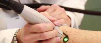
Laser removal procedure
- Local anesthesia in the affected area;
- an incision measuring 2 mm;
- a specialist inserts a laser source through the hole;
- the beam directionally evaporates the affected area and seals the blood vessels.
If the eyeball cyst is large or an abscess has appeared, then surgical removal under anesthesia is recommended. It takes place in several stages:
- limiting the surgical area with sterile materials;
- introduction of a staining solution to clearly define the boundaries of the lesion;
- fixation of the capsule;
- removal with a scalpel;
- rinsing the cavity with an antibacterial solution;
- connecting the incision using a continuous absorbable suture.
Rehabilitation is long and relapse is possible, unpleasant consequences such as hemorrhage, infection, suture dehiscence. In the postoperative period, antibiotics are prescribed, it is advised to reduce the load, and avoid contact with water. The applied bandage is not removed from the eye for five days. It serves as a barrier to dirt and bacteria. There are a number of restrictions on operations.
Preparation for surgery for pterygium and rehabilitation period
It is possible to prescribe anti-inflammatory, antiallergic drops or glucocorticosteroids (GCS) to reduce redness, swelling and pain.
Be prepared that it may take several weeks for you to fully recover from surgery. At first, the cornea will react sensitively to any external influences, and the image may be unclear - this is normal. To reduce the frequency of relapses, metabolites, corticosteroids and anti-VEGF drugs are used, which prevent the formation of new vessels, prevent the progression of the disease, relieve swelling and improve vision.