From birth, every person is endowed with a special color of skin, hair and eyes. This is due to genetics and is determined by race. Special cells are responsible for color - melanocytes, which produce the pigment melanin. The shade of the epidermis depends on its quantity. The more dye present in the epidermis, the darker the person’s color type.
However, throughout life, skin pigmentation disorders may occur, which are caused by various pathological processes. In some cases, the spots that appear go away without treatment, in other situations they remain for the rest of life and can only be removed surgically. It all depends on what causes the pigmentation of the skin. Darkening is caused by an excess of melanin, lightening by a lack.
Table of contents
- Etiology and pathogenesis
- Clinical manifestations
- Principles of treatment
Abnormal skin pigmentation on the face and body is a group of dermatological diseases/conditions that manifest as local or generalized changes in skin color, most often darkening.
In our company you can purchase the following equipment for the treatment of removal of abnormal skin pigmentation:
- M22 (Lumenis)
Etiology and pathogenesis of skin pigmentation
The main causes of abnormal skin pigmentation on the face and body:
- chronic exposure to ultraviolet radiation;
- endocrine disorders;
- pregnancy;
- taking medications;
- eating specific foods (for example, rich in carotene);
- environmental pollution;
- professional activity;
- tattoos;
- heredity.
Pathogenetic mechanisms of abnormal pigmentation (according to Fitzpatrick):
- abnormalities in the origin, migration and differentiation of melanoblasts;
- disturbances in melanocyte morphology;
- pathologies of tyrosinase biosynthesis;
- disorders of melanosome genesis;
- pathologies of melanin biosynthesis from tyrosine;
- dynamic anomalies that disrupt the structure of melanosomes and the color of the synthesized pigment;
- disruption of the transfer of melanin granules to keratinocytes.
Clinical manifestations of changes in skin pigmentation
Hyperpigmentation in thyroid diseases
This diffuse hyperpigmentation usually involves the sides of the trunk, lower back, and lower third of the abdomen ( Fig. 1 ). The color varies from light to deep brown with a brownish or grayish tint, which is very similar to the hyperpigmentation of Addison's disease. This change in skin pigmentation is usually observed when thyrotoxicosis lasts for more than 6–10 years.
With hypothyroidism, on the contrary, there is a diffuse weakening of pigmentation and pallor of the skin, less often - vitiligo and hyperpigmentation in the area of natural folds of the skin.
Chrysoderma
Persistent pigmentation with parenteral administration of gold preparations may increase with solar or ultraviolet irradiation of the skin (for example, in a solarium). The color is ash-gray, mauve, lilac and dark crimson. Sometimes a reticular type of pigmentation occurs. Chrozoderma occurs in patients with pulmonary tuberculosis who received injections of gold salts, as well as in patients with lupus erythematosus and some forms of cutaneous tuberculosis, also treated with gold.
Argyria
This change in skin pigmentation develops with long-term therapy with silver preparations or as an occupational disease in silver mine workers. The intensity of skin coloring is proportional to the duration of taking the medication or contact with silver. The skin and mucous membranes have different colors: from dirty gray to black-violet, the color is sharply expressed on the skin of the face, hands and large folds ( Fig. 2 ).
Bismuthia
It occurs during long-term therapy with bismuth preparations and clinically resembles argyria. Typically, pigmentation is limited to a "bismuth rim" on the gums, similar to the "lead rim" seen in lead poisoning. When exposed to ultraviolet light, the color of the lesions becomes more saturated.
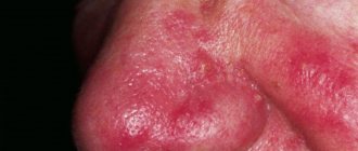
Caratinoderma (carotenosis)
Develops with excessive consumption of foods containing carotene - carrots, oranges, tangerines, apricots, pumpkin, etc. This condition can also occur with diabetes, xanthomatosis and myxedema due to metabolic disorders. It manifests itself as a yellowish or orange-yellow coloration of the skin of the palms, soles and nasolabial folds without any subjective sensations ( Fig. 3 ). In severe cases, generalized pigmentation is possible.
Tattoos and professional hyperpigmentation
Tattooing is the coloring of the skin with insoluble substances deliberately introduced into it ( Fig. 4 ). Occupational hyperpigmentation includes darkening of the skin in miners, gunpowder stains in shooters, etc.
Other causes of abnormal skin pigmentation on the face and body
With nephritis, especially those occurring with symptoms of uremia, diffuse melasma can be observed - the face acquires a characteristic dirty gray color. Parasitic melasma (with lice, chronic scabies) in the neck, shoulders, back, and buttocks have also been described.
Rice. 1. Hyperpigmentation (myxedema) in a 10-year-old child due to Hashimoto’s disease (Shroff A., Simpson G. Cutaneous manifestations of thyroid disease: A case of thyroid—induced myxedema. Case Rep Dermatol Med 2011: 386081)
https://www.researchgate.net/figure/Hyperpigmented-plaque-on-midback_fig2_233806853
Rice. 2. Paul Karason, who has a skin color characteristic of argyria, was nicknamed “human avatar” - by analogy with the famous film by James Cameron (www.epd.info)
Rice. 3. Carotenoderma (www.twitter.com)
Rice. 4. Tattoo as an example of abnormal pigmentation (YakobchukOlena / iStock Getty)
https://static.independent.co.uk/s3fs-public/thumbnails/image/2018/07/04/13/prison-tattoo-0.jpg?w968h681
Types of jaundice
The following types of jaundice are distinguished according to their nature:
- Physiological. It occurs most often in children if their liver tissue is immature. Usually occurs in a large percentage of newborns on the third or fourth days of life. Usually occurs in premature babies. This is due to the fact that the body has not yet had time to adapt to the habitable environment. Jaundice itself can disappear in seven to twenty-one days, and does not harm the child’s body.
- Hemolytic. It occurs when red blood cells are destroyed. Hemoglobin, when released, turns into bilirubin. This effect may occur due to anemia and changes in the composition of hemoglobin protein. It happens that red blood cells are destroyed due to the influence of poisons and various drugs on them. During pregnancy, this type of jaundice occurs due to conflict between the child and the expectant mother.
- Parenchymal (or hepatic). This type of jaundice occurs due to inflammatory processes in the liver tissue due to hypoxia, autoimmune diseases, toxins and hepatitis viruses, which are divided into A, B, C, D, E, F, G.
- Mechanical. This jaundice occurs due to disturbances in the bile flow from the liver to the duodenum. This jaundice can appear due to: stones in the ducts, inflammatory processes, narrowing of the ducts (the disease is dangerous due to complications), neoplasia of the pancreas and liver, roundworms and helminths in the gastrointestinal tract, due to which the bile ducts can be blocked and anemia develops.
Another classification of jaundice is based on its duration:
- Acute jaundice. Rapid simultaneous onset of symptoms;
- Chronic jaundice. Symptoms appear gradually, the speed of their manifestation is associated with the course of the disease that caused jaundice.
Principles of treatment
Treatment of abnormal skin pigmentation is based, first of all, on finding the cause that led to the development of this condition. However, in many cases, treating the underlying condition does not eliminate (or does not completely eliminate) skin discoloration. Therefore, you need to do the following:
- Normalize hormonal levels.
- Reduce skin sensitivity, “calm” it.
- Reduce inflammation.
- Restore microflora and barrier function of the epidermis.
- Strengthen antioxidant protection.
- Reduce melanin synthesis in pigment cells.
- Block the transfer of pigment from melanocytes to keratinocytes.
- Stimulate skin renewal.
- Protect skin from ultraviolet radiation and other negative factors.
Whitening ingredients and points of their application are presented in table. 1 .
Table 1. Ingredients and points of their influence on abnormal pigmentation
| Impact points | Ingredients |
| Inhibition of tyrosinase activity | Hydroquinone, resorcinol, kojic acid, arbutin, deoxyarbutin, ascorbic acid |
| Chelation of tyrosinase with copper | Ellagic acid, kojic acid |
| Suppression of glycolysis of tyrosinase | Glucosamine, N-acetin-glucosamine, tunicamycin |
| Melanosome transfer | Niacinamide, protease inhibitors |
| Inhibition of alpha-melanocyte-stimulating hormone binding to melanocytes | N-undecylenoyl-phenylalanine |
| Decreased tyrosinase activity | Retinoids |
| Antioxidant action | Vitamin C, vitamin E |
| Anti-inflammatory activity | Hydrocortisone, phytosterol, glycyrrhetinic acid, tranexamic acid, chamomile extract |
| Activation of cellular renewal | Retinoids, salicylic acid, alpha hydroxy acids, adenosine monophosphate |
Since the advent of devices operating on the principle of selective photothermolysis, they have become a reliable tool in the treatment of various forms of pigmentation, including abnormal ones.
If pigmentation is caused by the accumulation of melanin, IPL systems are successfully used, which selectively destroy areas of excess pigment accumulation due to their absorption of light energy.
In some cases, treatment of pigmentation is possible using an Nd:YAG Q-Switched laser. Its principle of action is instant heating of melanosomes with their destruction, which occurs very quickly and does not lead to a serious increase in temperature in the surrounding tissues. This method of therapy is suitable for types of hyperpigmentation that can be stimulated by intense heat. Fractional lasers are also successfully used to treat pigmentation, especially when the pigment is deeply embedded.
IPL, Nd:YAG Q-Switched and fractional lasers are included in the M22, which is the most versatile and convenient solution for treating pigmentation.
Questions from our users:
- abnormal skin pigmentation on the hands
- abnormal skin pigmentation on the hands treatment
- abnormal skin pigmentation laser removal
- abnormal skin pigmentation in the form of white spots
Diagnostics
Basically, pigmentation disorders do not pose a particular danger to the health and life of patients and only cause psychological discomfort from a cosmetic defect. However, if the affected areas are injured or exposed to ultraviolet radiation, melanomas can develop into malignant tumors.
Therefore, when age spots appear, it is necessary to undergo a comprehensive examination and determine the cause of their occurrence.
Diagnostics consists of:
- In external visual examination of the skin.
- In collecting anamnesis, which is often not limited to interviewing the patient, but also concerns his immediate family.
- In a dermoscopic examination, in which a device is used to repeatedly magnify and detail the pathological area of the skin.
- In computer diagnostics, which provides the study of affected areas and their comparison with reference samples.
- In histological analysis of tissue sections.
Based on the data obtained, treatment tactics are selected and internal medications are prescribed, as well as external means, for example, the use of photoprotective ointments, peelings, laser resurfacing and other methods of hardware therapy.