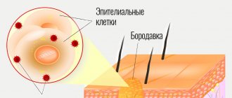
Atheroma can occur for a variety of reasons. The radio wave method of removing atheroma is the fastest and most painless. If you need atheroma removal in St. Petersburg, feel free to contact us. Atheroma is a cyst that appears due to blockage of the sebaceous gland duct. It is completely painless, has a round shape and soft consistency. Atheroma can appear in a variety of places in the body, but today we will talk about atheroma of the ENT organs, which most often appears on the earlobe and behind the ears. If a person has a large number of such neoplasms, doctors diagnose atheromatosis. The main cause of atheroma is blockage of the sebaceous glands. These are exocrine glands, which are located in the upper layers of the skin. The sebaceous gland includes the acini, which has a large number of alveoli, as well as an excretory duct. There are sebaceous glands that do not communicate with the hair follicle; their excretory ducts are located on the surface of the skin. The walls of the alveoli are made of cells that contain a small amount of fat. These cells are regularly renewed, and dead cells are excreted during normal metabolism along with fat. If the excretory channel becomes clogged with dead particles, the sebaceous gland begins to enlarge, thereby forming atheroma. Atheromas never appear in places where there are no sebaceous glands, that is, on the palms and soles. In some cases, atheromas are congenital; this occurs due to disturbances in the functioning of the sebaceous glands during embryogenesis.
Main symptoms of atheroma
Atheroma is a rounded neoplasm that rises above the skin and is completely painless. It should feel soft to the touch or slightly compacted. Atheroma can vary in size from a few millimeters to ten centimeters. Over time, it can grow, due to the fact that the sebaceous gland continues to work and secretes secretions. So, the symptoms of atheroma:
- The appearance of a new formation on the surface of the skin.
- The texture of the tumor may be firm or elastic.
- The contours of the neoplasm should be clear.
- The subcutaneous capsule must be mobile.
- There may be a visible excretory duct in the middle.
Sometimes atheromas can burst open on their own, which is accompanied by the release of contents and an unpleasant odor. Due to the fact that the contents of the atheroma are a nutrient medium, bacteria can begin to multiply in it, which can lead to its suppuration. In this case, redness, swelling, and increased body temperature appear.
Postauricular atheroma
Such a tumor appears very rarely, because the tissue behind the ear contains almost no fat. Such a cyst is differentiated from salivary gland atheroma using, in addition to visual examination, X-ray examination, ultrasound of lymph nodes, and computed tomography. If there are no signs of inflammation, the postauricular atheroma is excised in the “cold period”, and the extracted material is sent for histological analysis to confirm the diagnosis.
It is important to promptly distinguish atheroma from lipoma, which is quite difficult, since the symptoms of the diseases are similar. Both lipoma and atheroma of the postauricular space have a dense consistency and are painless. However, on the surface of the atheroma it is sometimes possible to discern a dark dot at the site where the gland duct exits to the outside. When inflammation of the postauricular atheroma occurs, pain appears and the temperature of the skin in the area of suppuration rises.
The transformation of atheroma into an abscess or phlegmon leads to symptoms of intoxication, and these are:
- Hyperthermia;
- Headache;
- Weakness, increased fatigue;
- Nausea.
When a postauricular atheroma spontaneously opens, the pus spreads through the subcutaneous tissue, spilling into the ear canal and the cartilaginous tissue of the auricle. Intoxication occurs and there is a risk of developing sepsis.
Removal of postauricular atheroma is complicated by the risk of damage to the postauricular lymph nodes and blood vessels.
The operation is painless and quick if modern technologies are used:
- Laser surgery;
- Radio wave surgery.
After their use, there are no scars left and no relapses occur.
Causes of atheroma formation
Most often, atheroma appears as a result of poor personal hygiene. There may also be a hereditary predisposition to the appearance of atheromas, but this theory has not yet been proven, since scientists have not been able to find the gene that is responsible for their appearance. Atheroma of the earlobe can also occur due to an unsuccessful ear piercing, as a result of which the duct of the sebaceous gland is injured. The causes of atheroma can be very different:
- The presence of rare genetic diseases.
- Incorrect use of cosmetics.
- Damage to the skin during the removal of acne and pimples.
- Incorrectly performed facial cleansing.
- Hormonal disorders.
- Hyperhidrosis.
Functions of the skin in children: protective, respiratory, resorption
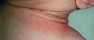
The functions of the skin are diverse, but the main one is protective . In children, this function is weakly expressed, as evidenced by the slight vulnerability of the skin, frequent infection due to insufficient keratinization of the stratum corneum and its thinness, immaturity of local immunity (children's immunologist - Markushka clinic) and abundant blood supply. These features make children's skin easily vulnerable and prone to inflammation , in particular with poor care (wet, dirty diapers).
The respiratory function of the skin in young children is more important than in adults. The skin actively participates in the formation of enzymes, vitamins, and biologically active substances. Excretory and thermoregulatory functions are closely related, which become possible only by 3-4 months. during the maturation of nerve centers. Before this age, poor regulation of body temperature is associated with a large relative body surface area and a well-developed network of blood vessels, which is why a newborn baby, especially a premature one, can easily overheat or become hypothermic if not properly cared for.
The resorption function of the skin in young children is increased (thinness of the stratum corneum, rich blood supply), and therefore medications in ointments, creams, pastes should be used carefully, since their accumulation can cause an adverse effect. The skin is an organ with numerous and varied receptors that provide tactile, temperature, and superficial pain sensitivity.
Possible complications
There are a number of consequences of atheroma:
- Risk of developing into phlegmon with an extensive abscess.
- Risk of relapse if opened independently.
- Risk of formation of suppuration and inflammation.
- The risk of postoperative scars that arise as a result of surgical removal of large atheroma.
- Inflammation of the scars may occur if the atheroma is removed in the clinic.
- As a result of incorrect diagnosis, a relapse may occur.
It is also worth noting that atheroma is a very rare disease; according to statistics, only 7-10% of the world's population gets sick with it. Atheroma is a benign neoplasm that never becomes malignant.
Diagnosis of atheroma
Diagnosis of atheroma is not difficult. This is usually done through a routine skin examination. Atheroma does not pose a danger to life. If there is no suppuration, then you can limit yourself to ordinary observation without undertaking any treatment. There is a high probability that the atheroma will open on its own. But if it has reached a very large size and begins to ache, then it is better to remove it. If the contents of the atheroma break out on their own, the inflammation may subside. Further prospects: atheroma may disappear completely, or it may continue to fester periodically. Despite the fact that atheromas are benign tumors, if you discover a neoplasm, it is best to consult a doctor. Let the doctor tell you what kind of tumor it is and whether treatment is required.
What does atheroma look like?
A cyst formed in the sebaceous gland can be recognized by characteristic changes in the skin. Atheroma looks like an elastic subcutaneous node, usually it has a soft or moderately dense consistency. The diameter of the atheroma depends on the duration of its existence, so it can vary from 1 cm or more. But atheromas are never gigantic in size, exceeding a few centimeters.
Atheroma has a pseudocapsule of connective tissue, which forms as the cyst enlarges. It is mobile and not fused to the surrounding tissues, but is fixed to the skin at one point. It corresponds to the exit of the gland duct.
The skin over the atheroma is of normal color, consistent with the surrounding tissues. There are no vascular changes around the atheroma.
Atheroma on the head
The first signs of atheroma may appear at the age of 20-30 years. But young people do not always go to the doctor when they find a small painless nodule at the base of the hair on the scalp. It slowly increases in size, sometimes the patient does not seek help for several months or years.
Atheroma on the head occurs more often than on other parts of the body. Sometimes several tumors appear. At the point where the duct exits onto the skin, which coincides with the hair shaft, the duct sometimes opens slightly and a cheesy content with an unpleasant odor comes out of the cyst.
But you cannot count on self-healing of atheroma; after emptying, sebum and dead cells will begin to accumulate in it again. The cyst will increase to its previous size.
With the long-term existence of atheroma on the head, the nutrition of the hair is disrupted. This leads to their loss and the development of baldness in the affected area, or alopecia. After treatment, hair restoration does not occur.
Atheroma on the back
Increased sweating and oily skin, poor hygiene leads to blockage of the sebaceous glands located on the back and the formation of atheroma. More often it occurs on the back of the neck, along the spine - in these areas there are more hair follicles with sebaceous glands.
Initially, atheroma on the back can be felt with your fingers while taking a shower. It doesn't hurt and feels like a small pimple. The cyst grows very slowly and does not cause concern.
But if the formation is located in an area of high friction, for example, on the neck, it can be injured. When a bacterial infection penetrates, the atheroma becomes inflamed and suppurates. It becomes swollen and painful. The skin becomes red.
Spontaneous opening of a suppurating atheroma is possible. Curd-like contents with an unpleasant odor are released. It contains cholesterol, cellular debris, lipids and fatty acids. The color of the discharge is white with a yellowish tint.
The danger of atheroma on the back is low. But after inflammation it can lead to the formation of an abscess. The spread of infection to adjacent tissues causes widespread inflammation.
Atheroma on the face
In areas of increased greasiness on the face, under certain circumstances, atheromas may appear. In men, sometimes this is the area of the chin and mustache. A small nodule that does not differ from the color of the skin is mistaken for acne or a pimple.
Atheroma on the face grows at a slow pace. It becomes a cosmetic defect, so patients see a doctor earlier than in other cases. If you try to squeeze it out yourself, there is a risk of introducing an infection into the gland.
Inflammation of the sebaceous gland on the face is especially dangerous in the area of the nasolabial triangle and the wings of the nose. Venous outflow is directed towards the cranial fossa and brain. Therefore, there is a risk of infection and the development of meningitis.
Regardless of the location, atheroma almost never degenerates into a malignant tumor. Literary sources describe isolated cases where skin cancer was diagnosed at the site of atheroma, but there is no evidence that it was a consequence of a sebaceous gland cyst.
Who treats atheroma of the ENT organs
If atheroma appears on the ears, then you should seek help from an ENT doctor
. This specialist specializes in the diagnosis and treatment of diseases of the ear, nose and throat. The study of these organs is united within the framework of the science of otorhinolaryngology. You should contact an ENT doctor if you or your family are concerned about diseases of the upper respiratory tract, runny nose, tumors in the ear area, pain when swallowing and breathing, as well as enlarged parotid lymph nodes. All these symptoms are known to many other doctors, but only an experienced otolaryngologist can prescribe the most effective treatment for you. Timely detection and treatment of diseases of the ENT organs will save you from complications that can spread to other organs.
Treatment of atheroma
An ENT doctor can offer the following treatment methods for atheromas:
- Medication.
- Surgical intervention.
If atheroma occurs behind the ear, it is recommended to immediately consult a doctor who will prescribe the necessary treatment for atheroma. The most effective way to treat this disease is surgery. And if the doctor directs you to undergo surgery, then you should not refuse; this operation is very easy to tolerate and is performed under local anesthesia. After the surgeon has completed his work, you will be able to forget about this disease forever. A good surgeon, when cutting the skin, acts as carefully as possible, without damaging the atheroma capsule. Then the capsule is removed from under the skin, and the cavity in which it was located is thoroughly disinfected with antiseptics. It will be much harder for those who waited until the inflammatory process appeared. If an atheroma tumor is detected, surgery is not performed. First, you will need to remove the inflammatory process so that the operation can be performed. Surgical intervention does not require an inpatient stay, but is performed on an outpatient basis. If the tumor is small in size, no sutures are applied. But if the atheroma has reached a large size, then there is a chance that you will have stitches, which will be processed once every 2 days. Recently, in surgery, instead of surgical intervention, laser and radio wave removal of atheroma have begun to be widely used. In this case, the contents of the atheroma are simply burned out or evaporated. As a rule, these methods are used in the early stages of atheroma development.
Atheroma in a child, treatment and removal in children.
Atheroma, sebaceous gland cyst, steatoma (atheroma) - occurs as a result of blockage of the excretory duct of the sebaceous gland and is found mainly on the scalp. In places where there are no sebaceous glands (the palmar surface of the hand, the plantar surface of the feet), atheromas cannot form. Etiology and pathogenesis.
Atheromas are understood as retention cysts of the sebaceous glands and congenital benign skin tumors that are inherited.
The first ones arise due to thickening of sebum with subsequent blockage of the excretory duct of the gland or when the acne shaft is incorrectly squeezed out, when part of it blocks the duct of the sebaceous gland. The second group of atheromas is of nevoid origin (from the appendages of the epidermis) and, unlike retention cysts (secondary or false atheromas), is called primary, or true, atheromas (steatomas). Symptoms of Atheroma
Despite the common clinical and morphological nature, some differences can be identified between these two types of atheroma.
Thus, retention cysts are more common in the face, neck and back in men; they have faster growth and can be single or multiple, of the same size, prone to merging into lumpy conglomerates and inflammation. True atheromas
are characterized by slower growth, are more common in women on the scalp, and reach very large sizes.
The atheroma capsule consists of connective tissue without papillae and is lined with 2-3 layers of flat epithelial cells with a characteristic arrangement of cells in the peripheral layer in the form of a palisade; intercellular bridges and keratinization phenomena are absent. Differential diagnosis
should be made with epidermal and dermoid cysts, which is sometimes possible only with the help of histological studies.
Epidermal cysts
, which are a consequence of the vicious development of the dermis, also have a connective tissue capsule.
differentiation from epithelioma of Malherbe. Large atheromaslocalized
on the head require a differential diagnosis with cerebral hernias using radiography of the skull bones.
Treatment of atheroma is possible in any phase. The opinion that only atheromas that have reached large sizes are subject to surgical removal should be considered erroneous. Why is it necessary to treat Atheroma?
As they increase in size, atheromas can cause cosmetic defects.
The most common reason for visiting a surgeon is the presence of a “bump” or “wen”. The next reason is the likelihood of atheroma suppuration, because The sebaceous secretion contained in atheroma is a favorable environment for the proliferation of bacteria. And with inflammation, an abscess is formed - a cavity with pus. This situation requires urgent surgical treatment: incision, drainage. The results of treatment of atheroma
without inflammation and with inflammation may differ both in cosmetic terms and in terms of relapse of the disease. Thus, the likelihood of re-operation is much higher after treatment in the inflammation stage.
Therefore, it is preferable to operate on atheromas in a “cold”, that is, non-inflammatory period. What is the essence of the operation?
Under local anesthesia, through a small incision or puncture, the atheroma is removed along with the capsule, which prevents the possibility of relapse.
After the operation, barely noticeable scars remain. Small, non-inflammatory atheromas
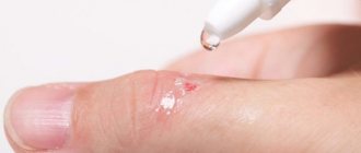
can be removed using electrocoagulation: a scalpel-like electrode is used to make a small incision in the skin through the atheroma capsule, and then squeeze out the contents with your fingers.
The capsule is removed through the resulting hole using tweezers and a spoon. The cavity is treated with a 5% iodine solution and an aseptic bandage is applied. Detachment of the capsule and painlessness of the procedure are ensured by preliminary infiltration of the tissues around the capsule with a 0.5% solution of novocaine. In case of inflammation of atheroma, UHF and anti-inflammatory ointments are prescribed locally at an earlier stage. In case of intoxication of the body - antibiotics (tetracycline, oleandomycin, etc.). 2-3 weeks after the disappearance of inflammatory phenomena, electrosurgery and tumor removal are indicated. Suppurating atheromas require a small incision of the abscess, through which the molten contents of the atheroma and the remains of the capsule are released. The remaining contents are removed with a spoon, and the cavity is treated with solutions of furatsilin, rivanol and a strip of antiseptic ointments (Vishnevsky ointment, furatsilin ointment, etc.) is introduced. Healing in these cases proceeds slowly by secondary intention. Sometimes an emulsion of hydrocortisone acetate is injected around the atheroma in a dose of 1 ml of a solution containing 25 mg of the drug for 10-12 days. Small inflamed atheromas located close to the surface of the skin are cauterized with liquid nitrogen. Atheromas are surgically removed after infiltrating the tissue around the tumor with a 1% solution of novocaine or trimecaine with the addition of a few drops of a 0.1% solution of adrenaline. A skin incision in the form of a spindle passes over the center of the tumor from the upper to the lower pole and in accordance with the lines of skin tension, which contributes to the formation of the most cosmetic postoperative scar. The area of skin above the atheroma is grabbed with tweezers or a hemostatic clamp and tissue detachment begins. Having reached the atheroma capsule, further manipulations should be carried out strictly under the control of the eye to prevent the opening of the capsule and the release of the contents into the wound. If the capsule is opened, you can try to move the clamp to the edges of the tear or, after releasing the contents, insert a thin gauze strip into the cavity of the atheroma, which promotes further detachment and better contouring of the tumor. Particular attention should
be paid to complete removal of the capsule, since the remaining part of the capsule contributes to the growth of atheroma.
After removing the atheroma, hemostasis is performed and the wound is sutured in layers with catgut, horsehair or thin silk. The stitches are removed after 5-7 days. Particular attention should be paid to complete removal of the capsule, since the remaining part of the capsule contributes to the growth of atheroma. After removing the atheroma, hemostasis is performed and the wound is sutured in layers with catgut, horsehair or thin silk. come back
Radio wave removal of atheroma
Radio wave removal of atheroma is considered the most effective and safe method. The procedure is carried out using special equipment that converts radio wave radiation into energy. Exposure to energy destroys the contents of atheroma without causing damage to healthy tissues. Today, such removal of atheroma is a very common and popular method. This type of atheroma removal has a large number of advantages, including:
- Completely eliminating the risk of relapse.
- Removal of atheroma is carried out without suturing.
- An almost completely painless procedure.
- Excellent cosmetic effect.
- Possibility to return to work immediately after surgery.
Moreover, this method of removing atheroma makes it possible to reduce the risk of scar tissue, but this cannot be promised with the intervention of a surgeon. Radio wave removal of atheroma lasts for twenty minutes using local anesthesia. First, the tumor is processed and then exposed to radio waves through a special device. The radiation completely “burns out” the tumor and allows the atheroma to be removed along with the capsule, leaving behind a small depression. After this, the doctor will treat the damaged area of skin with iodine and apply a bandage. If a large tumor is removed, in some cases a hospital stay may be necessary.
Subcutaneous fatty tissue in a child, children
Subcutaneous fatty tissue begins to form in the 5th month of intrauterine life, but is intensively deposited during the last 1.5-2 months. intrauterine life. In a full-term newborn, subcutaneous fatty tissue is well expressed on the cheeks, thighs, legs, forearms and weakly on the abdomen, and during the first 6 months. During life, it develops intensively on the face, limbs, and torso. Later, up to the age of 8, fluctuations occur in the formation of the fat layer, and then its growth begins again, more pronounced in girls. In young children, subcutaneous fat makes up about 12% of body weight, in adults this figure is more than 8%.
The composition of subcutaneous fatty tissue in children of different ages is different: in young children it contains more solid fatty acids (palmitic and stearic) and less liquid oleic acid, which causes denser tissue turgor in children 1 year of age, more a high melting point of fat and a tendency to form local compactions and swelling of the skin and subcutaneous tissue with the formation of sclerema and scleredema. It is important to note that the composition of the subcutaneous fat layer in infants is close in composition to the fats of human milk, so they are absorbed without being digested in the child’s gastrointestinal tract. Subcutaneous fat in different parts of a child’s body has a different composition, which determines the unique distribution and order of accumulation or disappearance of the fat layer during weight loss. So, when fat accumulates, it is deposited first of all on the face, then on the limbs, torso and then on the stomach (liquid fatty acids predominate here). Subcutaneous tissue disappears in the reverse order.
A peculiarity of young children is the presence of accumulations of brown adipose tissue in the posterior cervical region, supraileocecal zone, around the kidneys, in the interscapular space, around the great vessels. In a full-term newborn, its amount is about 1-3% of the total body weight. It provides a higher level of heat production due to the so-called non-contractile thermogenesis (not associated with muscle contraction).
A small amount of fatty tissue explains the greater displacement of internal organs in children under 5 years of age, since only by this age the amount of fat in the chest, abdominal cavities, and retroperitoneal space increases. Fat cells in young children are smaller and contain nuclei; with age they increase in size, and the nuclei, on the contrary, decrease. The roundness of girls' body shapes is due to the fact that more than 70% of adipose tissue is subcutaneous fat, while in boys it is only about 50%.
Recurrence of atheroma
During the operation, the atheroma is completely excised. Relapse can only occur if the sebaceous gland duct is poorly forgiven. The neoplasm must be removed completely, in some cases along with nearby tissues if the capsule has melted. The cause of relapse may also be the formation of a new cyst due to proximity to the postoperative scar. In some cases, relapse occurs due to incorrect diagnosis, when a lipoma or dermoid cyst is mistaken for atheroma. These tumors can also be treated surgically, but the operation is performed in a slightly different way. According to statistics, relapse of atheroma occurs in 15% of patients who have undergone surgery, and in 10% it is the result of opening an abscess cyst, when it is difficult to remove its contents due to purulent content. Such tumors must first be cured and removed after 2-3 weeks. It is recommended to remove atheromas in the winter season, when the cyst has just appeared and has not yet become inflamed. Relapse may also occur due to hyperhidrosis or a hereditary predisposition to obstruction of the sebaceous glands. In these cases, neoplasms appear not in the place where the operation was performed, but in the nearby excretory ducts of the gland.
Only radio wave removal of atheroma completely eliminates the risk of relapse. Before deciding on this procedure, try to find experienced surgeons to avoid possible unpleasant consequences due to an incorrectly performed operation. You can find out where to remove atheroma from reviews of people who have undergone this operation.
Pathogenesis of ear atheroma in children
A sebaceous cyst in a child may be congenital. It is always benign; according to the diagnostic standard, it is necessary to differentiate atheroma from lipoma, dermoid cyst, enlarged lymph node, and furunculosis.
If a child’s atheroma is not congenital, its appearance can be triggered by increased production of sebaceous gland secretions at the age of 5-6 years and then during puberty. Less commonly, the cause of atheroma formation in a child is injury to the epidermis due to incorrect haircuts or ignorance of hygiene rules.
Postauricular atheroma in children often does not manifest itself in any way; the child does not feel any pain or discomfort. Characteristic symptoms appear during inflammation and abscess formation. The abscess can be very large. If it is opened, the atheroma capsule remains and provokes relapses. Only surgery can radically get rid of it.
Small atheroma in children under 3-4 years of age is not removed; the tumor is monitored over time. After 4 years, the atheroma capsule is peeled off under general anesthesia. At the age of 7 years and older, local anesthesia is used. Surgery to remove atheroma in children lasts 30-40 minutes and is considered a simple procedure. This operation will avoid complications such as infection of the soft tissues of the head and the appearance of phlegmon.
There is a modern method of destruction of atheroma - its “evaporation” with a radio wave knife, which does not leave marks on the child’s skin. This treatment is very effective, as it eliminates the risk of relapse and does not lead to scar formation.
Advantages of visiting our clinic
If you need removal of atheroma using the radio wave method in a clinic in St. Petersburg, then you can safely contact us. Our clinic employs highly qualified specialists with extensive experience who can competently diagnose and prescribe effective treatment. During their work, our ENT doctors use the most modern equipment to treat atheroma. We try to find an individual approach to each patient in order to take into account the characteristics of his body as much as possible. Our center employs highly qualified surgeons who can successfully remove atheromas without the risk of relapse. So, by contacting us, you can enjoy the following benefits:
- Consultation with a doctor with extensive experience.
- Competent diagnostics.
- Correct prescription of treatment.
- Treatment of atheroma is carried out using the most advanced technologies and modern equipment.
- Individual approach to each client and polite treatment.
- Favorable prices.
In addition, our center also employs pediatric ENT doctors , during whose appointments your children will not be afraid. You can find out more detailed information about how atheroma is removed using the radio wave method, prices and much more by contacting our employees by phone.