Author
: Grachev Ilya Illarionovich
Editor
: Efremov Mikhail Mikhailovich
Date of publication: 09/24/2018 Date of update: 06/08/2020
Regular psoriatic plaques can be seen not only on the elbows. No less often they are located on the skin of the anterior (extensor) surface of the knee joint. In some patients they remain unchanged for years, while in others they spread to neighboring and distant parts of the body. There are other manifestations of psoriasis on the legs. When should they be treated and is it worth doing if the disease does not progress? The answer to this question will be given by the specialists of the Paramita clinic.
Why does psoriasis develop on the legs?
Psoriasis on the legs is a chronic skin pathology that has a hereditary predisposition. The development of the disease is facilitated by the influence of predisposing factors. Factors predisposing to the development of psoriasis on the legs:
- Limb injuries - abrasions, cracks, wounds. In people prone to this pathology, they often become inflamed, accompanied by itching, and scratching. Against this background, psoriatic rashes appear. These causes most often cause psoriasis in the knee joints in children, adolescents and athletes.
- Skin diseases and foot abrasions. The disease especially often develops against the background of fungal infections. It is important to wear tight shoes, in which the feet sweat and get injured.
- Metabolic disorders, obesity, diabetes mellitus, endocrine diseases, age-related hormonal imbalances, pregnancy. Any hormonal imbalance can cause the onset of the disease.
- Any chronic processes and foci of infection leading to intoxication of the body.
- High emotional, mental and physical stress.
- Improper, irregular diet, intestinal dysbiosis.
- Immunity disorders.
Psoriasis of any localization can spread throughout the body, so do not delay treatment.
See how easily the disease can be cured in 10-12 sessions.
Exposure to one or more predisposing factors causes a chain reaction in the body:
- metabolism is disrupted;
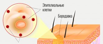
- biologically active substances are released that stimulate the rapid division of surface cells of the skin epithelium; the formation of scales and peeling begins;
- Antibodies to your own skin cells are formed in the blood, an autoimmune inflammatory process begins - characteristic papules appear.
Psoriasis on the legs can develop on different parts of the skin, but most often the rash appears on the knees and feet. It also looks different.
White spots on legs and arms: 5 reasons
Often women and others notice the appearance of small white spots on their legs and arms (hypopigmentation). Accordingly, this begs the question: What could it be and what actions should be taken?
This phenomenon is provoked by a number of factors, but it is worth identifying the most common:
- Vitiligo is a skin disease whose etiology is still unknown, and therefore cannot be cured. The only thing that can be done is to improve the condition of the skin as much as possible.
- Pityriasis versicolor is a fungal infection that is present on the dermis of any person and is activated in a favorable environment: prolonged exposure to ultraviolet radiation, increased sweating, and a humid climate. The fungus is treated with special drugs (Clotrimazole, Terbinafine and others).
- Guttate hypomelanosis is another disease, but it is genetic and usually inherited. The exact reasons for its origin have also not been identified, but since it occurs more often after 40 years, it is also associated with aging. Small spots sometimes itch and peel off.
- Lichen alba is a dermatological pathology characterized by changes in skin color. It most often occurs in preschool and teenage children living in regions with a warm and humid climate (you can clearly see this manifestation in the photo below). The skin becomes dry and very flaky. To be sure that it is lichen and to find out how to treat it, you should consult a doctor.
- An adverse reaction to certain pharmaceutical drugs - if they are taken for a long time, skin cells begin to produce pigment (melanin) poorly, resulting in an uneven tan. One such drug is birth control pills.
Discoloration of certain areas of the skin often occurs due to a lack of melanin in the body. Then white spots on the legs and other parts of the body appear after tanning naturally or in a solarium. They do not cause much discomfort.
Cosmetologist Rita Rakus
Representatives of the fair sex with extremely sensitive and pale skin and children under 13 years of age who have not yet fully developed their immune system are more susceptible to this disease.
Women also face a similar problem during pregnancy, after 40 years and during menopause. But this does not mean that others are immune from the appearance of such spots on the body.
Psoriasis on the knees
The manifestation of psoriatic rashes on the extensor surface of the knee joints is typical.
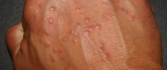
Psoriasis on the knees begins with the appearance of a small round or oval-shaped spot. The spot gradually turns into a papule with a raised, inflamed base. The early plaque is covered with silvery scales on top and is surrounded by a brighter corolla that rises above its surface and has no scales. If you scratch the papule, first the appearance of a stearin stain appears, and then a thin transparent film. Further exposure to the film is accompanied by the appearance of droplets of blood—blood dew—on its surface. In the initial stage of psoriasis of the legs (also called progressive), there may be one or two papules, then several more appear and they grow, merge with each other, forming large plaques of various configurations. The appearance of new rashes at the site of injury to the skin - scratches, abrasions, etc. is typical.
Psoriasis often stops at the initial stage and plaques in the knees, feet, and legs can be seen for several years and even decades. Such plaques are called duty plaques. With additional exposure to provoking factors, psoriasis can enter a progressive stage at any time, spreading from the legs to other parts of the body.
The next period is called stationary. The spread of the psoriatic rash stops and it looks the same all the time. After some time, the final regressive stage begins, when the rash gradually fades. The process can take place in two directions:
- from the center to the periphery - a pink corolla forms around the papule;
- from the periphery to the center - the corolla is white.
Gradually, white or brownish pigment spots form at the site of the rash. The process proceeds in waves: exacerbation is replaced by remission and vice versa.
Skin vasculitis
Skin vasculitis is a group of diseases of a multifactorial nature, in which the leading symptom is inflammation of the blood vessels of the dermis and subcutaneous tissue.
The difficulty in covering this topic is that until now there is no generally accepted classification or even agreed upon terminology of vasculitis. Currently, about 50 different nosological forms have been described, and understanding this diversity is not easy. The diversity of clinical manifestations and insufficiently studied pathogenetic mechanisms have led to the fact that under different names only a variant of the main type of skin lesion may be hidden. Also, in addition to primary vasculitis, which is based on inflammatory damage to the blood vessels of the skin, there are also secondary vasculitis (specific and nonspecific), developing against the background of a certain infectious (syphilis, tuberculosis, etc.), toxic, paraneoplastic or autoimmune (systemic lupus erythematosus, dermatomyositis etc.) process. It is possible to transform skin vasculitis into a systemic process with damage to internal organs and the development of severe, sometimes life-threatening complications.
Skin vasculitis is a polyetiological disease. The most common connection is with a focal infection (streptococci, staphylococci, mycobacterium tuberculosis, yeast, viruses, etc.). Hypersensitivity to a number of drugs, in particular to antibiotics and sulfonamide drugs, is of particular importance. Often, despite a carefully collected anamnesis and examination, the etiological factor remains unclear. Among the risk factors for vasculitis, one should take into account: age (children and the elderly are most vulnerable), hypothermia, excessive insolation, severe physical and mental stress, trauma, surgery, liver disease, diabetes, hypertension. The pathogenetic mechanism for the development of skin vasculitis is currently considered to be the formation of circulating immune complexes with their subsequent fixation in the endothelium, although this has not been definitively proven for all diseases of this group.
Skin vasculitis is a heterogeneous group of diseases, and their clinical manifestations are extremely diverse. However, there are a number of common features that unite these dermatoses:
1) inflammatory nature of skin changes; 2) symmetry of rashes; 3) tendency to edema, hemorrhage and necrosis; 4) primary localization on the lower extremities; 5) evolutionary polymorphism; 6) connection with previous infectious diseases, medication, hypothermia, allergic or autoimmune diseases, and impaired venous outflow; 7) acute or worsening course.
Skin lesions with vasculitis are diverse. These may be spots, purpura, nodules, nodes, necrosis, crusts, erosions, ulcers, etc., but the main clinical differential sign is palpable purpura (hemorrhagic rash that rises above the surface of the skin and is felt on palpation).
There is no generally accepted classification of vasculitis. Vasculitis is systematized according to different principles: etiology and pathogenesis, histological picture, severity of the process, features of clinical manifestations. Most clinicians use predominantly morphological classifications of cutaneous vasculitis, which are usually based on clinical changes in the skin, as well as the depth of location (and, accordingly, the caliber) of the affected vessels. There are superficial (damage to the vessels of the dermis) and deep (damage to the vessels at the border of the skin and subcutaneous tissue) vasculitis. Superficial ones include: hemorrhagic vasculitis (Henoch-Schönlein disease), allergic arteriolitis (polymorphic dermal angiitis), leukoclastic hemorrhagic Miescher-Storck microbiota, as well as chronic capillaritis (hemosiderosis): Majocchi's annular telangiectatic purpura and Schamberg's disease. To the deep: cutaneous form of periarteritis nodosa, acute and chronic erythema nodosum.
Hemorrhagic vasculitis is a systemic disease that affects small vessels of the dermis and manifests itself as palpable purpura, arthralgias, gastrointestinal (GIT) lesions and glomerulonephritis. It occurs at any age, but boys aged 4 to 8 years are at greatest risk. Develops after an infectious disease, after 10–20 days. The acute onset of the disease, with fever and symptoms of intoxication, is most often observed in childhood. The following forms of hemorrhagic vasculitis are distinguished: cutaneous, cutaneous-articular, cutaneous-renal, abdominal-cutaneous and mixed. The current can be lightning fast, sharp and protracted. The duration of the disease varies - from several weeks to several years.
The process begins symmetrically on the lower limbs and buttocks. The rashes are papular-hemorrhagic in nature, often with urticarial elements, and do not disappear with pressure. Their color changes depending on the time of appearance. The rashes occur in waves (once every 6–8 days); the first waves of the rash are the most violent. Articular syndrome appears either simultaneously with skin lesions or after a few hours. Large joints (knees and ankles) are most often affected.
One of the variants of the disease is the so-called necrotic purpura, observed during the rapid course of the process, in which necrotic skin lesions, ulcerations, and hemorrhagic crusts appear.
The greatest difficulties are caused by the diagnosis of the abdominal form of hemorrhagic vasculitis, since skin rashes do not always precede gastrointestinal phenomena (vomiting, cramping pain in the abdomen, tension and pain on palpation, blood in the stool).
The renal form is manifested by impaired renal activity of varying degrees of severity, from short-term unstable hematuria and albuminuria to a pronounced picture of acute glomerulonephritis. This is a late symptom and never occurs before the skin is affected.
The fulminant form of hemorrhagic vasculitis is characterized by an extremely severe course, high fever, widespread rashes on the skin and mucous membranes, viscerapathies, and can result in the death of the patient.
Diagnosis of the disease is based on typical clinical manifestations; in atypical cases, a biopsy is performed. In the abdominal form, surgical supervision is necessary. Observation by a nephrologist is recommended for three months after resolution of purpura.
The term “allergic arteriolitis” Ruiter (1948) proposed to name several related forms of vasculitis, differing in clinical manifestations, but having a number of common etiological, pathogenetic and morphological features.
The pathogenetic factors of the disease are considered to be colds and focal infections. The rashes are usually located symmetrically and are polymorphic in nature (spots, papules, vesicles, pustules, necrosis, ulcerations, telangiectasia, blisters). Depending on the predominant elements, three forms of the disease are distinguished: hemorrhagic type, polymorphic-nodular (corresponds to three-symptomatic Gougerot-Duperre disease) and nodular-necrotic dermatitis (corresponds to Werther-Dümling nodular-necrotic dermatitis). When the rash regresses, cicatricial atrophies and scars may remain. The disease is prone to relapse. Often before the rash, patients complain of malaise, fatigue, headache, and at the height of the disease - pain in the joints (which sometimes swell) and in the abdomen. Diagnosis of all types of the disease is difficult due to the lack of typical, characteristic symptoms. Histological examination reveals fibrinoid lesions of small-caliber vessels with the formation of infiltrative accumulations of neutrophils, eosinophils, lymphocytes, plasma cells and histiocytes.
The clinical course of hemorrhagic leukoclastic microbid Miescher-Storck A sign that makes it possible to distinguish this disease as an independent one is the presence of a phenomenon - leukoclasia (disintegration of the nuclei of granular leukocytes, leading to the formation of nuclear dust) during histological examination. Thus, hemorrhagic leukoclastic microbide can be interpreted as a dermatosis caused by chronic focal infection (intradermal tests with streptococcal antigen are positive), occurring with severe leukoclasia.
Chronic capillaritis (hemosiderosis), in contrast to acute purpura, is characterized by a benign course and is exclusively a skin disease.
Schamberg's disease is a lymphocytic capillaritis characterized by the presence of petechiae and brown purple spots, occurring most often on the lower extremities. Patients are concerned solely as a cosmetic defect.
Majocchi purpura is characterized by the appearance on the lower extremities of pink and liquid-red spots (without previous hyperemia, infiltration), slowly growing to form ring-shaped figures. In the central part of the spot, slight atrophy and achromia develop, and vellus hair falls out. There are no subjective sensations.
Periarteritis nodosa is characterized by necrotizing inflammation of small and medium-sized arteries of the muscular type, followed by the formation of vascular aneurysms and damage to organs and systems. Most common in middle-aged men. Of the etiological factors, the most important are drug intolerance (antibiotics, sulfonamides), vaccination and persistence of HbsAg in the blood serum. The disease begins acutely or gradually with general symptoms - increased body temperature, rapidly increasing weight loss, pain in the joints, muscles, abdomen, skin rashes, signs of damage to the gastrointestinal tract, heart, peripheral nervous system. Over time, polyvisceral symptoms develop. Particularly characteristic of periarteritis nodosa is kidney damage with the development of hypertension, which sometimes becomes malignant with the occurrence of renal failure. There are classic and cutaneous forms of the disease. Skin rashes are represented by nodules - single or in groups, dense, mobile, painful. The formation of nodes along the arteries is typical, sometimes they form strands. Localization on the extensor surfaces of the legs and forearms, on the hands, face (eyebrows, forehead, corners of the jaw) and neck. They are often not visible to the eye and can only be determined by palpation. Necrosis may develop in the center with the formation of long-term non-healing ulcers. Periodically, the ulcers may bleed for several hours (a symptom of a “bleeding subcutaneous node”).
Sometimes the only manifestation of the disease may be reticular or branched livedo (persistent violet-red spots), localized on the distal parts of the extremities, mainly on the extensor surfaces or lower back. It is typical to detect nodules along the course of livedo.
Diagnosis of the disease is based on a combination of damage to a number of organs and systems with signs of significant inflammation, fever, changes primarily in the kidneys, heart, and the presence of polyneuritis. There are no laboratory parameters specific for this disease. Dynamic clinical observation of the patient is crucial for diagnosis.
Acute erythema nodosum is a panniculitis that is characterized by the presence of painful pink nodules on the extensor surface of the lower extremities. Accompanied by fever, malaise, diarrhea, headache, conjunctivitis and cough. Among adults, erythema nodosum is 5–6 times more common in women, with a peak age of 20–30 years. The disease is based on hypersensitivity to various antigens (bacteria, viruses, fungi, neoplasms and connective tissue diseases). Half of the cases are idiopathic. Diagnosis is based on history and physical examination. A complete blood count, chest X-ray (detects bilateral adenopathy in the hilar region), a throat swab, or a rapid streptococcal test should be performed.
Chronic erythema nodosum is a group of different types of dermohypodermatitis nodosum. Women aged 30–40 years are most often affected. Nodes of various sizes appear on the legs with reddened skin over them, without a tendency to necrosis and ulceration. Inflammatory phenomena in the area of the rash and subjective sensations (arthralgia, myalgia) are mild. Clinical variants of chronic erythema nodosum have their own characteristics, for example, the tendency of the nodes to migrate (Beferstedt's erythema migrans) or the asymmetry of the process (Vilanova-Pinol hypodermitis).
Tactics for managing a patient with skin vasculitis
- Classify the disease (characteristic clinical picture, anamnesis, histological examination).
- Search for an etiological factor, but in 30% of cases it cannot be established (search for foci of chronic infection, microbiological, immunological, allergological and other studies).
- Assessment of the general condition and determination of the degree of disease activity: general blood and urine analysis, biochemical blood test, coagulogram, immunogram. Degree of vasculitis activity: I. Rashes are not abundant, body temperature is not higher than 37.5, general symptoms are insignificant, ESR is not higher than 25, C-reactive protein is not more than ++, complement is more than 30 units. II. The rash is abundant (extends beyond the lower leg), body temperature is above 37.5, general symptoms are headache, weakness, symptoms of intoxication, arthralgia; ESR is higher than 25, C-reactive protein is more than ++, complement is less than 30 units, proteinuria.
- Assessment of signs of systemicity (research according to indications).
- Determination of the type and regimen of treatment depending on the degree of activity: Art. I. — treatment on an outpatient basis is possible; II Art. - in the hospital. In all cases of exacerbations of skin vasculitis, bed rest is necessary, since such patients usually have pronounced orthostasis, which should be observed until the transition to the regressive stage. A diet excluding irritating foods (alcoholic drinks, spicy, smoked, salty and fried foods, canned food, chocolate, strong tea and coffee, citrus fruits) is recommended.
- Etiological treatment. If it is possible to eliminate the causative agent (drug, chemicals, infection), then resolution of the skin lesions follows quickly and no other treatment is required. But we must remember that when sanitizing foci of infection, an increase in the vascular process may be observed.
- Pathogenetic treatment.
- Preventive measures: medical examination, prevention of provoking factors (infections, hypothermia, insolation, stress, etc.), rational use of medicines, employment, physical therapy, sanatorium treatment.
Treatment of hemorrhagic vasculitis
- Glucocorticosteroids (prednisolone up to 1.5 mg/kg) alleviate the manifestation of skin-articular syndrome, but do not shorten the disease or prevent kidney damage. Prescribed in severe cases and under the cover of heparin, because they increase blood clotting.
- Nonsteroidal anti-inflammatory drugs (NSAIDs) in usual therapeutic dosages. The choice of a specific drug is not of fundamental importance (indomethacin, diclofenac, acetylsalicylic acid).
- Anticoagulants and antiplatelet agents. Heparin for a common process: 300–400 units/kg/day. The duration of the course should be at least 3–5 weeks. Under coagulogram control.
- Therapeutic plasmapheresis, when the manifestations of the disease are not eliminated by the listed means.
- Nicotinic acid in tolerable doses intravenously.
- You should not use: antihistamines (possibly only at the very beginning of the disease), calcium supplements, all vitamins.
Treatment of skin vasculitis
1) NSAIDs (naproxen, diclofenac, Reopirin, indomethacin, etc.); 2) salicylates; 3) Ca preparations; 4) vitamins P, C, antioxidant complex; 5) vasodilators (xanthinol nicotinate, pentoxifylline); 6) 2% solution of potassium iodide, 1 tbsp. l. 3 times a day (erythema nodosum); 7) anticoagulants and antiplatelet agents; detoxification methods IV drip; 9) glucocorticosteroids (GCS) 30–35 mg/day for 8–10 days; 10) cytostatics; 11) ultra-high frequency therapy, diathermy, inductothermy, ultrasound with hydrocortisone, ultraviolet irradiation.
External treatment. For erosive and ulcerative rashes
1) 1–2% solutions of aniline dyes; 2) epithelializing ointments (solcoseryl); 3) ointments containing glucocorticoids, etc.; 4) lotions or ointments with proteolytic enzymes (Chymopsin, Iruksol); 5) Dimexide applications;
For knots - dry heat.
Treatment should not end with the disappearance of clinical manifestations of the disease. It continues until laboratory parameters are completely normalized, and in the next six months to a year, patients are given maintenance treatment
Literature
- Adaskevich V.P., Kozin V.M. Skin and venereal diseases. M.: Med. lit., 2006, p. 237–245.
- Kulaga V.V., Romanenko I.M., Afonin S.L. Allergic diseases of the blood vessels of the skin. Lugansk: “Etalon-2”, 2006. 168 p.
- Berenbein B. A., Studnitsin A. A. et al. Differential diagnosis of skin diseases. M. Medicine, 1989. 672 p.
I. B. Mertsalova, Candidate of Medical Sciences RMAPO, Moscow
Contact information about the author for correspondence
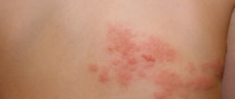
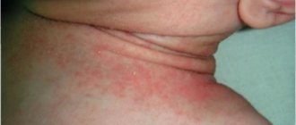
Atypical type of psoriasis on the legs
Psoriatic rashes may have uncharacteristic localization and appearance. This form of psoriasis is localized not on the extensor side, but on the flexor side of the leg joints, in the area of skin folds. Since the skin in this area constantly sweats, the scales on the rash are not always noticeable in the initial stage. Papules in the initial stage of the disease may look like ordinary prickly heat and be accompanied by severe itching.
Subsequently, the process spreads to surrounding areas and acquires a characteristic appearance. The stationary and regressive stages proceed in the same way as in a typical disease.
Plantar type of psoriasis
Psoriasis on the legs may first appear on the soles of the feet. In this case, the following may appear on the feet:
- typical rashes in the form of papules at the initial stage; over time, they merge with each other to form large plaques; this is a typical form, it proceeds in the same way as in other areas;
- the skin of the soles becomes covered with flaky itchy spots, it is dry, thickened (psoriatic calluses), and covered with cracks; this is the so-called horny form of the disease; cracks in the affected areas of the skin of the feet are very painful, which creates great problems when wearing shoes;
- pustular (purulent) rashes appear on the soles, merging into large lakes - this is Barber's pustular psoriasis; after the ulcers dry out, brown crusts form, at the site of which red spots remain; the toes may also be affected;
Treatment methods for psoriasis on the legs
Treating psoriasis on the legs is a difficult task. Psoriasis should be treated comprehensively. It is recommended for patients with psoriasis on the legs:
- optimization of the daily routine, combining an active lifestyle with rest and healthy sleep at night;
- avoiding smoking and drinking any types of alcoholic beverages;
- eliminating stress;
- avoiding high physical activity with associated sweating;
- exclusion of contact (wrestling, boxing) sports;
- proper regular nutrition; Salty, sour, spicy foods, as well as vegetables containing essential oils that irritate the skin are not recommended; limited consumption of meat, consumption of large quantities of vegetables and fruits; drink plenty of fluids: drink plus 6 glasses per day added to the rest of the liquid;
- wearing shoes made of genuine leather or textiles with low heels that do not compress the feet;
- when playing sports, try not to injure the knee area;
- timely elimination of fungal, bacterial and viral infections of the feet;
- regular treatment under the supervision of a physician of all chronic pathologies, elimination of foci of infection;
- daily foot washing using hypoallergenic gels or baby soap; cutting toenails; after this, a moisturizing hypoallergenic cream should be applied to the skin;
- wearing socks made of cotton fabrics in light colors (dyes can provoke an exacerbation of psoriasis).
Red spots on the skin after stress
Diagnosis of skin diseases
Drug therapy
The choice of medications depends on the nature of psoriasis, the stage of the disease, its course and spread, the age of the patient, and the presence or absence of concomitant pathology. The goal of drug therapy is to eliminate the symptoms of the disease, which leads to an improvement in the patient’s quality of life. With the right approach to the treatment process, you can constantly maintain psoriasis in a state of remission, but you cannot guarantee its complete cure.
Preference is given to step-by-step external treatment with alternation of modern topical drugs belonging to different groups. For severe and widespread forms of psoriasis of the legs, individually selected combination treatment with general and local drugs is prescribed.
Local treatment
For local therapy the following are used:
- Ointments, creams, solutions based on glucocorticoid hormones (GCS). These remedies are widely used in the treatment of all forms of psoriasis of the legs. They perfectly relieve inflammation, swelling, itching, suppress proliferation (rapid division) of epithelial cells, inhibiting peeling. Combination ointments are also used, which, in addition to GCS, contain keratolytics - substances that dissolve keratinized epithelial cells and ensure rapid penetration of GCS to the cells (Diprosalik). But if the rash spreads quickly and aggressively, keratolytics can irritate the skin, so they are used with caution.
- Ointments, creams and lotions based on a synthetic analogue of vitamin D (Davonex). The drug actively affects the metabolism in cells, suppressing processes that contribute to the occurrence of disorders. Recently, a drug has been produced that, in addition to a synthetic analogue of vitamin D, includes an active substance from the GCS group (Daivobet). Modern step-by-step therapy includes the use of Daivonex first (in the early stages), and then Daivobet (during follow-up treatment).
- Keratolytic agents (salicylic acid) - soften rough layers of dead cells, improve access to cells of other drugs. Suitable for the treatment of horny psoriasis of the soles.
- Ointments and pastes based on activated zinc - have an anti-inflammatory and antiseptic effect, well suited for eliminating psoriasis of the feet and atypical forms.
- Ointments, creams, gels based on tar - have an anti-inflammatory, absorbable, antiseptic, analgesic effect. Suitable for eliminating the symptoms of plaque psoriasis on the feet in the stationary stage.
General treatment
General therapy is prescribed for a widespread process, as well as for the development of severe pustular forms of the disease, when local treatment is ineffective. Patients are prescribed:
- In order to eliminate intoxication and suppress allergies:
- calcium supplements, hemodez;
- antihistamines – Suprastin;
- In order to suppress the rapid proliferation (division) of cells in the lesions:
- drugs based on corticosteroids - administered in the form of infusions, injections or taken orally;
- cytostatics (Metoltrexate);
- aromatic retinoids (Acitretin).
- In order to suppress the excessive sensitivity of the immune system - immunosuppressants (Cyclosporin-A);
- In order to eliminate the symptoms of inflammation and tissue swelling, itching and pain:
- drugs based on corticosteroids;
- biological drugs (Remicade) - contain proteins that recognize and neutralize substances that support inflammation.
- In order to eliminate neuropsychic symptoms:
- sedatives (sedatives (Novopassit).
Tanning rules (recommendations)
What to do if, after a thorough medical examination, no dermatological problems are identified, but the tan still does not lie evenly.
It is recommended to change your approach to skin care and tanning sessions. Here are some useful tips:
- Avoid low-quality and cheap cosmetics. It is better to buy one cream or lotion, but such that it fully provides the dermis with all the necessary components. It is especially important that the composition contains vitamins (A, E) and minerals, as well as natural oils.
- Drink plenty of water. Dehydration negatively affects the skin, increasing its sensitivity to UV radiation. It is advisable to exclude pickles and smoked foods from your daily diet, which retain water in the body.
- Clean your skin regularly. Dead skin cells, clogged pores, sebum and other impurities cause uneven tanning. Therefore, take wet wipes to the beach and exfoliate more often.
- Follow basic hygiene rules. In the solarium, make sure that the cabin is pre-treated with disinfectants. During the session, use personal household items: mat, footprints, glasses. This way you can protect yourself from contracting any infection.
- Use special SPF creams with a protection index of more than 30. It is recommended to renew the cream layer several times a day and each time after leaving the water. Use such creams in urban areas with the onset of summer.
- Stick to a proper diet. If the skin is healthy, then the tan will lie evenly on it and the risk of pigmentation is minimal.
Do not sunbathe for more than 2 hours, especially during the sun's peak activity (from 11 a.m. to 4 p.m.). If the spots do not disappear, but, on the contrary, their number and size increase, then completely avoid sunbathing and visiting the solarium.
Dermatological surgeon Karin Litani
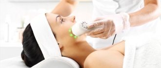
Effective methods for treating psoriasis on the legs
The following methods used in our clinic have been proven to be highly effective:
- PRP therapy - the essence of the method is that the patient is injected with his own blood enriched with platelets; Initially, blood is taken from a vein and injected pointwise into the affected areas of the skin of the legs; this leads to improved metabolic processes and suppression of the spread of psoriasis; helps well with all forms of disease;
- autohemotherapy - intramuscular injection of blood taken from the patient’s vein; has a general stimulating effect on the body;
- herbal medicine – the use of herbs and other medicinal plants. They are used as part of complex therapy to reduce the drug load on the patient’s body;
- acupuncture (acupuncture) is one of the methods of reflexology, the oldest method practiced by Chinese doctors; influence on special acupuncture points (AP) on the body and through them on various organs and tissues; specialists who are fluent in this technique can eliminate psoriatic rashes without the use of medications;
- vacuum therapy – exposure of the AT to rarefied air; for this purpose, special jars or devices are used;
- other methods of reflexology - cauterization with wormwood cigarettes, acupressure, auriculotherapy (impact on reflex points on the ear), etc.
How is leg psoriasis treated at Paramita Clinic?
A special feature of the Paramita clinic is the use of only effective methods of treating psoriasis on the legs at an affordable price. Both the most advanced European techniques and traditional oriental ones are used.
The correct choice and application of one or another method of therapy is possible only if the doctor has appropriate training and clinical experience. The clinic's doctors are fluent in all known methods of treating psoriasis, so it always takes place without side effects. All of them were trained in traditional healing methods in China and Tibet.
To avoid relapses, it is necessary to eliminate the cause of the disease.
Read more about our unique method of treating psoriasis
You can learn about the quality of treatment provided by Paramita specialists from reviews, as well as simply by asking visitors, because many of them are systematically treated at the clinic, maintaining a state of remission for years.
Other reasons
If we exclude common causes, the appearance of white spots on the skin may be associated with the following situations:
- use of low-quality cosmetics (creams, tonics, lotions);
- after shaving the skin;
- leading a sedentary lifestyle (for example, when you stand on your feet or sit for a long time), which leads to poor circulation in the lower part of the body;
- wearing tight clothing, which causes excessive sweating;
- unbalanced diet, lacking vitamins and microelements.
Any of these factors can lead to a decrease in melanin synthesis, which inevitably causes hypopigmentation.
Do not immediately panic when you discover white spots on your legs and arms. The first thing you need to do is visit a dermatologist to rule out any serious diseases after the examination.
Only after finding out the cause can you begin treatment, which will be prescribed by a specialist.
Separately, it is worth highlighting information about the appearance of white spots on the legs and other parts of the body in children. In most cases, this is caused by the following diseases:
- Hypomelanosis . It makes itself felt in the first years of life after infectious diseases.
- Tumorous sclerosis . It is a signal of more serious problems in the body (epilepsy, mental retardation, damage to some internal organs).