There are a huge number of types of moles. According to the World Health Organization (WHO) classification there are several hundred of them. How can an ordinary person who has nothing to do with medicine figure out how dangerous his mole is? At least roughly? There are several signs that we will talk about today.
I would like to point out right away that this article is not intended to help you diagnose yourself. Its main purpose is to reduce any tension you may have that has arisen after independently studying information about melanoma on the Internet.
Bad Signs
Rapid growth of a mole
What does "fast" mean? Which one is slow then? Without answers to these questions, almost any mole on the body can be safely classified as melanoma, because almost every one of them increases by a couple of millimeters over the course of a lifetime.
By fast, I mean an increase in the size of a mole faster than 1-2 mm per year. This is a very conditional border, because There are melanomas that grow more slowly, however, in my opinion, most of these tumors will show a growth rate that exceeds this value.
Color change
The production of pigment by the cells of a benign mole can normally lead to its darkening. On the other hand, proliferation of melanoma cells can also occur. How to distinguish one from the other? In my opinion, there are 2 ways.
The first is the speed at which the color change occurs. A benign mole has the right to darken a little over the course of several years.
The second is melanoma, unlike the latter, it will most likely have more than 2 colors, for example, light brown, black, red, white.
3.Change shape
There are a huge number of irregularly shaped moles, but only a very small proportion of them will turn out to be melanoma. Irregular shape usually means asymmetry along 2 axes in combination with an edge in the form of a “coastline on a map.”
I would not like to dwell in more detail on the bad signs. Everyone who could has already written about them. Today we will dwell in more detail on what types of
Where do moles appear?
Moles can appear on the skin of any part of the human body. Nevi occur on the trunk, limbs, scalp, face and neck, as well as on the palms and soles.
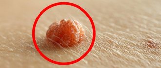
Hanging moles often appear on the head, neck and armpits due to the thinness of the skin and the lack of pronounced subcutaneous fat in these places. Hair may grow on moles. Moles from which hair grows are benign. Convex moles shown in the photo also rarely undergo malignancy (degeneration into cancer).
Good signs
A pedunculated mole, the thickness of which is no more than 2-3 mm
A distinctive feature of malignant tumors is the unlimited division of their cells. This property does not allow such neoplasms to have a “leg”. Unfortunately, this sign cannot guarantee that the formation is benign. In the literature, about once every 10 years there are indications of pedunculated melanoma.
Long-term existence of a mole without changes
If a mole exists from birth or 5, 10, 20, 30 or more years and at the same time! nothing! has not changed - with a high probability, everything is fine with her. This sign is not unconditional. It must be remembered that a long-existing mole is not immune from turning into melanoma, but its chances are, in any case, less.
Presence of hair on the surface of the mole
In fact, hair does not grow from a mole, but grows through it. Hair roots (hair follicles) are usually located deeper. If hair grows from a mole, this means that the cells of the mole still retain their functions. They can form “channels” for hair growth and “pass” it through the mole. This sign also does not 100% “insure” that the mole is actually melanoma. Fortunately, in my practice there was only one exception to this rule.
The skin pattern on the surface of the mole is preserved
This sign of benignity has the same roots as the previous one. It means that the mole is covered by normal skin cells, not tumor cells. Unfortunately, like all previous signs, it is NOT always reliable.
Flesh-colored mole
Most melanomas are brown or black. Therefore, if the color of a mole is flesh-colored, it is highly likely to be benign. Answering a possible question about the non-pigmented form of melanoma, I will say that it is! Disappearing! a small part of the total number of this type of tumor.
Soft consistency
If a mole is softer than the surrounding skin, it is most likely benign. This is due to properties common to all malignant tumors. If cells divide uncontrollably, in most cases they form a compaction that stands out against the background of the surrounding tissues.
Symmetry along at least one axis, smooth edge.
More than enough has already been said about these signs; I will not repeat them.
Types of moles
The skin is the largest organ in the human body. The skin consists of the epidermis (surface layer), dermis (inner layer of the skin, which contains blood vessels, nerves, hair follicles) and subcutaneous fat.
Moles can be superficial (epidermal) or located in the deep layers of the dermis. Depending on the color and location on the skin, the following types of moles are distinguished:
- transitional nevi (flat or slightly convex moles at the border of the epidermis and dermis);
- complex nevi (convex moles in the epidermis and dermis);
- intradermal nevi (intradermal moles that are located in the dermis and protrude above the surface of the skin in the form of a nodule covered with epidermis);
- halonevus (a discolored white spot forms around the mole, the causes of which may be autoimmune diseases and sunburn);
- blue nevi (moles are gray-blue in color and located in the dermis).
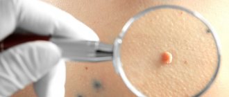
Red and pink moles are not nevi and do not consist of melanocytes. Red moles are growths of small blood vessels. Vascular moles are called angiomas.
Briefly about the main thing:
You should not try to make a diagnosis yourself; this should still be done by an oncologist. On the other hand, it’s also not worth making a mountain out of nothing out of the blue.
If your mole does not have a single “bad” sign, but has several good ones, you should still see an oncologist. Calmly, without haste and fuss, as time permits.
The main point of this article is not for you to be able to understand for yourself whether a mole is malignant or not. My goal is to help you not go crazy because of the horror stories you have previously read on the Internet.
If doubts still overcome you, you can ask me any question in the format of an online consultation.
Why do moles appear?
Most melanocytes are evenly distributed throughout the skin of the entire body. Nevi form when clusters of melanocytes form. Melanin acts as a natural screen that protects the human body from exposure to ultraviolet rays.
The activity of melanocytes is normally regulated by the work of the pituitary gland, a gland that regulates the production of all hormones in the body (melanocyte-stimulating hormone of the pituitary gland enhances the production of melanin). The amount of melanin also increases under the influence of sunlight. The appearance of moles is promoted by:
- insolation (solar radiation);
- changes in hormonal levels;
- hereditary predisposition.
Under the influence of ultraviolet radiation, not only the formation of a new nevus can occur, but also the degeneration of an existing mole into an atypical mole, from which skin cancer can develop. Heredity and hormonal levels also play a role in the development of melanoma (skin cancer).
How does this happen?
With intense tanning, the cells of the birthmark, containing scatterings of melanocytes, absorb quantum particles, i.e. significant amount of energy. The photon energy is transferred to the place where all hereditary information is stored - the cell nucleus. Processes of mutation and rearrangement begin in DNA and chromosomes, which include the mechanism for the development of a malignant process.
When a person has a strong immune system, that is, it is possible to neutralize the growth of cancer, DNA will also begin to recover. In the opposite case, the cells of the speck will begin to develop into low-quality tumors.
Concept
Moles are formations that arise as a result of increased work of melanocyte cells. They are benign and do not cause much discomfort. They can have different colors, from beige to dark brown, black.
The shape of condylomas also varies. Moles can be flat (birthmarks), convex, hanging (have a stalk). The most common pezhins are round in shape.
Nevi appear for various reasons. Experts identify several main factors that provoke their appearance.
Factors:
- Heredity,
- Hormonal changes in the body,
- Frequent and prolonged exposure to the sun or solarium,
- Constant stress, nervous shock,
- Diseases of the gastrointestinal tract,
- Diseases of the cardiovascular system.
Forecast
Congenital nevi are usually not harmful. The prognosis worsens if they grow rapidly or change their external characteristics. Benign formations do not pose a threat to life, but can be removed for aesthetic reasons.
It is impossible to predict the behavior of a malignant tumor. The earlier the diagnosis is made, the better the prognosis. However, melanoma is distinguished by its aggressiveness among all cancers⁴, as it progresses quickly and metastasizes to other organs early, so it is important to conduct regular self-examinations and be observed by a dermatologist in case of suspicious moles.
Important!
Tattooing on birthmarks and nearby skin is prohibited. Needles severely damage the tumor, which can cause dangerous complications: infection, allergic response, improper healing, provocation of oncological degeneration of cells.
Problems and discomfort
Any pigmented nevus, when damaged, can cause discomfort - bleed, fester when infected, that is, behave like any wound. Doctors have not identified a relationship between permanent trauma to a mole and its degeneration into cancer. If it is located in such a place that you can repeatedly cut, tear or squeeze it, then you should think about removing it from the point of view of convenience and comfort.
Another situation is when the “old” naevus began to bother you for no reason. What does it mean if it suddenly bleeds, itches or hurts and what to do in such cases? We recommend that you immediately contact an oncodermatologist. The occurrence of such symptoms may indicate the formation of an oncological process, which requires timely treatment.
Is it possible to remove moles?
Dermatologists diagnose and remove dangerous moles. Moles can be removed by surgical removal using minimally invasive or conservative methods.
Atypical nevi and moles that are subject to frequent trauma are subject to removal (moles on the face of men are injured during shaving, and moles located under bra straps or under the elastic band of trousers can be damaged by clothing). You can also get rid of moles that cause aesthetic discomfort. You cannot remove moles at home, as this can cause bleeding or the spread of metastases (tumor elements) if the mole was malignant.
What is melanoma?
Melanoma is a type of skin cancer. It accounts for only about 1% of all cases of dangerous tumors of this important organ, but it is also the cause of most deaths from them.
It all starts with the appearance of one altered cell, different from other normal ones. Instead of working as it should, that is, being born, living and dying in a strictly designated place in the tissue, it multiplies at an accelerated pace. Gradually, where it reproduces its own kind, a tumor appears, which grows deep into the skin and extends beyond its limits, and the dangerous cells themselves literally crawl throughout the body, getting into any of its corners with the bloodstream or lymph - containing proteins, water, salts and waste products cell fluid. They do not just travel, but become established in other parts of the body, where they create new foci of cancer - metastases, destroying all vital organs and killing a person.
Children's pigmented neoplasms
At the moment the baby is born, there are no pigment spots on his body. They actively appear on the skin of children by the age of four.
The types of moles in children and adults differ.
Children's nevi are divided as follows:
Borderline. Nodules rising above the skin with a clear silhouette of chocolate, dark and lilac colors. Painless on palpation. The dimensions of such a neoplasm range from several millimeters to five centimeters. There is no hair.
Intradermal, Various in size and color. Some species resemble blackberries, others grow on a stalk.
Complex. When viewed, they are dense, spherical or dome-shaped. Colors are dark shades of wine and chocolate, even black. Hairs grow in complex nevi. Requires monitoring by a dermatologist.
Congenital. The most common pigment formations among babies. They develop as a result of a failure in the process of converting the skin of the embryo into melanin.
To prevent an ordinary mole from causing trouble, you must follow some simple rules:
- sunbathe at the right time - before 10 am and after 6 pm
- use high-quality creams and lotions that protect from bright sun rays
- do not visit the solarium
- do not injure the mole with belts, straps, chains and bracelets
- visit a skin doctor
- if necessary, visit a medical institution specializing in the removal of skin tumors and perform surgery
Diagnostics
Dermatologists and oncologists are involved in determining the type of nevus.
Using a dermatoscope, the doctor examines the formation and determines its nature (benign or malignant). Sometimes a histological examination (scraping method) is required.
Biopsy (tissue sampling) for nevi is not used due to their trauma during this procedure. And as you know, it’s better not to touch moles again!
Sources
- Blue nevus / A.V. Kozlova [and others] // Russian Journal of Skin and Venereal Diseases. 2015. No. 6. pp. 18-20. URL: https://rucont.ru/efd/399918
- Congenital nevi in children // Degtyarev O.V., Dumchenko V.V., Shashkova A.A., Alieva E.R., Khairulin Yu.Kh. Russian Journal of Skin and Venereal Diseases. 2013.
- Melanocytic nevi and cutaneous melanoma. Author: Molochkov V.A. Publisher: Litterra. year 2012. ISBN 978-5-4235-0057-3.
- Clinical recommendations of the Ministry of Health of the Russian Federation. Melanoma of the skin and mucous membranes. 2022
- Algorithm for examining patients with skin tumors / A. A. Kubanov, T. A. Sysoeva, Yu. A. Gallyamova, A. S. Bisharova, I. B. Mertsalova. Attending physician No. 3/2018; Issue page numbers: 83-88


