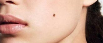
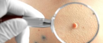
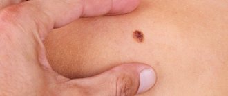
Before removing any age spots, you need to consult a doctor and get a competent opinion about what kind of formation it is, whether it can be removed and how exactly. It is best to consult a dermatologist. Different types of nevi are suitable for different excision methods.
Benign nevi themselves are not dangerous as long as they maintain a clear shape, do not change color and remain flat. Some of them may begin to grow over time. Some skin lesions are not actually moles, but warts or seborrheic keratosis.
Under no circumstances should you remove a mole yourself at home, even if you are absolutely sure that it is benign.
A dermatologist can remove nevi that are dangerous from a health point of view only surgically so that a histological examination of the tissue can be carried out. For this there must be special indications:
- the appearance of itching in a mole;
- increase in size;
- change of form;
- color change (pallor, discoloration, streaks, redness);
- bleeding
Laser mole removal
This innovative method is considered the most painless and fastest healing. Thanks to local anesthesia, the patient does not feel anything. The excision process takes place in stages: a directed laser beam removes the mole layer by layer.
However, after the procedure, a hole remains, which does not always completely disappear during healing. The beam evaporates the entire tissue of the mole, so it is not suitable for cases where an oncological study of the formation is necessary.
Pigment spots on the face require special attention. Since a healed scar is extremely important here, the specialist also needs to check whether keloid scars remain on the skin.
After removing a mole, the patient needs to protect the area affected by the laser from direct sunlight; this area on the face can be covered with a plaster. When the rehabilitation period is over, the protection can be removed.
Briefly about the main thing:
The safety of mole removal is ensured not so much by a specific method as by preserving the mole for histological examination. The only method that does not allow this to be done is removal with liquid nitrogen (cryodestruction). As an oncologist, I do not recommend removing moles this way.
Other methods - scalpel, electrocoagulation, laser, radio wave surgery can be used so that the mole is not completely destroyed. Unfortunately, not all doctors who perform mole removal have the necessary removal techniques.
If you decide to remove a mole using electrocoagulation, laser or radio wave surgery, before starting the procedure, clearly indicate to the doctor the need for histological examination.
If you have any questions, ask me through the online consultation form.
Removing moles using the radio wave method
Removal of moles by this method is usually performed using a radio wave installation of the Surgitron type. The method is painless and effective; with not very large areas of treatment, it leaves almost no marks on the skin.
High-frequency radio wave vibrations are transmitted through tissue and cause minimal damage to the surrounding skin surface, less than a surgical scalpel. After radio wave treatment, a small wound remains on the body. After some time, it heals, leaving a small, gradually disappearing spot. The healing process takes from 3 days to 2 weeks, depending on the size of the treatment area.
The radiosurgical method is used to eliminate deep formations on the skin:
- warts,
- keratosis,
- fibroids,
- moles,
- facial capillaries or spider veins,
- papillomas,
- scars after acne.
What types of moles are there on the head?
The most common type of moles growing on the scalp are pigmented nevi with papillomatosis
. They appear in childhood or adolescence and slowly increase in size. At first they appear as a small flesh-colored bump that protrudes at the level of the surrounding skin and is covered with hair. Then, as it grows, the mole becomes more and more elevated and acquires the appearance characteristic of this type of nevus.
At later stages of development, this type of nevus becomes covered with something like small papillae. This is where its name comes from - nevus with papillomatosis
(from Latin papilla - nipple). These moles may appear large, but at the same time, they are in contact with the scalp over a much smaller area than it appears from above (see picture below). This makes them easier to remove - the smaller the contact area, the easier it is to remove the mole.
Removal of moles using electrocoagulation
During the electrocoagulation procedure, tissue is removed using electric current for therapeutic purposes. This method allows you to visually control the depth of removal.
An electric current causes thermal damage to the mole and the area around it, which then forms a dry crust. During final healing, a noticeable mark may remain, so this method is not recommended for removing formations on the face.
But using this method, you can remove a mole in 1 session; moreover, the excised tissue is preserved and can be sent for histological examination. Electrocoagulation is carried out using direct and alternating current devices designed for this purpose. Different electrode shapes are used for different situations.
“Will you take a test from the mole the day before?”
The only analysis that can reliably determine the nature of the formation is histological examination. The formation is removed entirely and sent for pathomorphological examination. No “plucking” is performed in advance before removing moles.
It is possible to perform a cytological examination in advance if there is discharge, ulceration or trauma to the formation. This study allows you to establish a preliminary diagnosis and is performed when it is difficult to make a preliminary diagnosis. Often, an external examination or dermatoscopy is sufficient to make a diagnosis. For example, in relation to fibroepithelial polyps (papillomas), keratomas, fibromas, viral warts, a fairly large group of nevi (intradermal, warty, non-pigmented, etc.).
Mole removal with nitrogen
Cryodestruction is the instant freezing of a piece of tissue with liquid nitrogen. The substance, located at very low temperatures (from -100 to -180 ° C), causes freezing, destruction and rejection of tissue with a rapid recovery period.
The main advantage of cryodestruction is that dead tissue is not removed after destruction, but serves as a kind of bandage that protects the wound from infections. The wound heals painlessly. First, a crust forms, under which new, healthy tissue begins to grow.
However, the wound does not need to be treated with anything additional during the entire recovery period. This method is least suitable for the face, and the wound healing time here is much longer than with laser or radio wave removal.
Initially, after the scab falls off, the nitrogen-frozen area of skin will be lighter than the rest of the skin. But later everything will return to normal and traces of cryodestruction will become invisible.
However, the doctor is not in all cases able to control the depth of nitrogen exposure. Based on this, there is a possibility of frostbite of the tissues around the area, which can lead to the appearance of a scar. It is also possible that the mole will not be completely removed and the procedure will have to be repeated.
What ensures safe deletion?
There are several ways to evaluate a mole for health risks. Unfortunately, the accuracy of the methods used before removal (scraping/puncture and dermatoscopy) does not exceed 95%. There is only one option to find out whether a mole was 100% benign or not - a histological examination. It is this that allows us to determine the nature of the mole and, if it turns out to be malignant, to carry out the necessary treatment.
I’ll specifically focus on a simple “eye” inspection. I often read stories on the Internet like this: “ The doctor quickly looked at the mole, said that it was 100% benign and removed it without histology, and said that there was no point in paying for it. Now I’ve read a lot on the Internet, they write everywhere that histology is needed. Now I can’t sleep, I can’t eat, I don’t know what to do next, please help!”
An examination with the “eye” of even an experienced doctor may not be accurate.
According to clinical studies, the accuracy of such diagnostics does not exceed 80%.
IMPORTANT!!! Diagnostics “just looking” is a very inaccurate tool. When removing flat moles, always request a histological examination.
Surgical removal of a mole
Excision of the formation with a scalpel is considered the most painful. If the mole is on the face, this method is not advisable.
After surgery, the temperature may increase.
Surgical removal of moles requires not only the surgeon's qualifications, but also special care for the excision site after surgery. During the rehabilitation process, the patient will have to apply gauze bandages with an antibiotic or other healing ointment, changing them regularly (2 times a day). During the entire healing period, it is prohibited to swim or wet the removal site.
Are moles in hair dangerous?
Moles located on the scalp are very rarely dangerous. As a rule, they accompany a person for a significant part of his life and hardly change at all, with the possible exception of slow growth, no more than 1-2 mm per year. Sometimes even benign nevi of this type can become covered with small crusts. These crusts periodically fall off and appear again. It is important to note that this is a normal occurrence for pigmented nevus with papillomatosis. If it is not accompanied by bleeding, weeping of the surface of the mole, change in color or rapid growth, such formations are most likely not dangerous.
Consequences of mole removal
The consequences of removing moles on the body can be very different. Depending on the individual characteristics of the body, the chosen method and the equipment used, patients may quickly forget about the removed spot and not even recognize the remaining traces or suffer from complications that arise.
Removing skin lesions at home is absolutely unacceptable, even if the patient purchases special equipment for this. The possible consequences can only be minimized by a doctor in a specialized clinic.
You should immediately contact a specialist if after the procedure you notice the following symptoms:
- the appearance of suppuration;
- temperature increase;
- excessive secretion of lymph;
- strong pain;
- bleeding;
- negative changes in mental state.
Consequences of removing a mole on the face
There are many blood and lymphatic vessels on the face, and the skin on it is delicate and thin. Therefore, the removal of any formations on the face must be performed very carefully and professionally, using the most gentle methods possible.
The consequences of removing a pigment spot on the face may depend on:
- from the professionalism of the doctor;
- on the shape and size of the formation;
- on the selected removal method;
- on the individual health characteristics of the patient;
- from the patient’s thorough compliance with the recommendations of the rehabilitation period.
Proper care of the excision site largely determines how aesthetically pleasing the skin in this area will look.
The damaged area of skin must be treated with antiseptics. The crust that has formed on the surface of the wound cannot be peeled off, otherwise the wound will take a long time to heal, and an unsightly scar will form in its place. Sooner or later the crust will fall off on its own.
During the healing period, it is forbidden to take a bath, go to the sauna, swim in ponds, apply cosmetics to damaged tissues, or stay in the sun for a long time.
After the procedure, the doctor will definitely tell you about the mandatory rules that you need to remember and follow to avoid complications.
Consequences of removing moles with liquid nitrogen
The procedure for removing nevi using liquid nitrogen does not provide any guarantees, although in general the results are positive. The fact is that when treating fabrics with a substance at such a low temperature, it is impossible to accurately calculate the depth of freezing penetration. This often leads to incomplete removal of the formation, so sometimes additional procedures may be required.
The healing process after cryodestruction is long, and after it there are burn marks - scars. Tissue restoration sometimes takes several months.
During the process of cryodestruction of moles, healthy nearby tissues may also accidentally be damaged. This type of damage usually looks like a burn and takes longer to heal than usual.
Consequences after laser mole removal
Laser removal of formations is currently considered the safest, does not require additional time for rehabilitation, and after the procedure there are no scar changes on the skin.
A laser removal session lasts several minutes, there is no cutting of tissue, and there is no risk of blood poisoning or bleeding.
Complications after laser mole removal are extremely rare. The only consequence is a thin, dry crust that disappears on its own after 7-10 days. After the skin is restored, almost no trace remains on it.
Consequences of mole removal by electrocoagulation
In most cases, electrocoagulation leaves a fairly noticeable scar, so the procedure is not recommended for the face and open parts of the body. However, there is no risk of bleeding.
How are moles located under the hair removed?
The type of operation and what happens after it depends on one single factor - the area of contact of the mole with the skin:
- If the area is no more than 15 mm.
These moles are the easiest to remove.
Modern high-tech methods are suitable - laser and radio wave surgery. With this type of removal, no preparation is required and the mole is removed in one visit,
within 10-15 minutes
.
It is as if it is “cut off” from the skin and the wound heals like a simple deep abrasion. - From 15 to 20 mm.
To remove such a mole, you need a scalpel and preliminary tests, the application and removal of sutures, regular medical supervision and dressings are required. - More than 20 mm.
Skin grafting may be required to remove such a mole. This is because it is very difficult to match the edges of the wound on the head for suturing. The defect has to be covered with a “patch” made from the skin of the owner of the mole. The skin for the “patch” is taken from another area of the body (most often, the inner surface of the shoulder or lower abdomen).
Contraindications to burning moles
New growths on the skin can be benign or malignant. Removing moles with a coagulator is considered safe provided that the nevus does not show signs of developing a cancerous tumor. The doctor determines the primary signs during a visual examination. You cannot remove a nevus using an electric knife if you suspect the formation of a malignant process. Also contraindications include:
- bleeding disorders
- intolerance to electrical procedures
- allergic reactions to local anesthetics
- herpetic infection in the acute phase
- an inflammatory skin disease at the site where a mole was burned out with an electric knife
Any such case must be eliminated before electrocoagulation is performed.
Excision is the safest procedure for treating moles
Freeze and burn is suitable for viral warts, advanced warts or senile epithelial warts. When it comes to moles, the only professionally accepted method for removing birthmarks is excision.
Mole treatment procedure
Thanks to a special electric loop, the dermatologist precisely follows the line of the birthmark, removing it to the desired depth and without touching healthy skin. It also does not destroy mole tissue, which can cause malignant degeneration.
After the procedure, a histological examination of the removed skin tissue is performed. If cancer is detected, which is unfortunately not uncommon, further treatment is prescribed.
The mole is excised under local anesthesia; the procedure takes from 10 minutes to half an hour. Next, a sterile bandage is applied to the wound. After a couple of weeks, you will need to show the removal site to a dermatologist, who should make sure that everything is in order.
ONLINE REGISTRATION at the DIANA clinic
You can make an appointment by calling the toll-free phone number 8-800-707-15-60 or filling out the contact form. In this case, we will contact you ourselves.
Indications for resection
Moles that have the following properties are subject to removal:
- frequent mechanical damage due to rubbing with clothing, jewelry, other accessories, and surrounding objects;
- localization on open areas of the skin that are exposed to direct sunlight for a long time;
- localization in hazardous areas, which are most susceptible to injury;
- severe mechanical damage;
- frequent infectious processes, bleeding;
- gradual increase in size with the risk of developing a malignant process;
- multiple types, that is, if additional moles form near the location;
- the presence of an inferiority complex due to the presence of an overly large, ugly mole that bothers a person.
The danger comes from formations that are located in the following parts of the body:
- back of the hand;
- head area with hair;
- bends of limbs;
- crotch;
- face, neck, back.
Each mole requires constant monitoring. If active growth occurs in it, the concentration of melanocytes may gradually increase. They degenerate into a malignant form, which is why melanoma is formed. This is a malignant skin disease that can gradually form metastases that spread through the bloodstream to other organs.
A bad head gives no rest to your hands
On average, a dozen moles can be found on most people's skin. In some cases, they may look unaesthetic, so they haunt their owners. It is not surprising that in such cases the idea of trying to remove an annoying mole without medical help pops into your head. And here the Internet comes to the rescue, with supposedly safe methods of getting rid of warts, moles and everything your heart desires.
But before using such advice, it would be nice to think carefully. Any sane person should understand that forums and websites are mainly run not by doctors, but by specialists who rewrite other people’s thoughts - rewriters. These could be schoolchildren, students, mothers on maternity leave - anyone except dermatologists. So should you trust them, risking your health?


