Nail fungus, medically known as onychomycosis, is a nail infection caused primarily by a class of fungi called dermatophytes. These are the same fungi that cause shingles. The infection can affect any part of your nail, including the nail plate, nail bed, and root. However, 85% of fungal nail infections occur under the nail plate, on the nail bed. Fungal nail infections are seven times more common on toenails than on fingernails.
Fungal nail infections are highly contagious. They are easily caught in areas where you walk barefoot, such as locker rooms or swimming pools, or from unsterilized equipment in nail and beauty salons. If you touch someone who is infected and forget to wash your hands, the infection will quickly develop. Sharing contaminated items such as towels, sheets and clothing can also spread the infection.
Treating a fungal nail infection can be a lengthy process because nails grow slowly, so you may only see improvement as a new nail grows. It is also common for nail infections to return. To avoid recurring fungal nail infections, it is important to take proper care of your feet and nails. Make sure you treat the infection, whether it's shingles or onychomycosis, as soon as you start noticing symptoms.
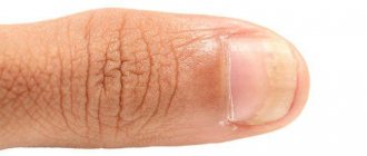
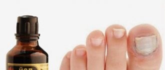
Fact 7: “When treating nail fungus, you should not paint your nails”
This myth is also a thing of the past with the right treatment method. For example, nails treated with loteryl can be painted with cosmetic varnishes if desired.
Wait ten minutes after applying the medicine and then coat the nail plate with your favorite color of polish. This way, you won’t have to hide your nails even during treatment!
ONLINE REGISTRATION at the DIANA clinic
You can sign up by calling the toll-free phone number 8-800-707-15-60 or filling out the contact form. In this case, we will contact you ourselves.
Top tips for preventing infections at home:
- Throw away old shoes, especially those that you used during exercise.
- If you suffer from shingles, make sure you treat it as the fungus can also affect your toenails. Don't use the same nail accessories or tools on normal and infected nails - buy two sets to prevent contamination
-Wash and dry your hands thoroughly after handling any fungal infection or treatment
-Take care of your toenails - keep them trimmed and clean. Nails should be cut straight across (not rounded or V-shaped)
- Wear shoes that fit your feet and legs - high heels and shoes with narrow toes can damage the natural skin between the nail and the skin underneath, allowing fungus to spread underneath the nail
— Make sure other family members are promptly treating their fungal infections and taking similar precautions
— Disinfect your hands and feet after contact with infected areas, as well as bedding and towels of an infected person. Disinfecting soap is good for everyday use. .
— If someone in your household is diagnosed with nail fungus, it is not enough to simply avoid contact with the affected areas. It is imperative to disinfect surfaces, especially those in a humid environment, in which microorganisms multiply especially quickly. First of all, we are talking about the bathroom. To disinfect small surfaces, you can use a spray that kills microorganisms and bacteria.
Dangerous fungi
Fungi that parasitize us are dangerous. They are microorganisms of plant origin. Once in a healthy human body, the fungus begins to multiply quickly. It spreads through small vessels and arteries with the blood flow. True, sometimes ten years pass before clinical symptoms of the disease appear. But the fungus does not stop its harmful work for a minute.
Mycology, that is, the science that studies fungal diseases, knows more than five hundred species of pathogenic fungi. But most often doctors encounter about a hundred. Moreover, the whole family is usually affected by one type of fungus. After all, infection easily occurs through shared beds, toiletries, and shoes.
Unfortunately, fungal disease is not just spots, peeling, ingrown and deformed nails. The fungus affects the entire body. In old age, these microorganisms can even penetrate very deeply into various organs. They can aggravate existing diseases and cause severe allergies.
Myth 3. If you find a fungus, you need to throw out all your shoes.
No, it's enough to process it. There is a special device that fits into shoes, emits ultraviolet light and has an antifungal effect. The sun and radiator, by the way, do not kill mushrooms. You can treat the inside of shoes with a 40% vinegar solution (for this, the vinegar essence is diluted with water) or buy inexpensive domestic products for treatment at the pharmacy. For example, Samarovka liquid or 1% chlorhexidine digluconate (gibitan). Wipe the inside of your shoes with them and keep them in a sealed bag for 2-3 hours. Another option is to treat your shoes with 25% formaldehyde. Afterwards, you should wipe your shoes with ammonia - this reduces the risk of allergies. It is also better to disinfect underwear and bed linen, stockings, socks, tights, wash them and, if possible, iron them damp.
When should you see a doctor?
You should see a doctor if you see green-yellowish or black pigmentation on your nail or under your nail. If you have diabetes or are pregnant, consult your doctor before starting any treatment.
Did you know that?
…Biting your nails and using artificial nails can increase your risk of developing a fungal nail infection on your toenails!
...fungal nail infections affect 10% of the world's population!
Fungal nail infections (onychomycosis): a never-ending story?
Is onychomycosis still a dangerous problem?
Most superficial fungal infections are caused by dermatophytes belonging to one of three genera (Trichophyton, Epidermophyton, and Microsporum), with T. rubrum being the best known cause of fungal nail infection (Figure 1). Table 1 shows the prevalence of various superficial fungal infections in different geographic areas [1]. Among superficial fungal infections, nail onychomycosis is by far the most difficult to treat (Figure 2). As reported above, the prevalence of onychomycosis is more than 23% in Europe [2] and 20% in East Asia [3]. In North America, the incidence of onychomycosis is approximately 14% [4], with fungal infections causing up to 50% of all nail diseases [5]. With millions of dollars spent annually on oral and topical medications, laser treatments, over-the-counter medications and home remedies, it is clear that toenail fungal infections continue to plague the public and are determined to get rid of them. Unfortunately, this is easier said than done. Successful treatment of nail onychomycosis requires long-term therapy, which can last a whole year. Even then, complete cure includes both clinical cure (meaning clearing (sanitation) of nails) plus mycological cure (obtaining negative results both by microscopy and by sowing and growing dermatophyte cultures on nutrient media), which is often unattainable.
Picture 1 . View of a Trichophyton rubrum colony under light microscopy (40x).
Figure 2 . Distal subungual onychomycosis of the big toe.
Table 1 . The most common dermatophytosis in different regions of the world.
| Region | Dermatophytosis | Pathogen |
| Northern/South Africa | Dermatophytosis of the foot, onychomycosis | T. rubrum |
| Western Europe | Dermatophytosis of the foot, onychomycosis | T. rubrum |
| Russia | Onychomycosis | T. rubrum |
| Mediterranean (Italy/Greece) | Dermatophytosis of the trunk, dermatophytosis of the scalp | M. canis |
| Türkiye | Dermatophytosis of the foot | T. rubrum |
| North Africa/Tropical Africa | Dermatophytosis of the trunk | T. violaceum, M. audouinii |
| China/Japan | Dermatophytosis of the foot, onychomycosis | T. rubrum |
| India | Dermatophytosis of the trunk | T. rubrum |
| Asia | Dermatophytosis of the foot, onychomycosis | T. rubrum, T. mentagrophytes |
| Australia | Dermatophytosis of the foot, onychomycosis | T. rubrum, T. mentagrophytes |
What are the risk factors for onychomycosis of the big toes?
The most common predisposing risk factor for the development of onychomycosis is reported to be advanced age, which is 18.2% in patients 60–79 years of age compared with 0.7% in patients under 19 years of age. Additionally, men are three times more likely to develop onychomycosis than women, although the reasons for these gender differences are unclear [6]. In addition, the prevalence of infection in people whose spouses have been infected with onychomycosis is low compared with the prevalence of the disease among children of affected parents, suggesting a genetic risk factor [7]. Although extremely rare, one study reported the identification of four members of the same family among seven unrelated groups of individuals sharing a common genetic trait (autosomal recessive CARD9 deficiency) who developed a deep tissue dermatophyte infection that was fatal [8 ].
Other risk factors include diabetes and conditions that contribute to poor peripheral circulation [9]. In fact, onychomycosis may represent an important predisposing factor for the development of diabetic ulcers and diabetic foot syndrome [10]. Immunocompromised patients, such as those with HIV infection or undergoing chemotherapy for cancer, are also predisposed to fungal nail infections [11]. There is at least one case in the literature of a fungal nail infection caused by a Fusarium fungus (non-dermatophyte) causing a fatal systemic generalized infection in a patient with lymphoma following a bone marrow transplant [12].
Several non-clinical risk factors also influence a person's chance of developing fungal nail infections. Onychomycosis of the nails is not common in tropical climates, apparently because people in these regions do not wear tight, closed shoes, which create a warm, moist environment for the fungal infection to spread. In addition, the spread of foot infections, including tinea pedis (athlete's foot), can occur in areas such as showers, bathrooms, or locker rooms where the floor surface is often wet and people walk barefoot [13]. Nail trauma also increases the risk of fungal infection of the affected nail, especially in older adults [11].
A recent study by our research team used regression analysis to show that a history of tinea pedis, as well as three clinical situations—onychomycosis, increasing (scaling) of the skin pattern on the plantar surfaces (a clinical sign of tinea pedis), change Nail colors (a clinical sign of onychomycosis, usually indicating the severity of nail infection) were statistically associated with the prevalence of infection in families with multiple infected family members (P≤.044) [14].
How to treat onychomycosis?
Treatment of onychomycosis includes chemical or surgical removal of the affected nail, the use of systemic or local drugs, pulse therapy, or a combination of these methods. Table 2 shows treatment regimens using oral and topical medications; and, as can be seen from the table, the course of treatment for a fingernail infection is shorter than the course of treatment for a toenail infection. Treatment of onychomycosis has improved significantly over the past few decades with the introduction of the antifungal drugs terbinafine and itraconazole. However, these drugs may have side effects such as liver damage or adverse drug interactions, which is especially true in older adults [15]. Additionally, nail infections caused by non-dermatophyte fungi, such as Fusarium, are particularly difficult to treat [16].
Table 2 . Treatment of onychomycosis with antifungal agents.
| A drug | Dose | Duration |
| Terbinafine | 250 mg | Toenails: once a day for 12 weeks |
| Fingernails: once a day for 6 weeks | ||
| Itraconazole | 200 mg | Toenails: once a day for 12 weeks |
| pulse therapy | Toenails: 200 mg twice daily for 1 week/off treatment for 3 weeks. Repeat the course within 3-4 months | |
| Fingernails: 200 mg twice daily for 1 week/off treatment for 3 weeks. Repeat course within 2 months | ||
| Fluconazole | 300-450 mg | Toenails: once a week for 9-12 months |
| 150-300 mg | Fingernails: once a week for 4-6 months | |
| Nail polish ciclopirox | apply once a day | Remove varnish once a week. Treatment up to 48 weeks |
| Nail polish amorolfine | apply once or twice a week | Remove varnish before each new use. Toenails: 9-12 months. Fingernails: 6 months |
Why can't topical antifungals work better?
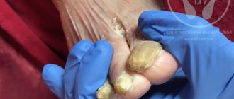
Unfortunately, currently available topical agents such as amorolfine 5% and cyclopirox 8% have low efficacy (approximately 5%-12%) [17, 18]. Low effectiveness may be due mainly to the inability of the drug to penetrate through the nail plate to the nail bed, where the infectious agents are located [19]. Thickened nails, extensive involvement of the entire nail, extension of the disease to the lateral ridges around the nails, and yellow nail spikes contribute to poor results of local treatment [11]. Figure 1 shows an example of distal subungual onychomycosis; nails are specially trimmed to show their thickening.
Further complicating the clinical situation is the fact that some antifungal drugs bind to the matrix of the nail plate and, therefore, do not reach the site of infection, which is localized in the nail bed. For example, terbinafine has been shown to rapidly concentrate in the nail, reaching a maximum concentration of 0.39 mg/g and persisting for up to 2 months after the end of treatment [20]. In this regard, Ryder and co-workers developed an in vitro nail model in which it was shown that the fungicidal effect of terbinafine when testing established dermatophyte infections on the human nail was actually less effective than in conventional microplate assays where crushed powder on affected nails [21].
Recently, many attempts have been made and various approaches have been developed to solve the problem of drug penetration into the nail plate. For example, there have been attempts to develop permeation enhancers to facilitate drug delivery through the nail plate, such as the addition of dodecyl-2-N,N-dimethylaminopropionate hydrochloride (HCl DDAIP, trade name NexACT-88; NexMed) and terbinafine to nail polish [19]. ]. Another method was to add terbinafine to nail polish as part of transfersome-ultradeformable lipid vesicles that can pass through intercellular spaces and are used as carriers for non-invasive targeted delivery and sustained release of drugs [22]. In addition, a new small molecule antifungal agent, oxaborole (AN2690), has been recently developed to better penetrate the nail plate [23]. However, to date, none of these topical preparations have been commercialized.
Therefore, regulatory approval of drugs for the topical treatment of onychomycosis and their recommendation for use in clinical practice may be hampered by the negative impact of an overly strict definition of complete cure, which includes nail debridement plus mycological cure (negative microscopy and culture results). . A review of data from several international clinical trials of antifungal drugs by Ghannoum and co-workers concluded that re-evaluating the definition of treatment for onychomycosis is critical [24]. In these trials, large numbers of toenail samples were collected from patients at the end of the clinical trial and contained visible fungal hyphae that, when further cultured, failed to grow on culture media. However, current technology does not differentiate between "live" and "dead" fungi, so even if these samples were to be classified as positive by microscopy, the infection may in fact be cured. The authors suggest that for clinical trials of topically applied topical medications, the treatment duration should be extended to 18 months, followed by a longer washout period of 3–6 months before initial assessment of effect to achieve both removal of residual drug in the nail , and non-viable fungal cells. Thus, the absence of clinical signs after an adequate period of elimination, combined with negative fungal culture results, with or without negative microscopy results, should be considered cured of onychomycosis. These changes in definition may allow more medicinal antifungal drugs to prove their clinical effectiveness for topical use.
What's new in the treatment of onychomycosis?
Recent devices developed for the treatment of onychomycosis include laser devices, photodynamic therapy devices, iontophoresis and ultrasound. Laser treatment has been approved for cosmetic treatment only, but the effectiveness of treatment in eradicating fungal infection needs to be determined in additional randomized controlled trials [25]. There are isolated reports in the literature about the treatment of onychomycosis using phototherapy, which involves irradiation of accumulated protoporphyrin within the fungal hyphae, which leads to subsequent damage to the hyphal cells [26]. The availability of iontophoresis and the use of electric current (0.5 mA/cm2) facilitates the passage of the drug through the nail plate, which has been proven in studies on human cadaveric nails, but corresponding clinical studies have not yet been completed [27]. Finally, although ultrasound therapy has previously shown fungistatic activity against nail lesions [28], the device itself appears to be too complex with ultrasound transducers and drug delivery compartments required for each nail and requires the development of a special computer program interface, making this method only available in a doctor's office and probably very expensive [29].
Why do patients relapse so often?
There are several factors that may contribute to a high risk of recurrent fungal nail infections. Patients with a genetic predisposition to onychomycosis, those with immunodeficiency or diabetes mellitus may develop relapses and may never achieve permanent recovery from the fungal infection [11].
This may be due to either failure of eradication or reinfection with a new fungal strain upon subsequent exposure. Arthrospores, which are chains of conidia in fungi, are formed when the integrity of fungal hyphae is disrupted and are considered the main agent causing nail damage. These arthrospores have been shown to have thicker cell walls than conidia formed in vitro and are more resistant to antifungal drugs and thus may remain in the nail bed as a reservoir for disease recurrence [30]. However, the level of innate resistance among dermatophytes is low. In vitro susceptibility testing was performed at our center on 140 consecutive strains from subjects who failed topical terbinafine in clinical trials. In all cases, the minimum inhibitory concentrations (MICs) of terbinafine for each patient in the kit or within a single in vitro dilution were identical and there were no cases of resistance development. The same results were obtained within each set with fluconazole, itraconazole, griseofulvin (with the exception of one isolate having a 3-fold increase in MIC). This provides further evidence that there was no cross-resistance between antifungal agents [31]. The study found that the case of treatment failure for infected nails may be due to multiple/familial factors.
Even in cases where the fungal infection has been completely eradicated after antifungal therapy, there is still a risk of re-infection. As mentioned above, people are exposed to dermatophytes in water bodies through many repeated day-to-day activities, including trips to the gym and increased travel. General preventive measures, such as not walking barefoot in public showers or hotel showers, will help avoid unnecessary infection. This is one of the most common routes of infection in the home. It has long been suspected that nail infections are more common in close contact with family members. However, this claim was not confirmed until recently, so our research team used molecular techniques to prove that individuals in the same family were infected with the same strain of T. rubrum [14]. For those trying to avoid reinfection, measures such as treating shoes with a spray containing antifungal agents or disinfecting shoes in a commercial ultraviolet device [32] after each use are recommended. Thus, the patient not only must treat the infection, but also break the vicious circle of reinfection.
Literature
1. Havlickova A, Czaika VA, Friedrich M (2008) Epidemiological trends in skin mycoses worldwide. Mycoses 51 S4:2–15. doi: 10.1111/j.1439-0507.2008.01606.x
2. Haneke E, Roseeuw D (1999) The scope of onychomycosis: epidemiology and clinical features. Int J Dermatol 38 Suppl 2:7–12. doi: 10.1046/j.1365-4362.1999.00015.x
3. Ogasawara Y (2003) Prevalence and patient's consciousness of tinea pedis and onychomycosis. Nihon Ishinkin Gakkai Zasshi 44:253–260. doi: 10.3314/jjmm.44.253
4. Ghannoum MA, Hajjeh RA, Scher R, Konnikov N, Gupta AK, et al. (2000) A large-scale North American study of fungal isolates from nails: the frequency of onychomycosis, fungal distribution, and antifungal susceptibility patterns. J Am Acad Dermatol 43:641–648. doi: 10.1067/mjd.2000.107754
5. Scher RK (1994) Onychomycosis is more than a cosmetic problem. Br J Dermatol 130: 15. doi: 10.1111/j.1365-2133.1994.tb06087.x
6. Gupta AK, Jain HC, Lynde CW, MacDonald P, Cooper EA, et al. (2000) Prevalence and epidemiology of onychomycosis in patients visiting physicians' offices: a multicenter Canadian survey of 15,000 patients. J Am Acad Dermatol 43:244–248. doi: 10.1067/mjd.2000.104794
7. Faergemann J, Correia O, Nowicki R, Ro BI (2005) Genetic predisposition—understanding the underlying mechanisms of onychomycosis. J Eur Acad Dermatol Venerol 19:17–19. doi: 10.1111/j.1468-3083.2005.01283.x
8. Lanternier F, Pathan S, Vincent QB, Liu L, Cypowyj S, et al (2013) Deep dermatophytosis and inherited CARD9 deficiency. N Engl J Med 369:1704–1714. doi: 10.1056/nejmoa1208487
9. Saunte DM, Holgersen JB, Haedersdal M, Strauss G, Bitsch M, et al. (2006) Prevalence of toe nail onychomycosis in diabetic patients. Acta Derm Venerol 86:425–428. doi: 10.2340/00015555-0113
10. Nenoff P, Ginter-Hanselmayer G, Tietz HJ (2012) Fungal nail infections - an update: Part 1 - Prevalence, epidemiology, predisposing conditions, and differential diagnosis. Hautarzt 63: 30–38. doi: 10.1007/s00105-011-2251-5
11. Scher RK, Baran R (2003) Onychomycosis in clinical practice: factors contributing to recurrence. Br J Dermatol 149:5–9. doi: 10.1046/j.1365-2133.149.s65.5.x
12. Arrese JE, Pierard-Franchimont C, Pierard GE (1996) Fatal hyalohyphomycosis following Fusarium onychomycosis in an immunocompromised patient. Am J Dermatopathol 18:196–198. doi: 10.1097/00000372-199604000-00014
13. Aly R (1994) Ecology and epidemiology of dermatophyte infections. J Am Acad Dermatol 31:S21. doi: 10.1016/s0190-9622(08)81262-5
14. Ghannoum MA, Mukherjee PK, Warshaw EM, Evans S, Korman NJ, et al. (2013) Molecular analysis of dermatophytes suggests spread of infection among household members. Cutis 91:237–246.
15. Elewski B, Tavakkol A (2005) Safety and tolerability of oral antifungal agents in the treatment of fungal nail disease: a proven reality. Ther Clin Risk Manag 1:299–306.
16. Tosti A, Piraccini BM, Lorenzi S (2000) Onychomycosis caused by nondermatophyte molds: clinical features and response to treatment of 59 cases. J Am Acad Dermatol 42:217–224. doi: 10.1016/s0190-9622(00)90129-4
17. BlueCross BlueShield of Northeastern New York (2006 November 28) Drug Therapy Guidelines: Antifungal Agents Lamisil (terbinafine), Sporanox (itraconazole), Penlac (ciclopirox), Vfend (voriconazole). Drug P&T Newsletter.
18. Lauharanta J (1992) Comparative efficacy and safety of amorolfine nail lacquer 2% versus 5% once weekly. Clin Exp Dermatol 17:41–43. doi: 10.1111/j.1365-2230.1992.tb00277.x
19. Ghannoum MA, Long L, Pfister WR (2009) Determination of the efficacy of terbinafine hydrochloride nail solution in the topical treatment of dermatophytosis in a guinea pig model. Mycoses 52:35–43. doi: 10.1111/j.1439-0507.2008.01540.x
20. Faergemann J, Zehender H, Millerioux L (1994) Levels of terbinafine in plasma, stratum corneum, dermis-epidermis (without stratum corneum), sebum, hair and nails and after 250 mg terbinafine orally once daily for 7 and 14 days. Clin Exp Dermatol 19:121–126. doi: 10.1111/j.1365-2230.1994.tb01138.x
21. Osborne CS, Leitner I, Favre B, Ryder NS (2004) Antifungal drug response in an in vitro model of dermatophyte nail infection. Med Mycol 42:159–163. doi: 10.1080/13693780310001656803
22. Ghannoum M, Isham N, Herbert J, Henry W, Yurdakul S (2011) Activity of TDT 067 (Terbinafine in Transfersome) against Agents of Onychomycosis, as Determined by Minimum Inhibitory and Fungicidal Concentrations. J Clin Microbiol 49:1716–1720. doi: 10.1128/jcm.00083-11
23. Alley MR, Baker SJ, Beutner KR, Plattner J (2007) Recent progress on the topical therapy of onychomycosis. Expert Opin Investig Drugs 16:157–167. doi: 10.1517/13543784.16.2.157
24. Ghannoum M, Isham N, Catalano V (2013) A Second Look at Efficacy Criteria for Onychomycosis: Clinical and Mycological Cure. Br J Dermatol. E-pub ahead of print. doi:10.1111/bjd.12594.
25. Gupta AK, Simpson FC (2013) Laser therapy for onychomycosis. J Cutan Med Surg 17:301–307.
26. Harris F, Pierpoint L (2012) Photodynamic therapy based on 5-aminolevulinic acid and its use as an antimicrobial agent. Med Res Rev 32:1292–1327. doi: 10.1002/med.20251
27. Delgado-Charro MB (2012) Iontophoretic drug delivery across the nail. Expert Opin Drug Deliv 9:91–103. doi: 10.1517/17425247.2012.642364
28. Silva JL, Doimo G, Faria DP (2011) The use of high frequency waves to treat onychomycosis: preliminary communication of three cases. An Bras Dermatol 86:598–600.
29. Abadi D, Zderic V (2011) Ultrasound-mediated nail drug delivery system. J Ultrasound Med 30:1723–1730.
30. Yazdanparast SA, Barton RC (2006) Arthroconidia production in Trichophyton rubrum and a new ex vivo model of onychomycosis. J Med Microbiol 55:1577–1581. doi: 10.1099/jmm.0.46474-0
31. Bradley M, Leidich S, Isham N, Elewski B, Ghannoum M (1999) Antifungal susceptibilities and genetic relatedness of serial Trichophyton rubrum isolates from patients with onychomycosis of the toenail. Mycoses 42:105–110.
32. Ghannoum MA, Isham N, Long L (2012) Optimization of an infected shoe model for the evaluation of an ultraviolet shoe sanitizer device. J Am Podiatr Med Assoc 102:309–313. doi: 10.7547/1020309
Best general tips for preventing infections:
- Avoid walking barefoot in public - wear flip-flops or sandals in public locker rooms, showers and the area around the pool
— If walking barefoot is unavoidable (for example, in some sports and wrestling on mats), as soon as possible, disinfect your skin and nails with disinfectant soap . If there is no access to water, then use skin antiseptic or disinfectant wipes for primary disinfection. Then treat the skin with an antifungal drug.
- Bring your own nail care accessories to your nail salon, including scissors, clippers, files, shaving tools.
— Make sure that the tools in your nail salon are properly cleaned and sterilized after the previous client
Avoid spreading infection
Fungal nail infections are contagious, so keep this in mind while you are infected. It's best to protect your feet and avoid sharing clothes, shoes, towels or sheets. To prevent the spread of infection, always wash your hands after applying treatment and remember to use different towels for your feet and body. It's a good idea to tell your loved ones about fungal nail infections so they remember to take similar precautions.
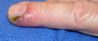
Signs of fungus damage to the nail plate
If you notice the following manifestations on your nails, then it’s time to sound the alarm:
- Yellowish tint
- Delamination, destruction or thickening
- Blotches of yellow or white spots
- Unpleasant odor, pain
- Changing shape
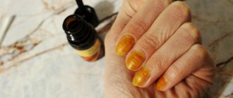
Given the variety of types of fungus, the infection in its first manifestations may look different.
For example, yeast fungi make the tone of the nail plate darker and add a yellowish color. The nail becomes deformed and takes on a wavy shape. Redness forms on the skin around the nail and even with slight exposure painful sensations appear.
Dermatophytes, affecting the nail, first appear as small spots along the edges of the plate of yellow, white and gray color. Subsequently, the nail completely turns yellow and moves away from the base.
Molds most often attack against the background of reduced immunity. It is difficult to confuse lesions with this species due to the rich palette of colors on the nails: green, blue, black, brown, yellow. Sometimes, already at the first stage, peeling and separation of the nails from the base are observed.
How to prevent infection?
The best cure for nail fungus is prevention.
To protect yourself from infection you must:
- use medical disinfectants with the inscription GOST (capable of killing onychomycosis, tuberculosis, hepatitis, HIV);
- monitor the condition of the feet and nails, the main rule is that the feet should be clean and dry;
- take care of shoes, regularly disinfect and dry them; it is convenient to use electric dryers for drying;
- treat your feet with drying agents or antiperspirants if your feet sweat a lot;
- in the pool, sauna, and gym locker rooms, do not walk barefoot, but wear rubber flip-flops with closed toes;
- wear comfortable shoes made of natural, breathable materials;
- Do not use other people’s towels, shoes or manicure accessories.
These tips will help you prevent onychomycosis and keep your nails healthy and well-groomed. They are effective not only for the prevention of fungus, but also other problems: ingrown toenails, calluses, corns.