Lipoma - what is it?
Lipoma is a benign fatty tumor. Lipoma can be limited capsularly from other tissue, or it can be diffusely present in tissues.
This tumor occurs in females aged from thirty to fifty years (middle age).
A tumor forms in areas of the body that are rich in adipose tissue. This can be: subcutaneous tissue, skin, adipose tissue near the kidneys, fat behind the peritoneum, muscle areas of adipose tissue, intestines, mammary gland, as well as the meninges, lungs, myocardium, bones and nerve trunks. Often the latter are formed from embryonic tissue if the tissue falls into the wrong area during formation and development.
Types of lipomas
There are several main classifications of lipomas that are actively used in medical practice. Depending on the type of tissue that is involved in the pathological process, the following types of neoplasms are distinguished:
- perineural - localized around nerve trunks;
- intermuscular – located between the muscles of the body;
- lumbosacral - grow near the vertebrae or in the spinal canal;
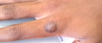
- soft tissues - located on the surface of the skin, less often subcutaneous;
- joints - are located in the synovial membrane or vagina of the joints.
The formation can appear in almost any part of the body and internal organs. Depending on the location of the compaction , the following types of lipomas are most often diagnosed:
- mammary gland - forms in the glandular tissue and deforms the shape of the breast as it grows;
- breasts - a soft and mobile formation that appears in the subcutaneous fatty tissues;
- head - a frequently occurring pathology, which is mainly formed as a result of insufficient hygiene;
- back - one of the most common neoplasms, characterized by extremely slow development;
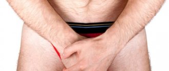
- neck is a hereditary disease that, during development, can impair the airways, cause weakness and angina.
There are also other, less common places where pathology forms, which include the brain, limbs, skin, peritoneum, eyes, lips and face.
Also, these compactions are divided into two large groups: single and multiple. The first represent a single formation in any part of the body. The latter, accordingly, are characterized by multiple manifestations in different areas of the body and are much less common.
Material and methods
At the Pediatric Surgery Clinic named after. prof. N.L. During the period from 1991 to 2010, 222 children aged from 3 months to 17 years were treated for LP. There were 118 (53.1%) females, 104 (46.9%) males. Benign tumors (lipomas) were detected in 216 (97.3%) patients, malignant tumors (liposarcoma) - in 6 (2.7%) patients. In the vast majority of children (217 observations - 97.7%), the tumor was externally localized. Among them, subcutaneous lipomas were identified in 170 (78.3%) cases, subfascial lipomas in 28 (12.9%) cases, and intermuscular lipomas in 19 (8.8%) cases. In 5 patients, internal localization of the lymphoma was observed, when the neoplasm was located in the mediastinum (2 children - 0.9%), in the abdominal cavity (2 patients - 0.9%), in the retroperitoneal space (1 patient - 0.5%). Single lipomas were observed in 198 (89.2%) children, multiple - in 24 (10.8%) patients.
Among patients with lipomas, the tumor was most often localized in the back - 45 (20.8%) patients, hips - 33 (15.3%), necks - 20 (9.3%), buttocks - 20 (9.3%) , shoulder - 18 (8.3%), abdominal wall - 16 (7.4%). Less commonly, the lipoma was located in the groin region - 10 (4.6%), in the sternum - 9 (4.2%), on the forearm - 9 (4.2%), in the axillary region - 8 (3.7%), on the hand - 7 (3.2%), on the foot - 7 (3.2%), on the scrotum - 6 (2.8%), in the popliteal region - 5 (2.3%), in the abdominal cavity and retroperitoneal space - one observation (0.5% each, respectively).
A pathohistological study of removed LM among our patients revealed that lipoma occurred in 79 (35.6%) children, fibrolipoma in 105 (47.3%), angiofibrolipoma in 32 (14.4%), liposarcoma in 6 (2.7%).
In contrast to benign lymph nodes, liposarcoma in children was much less common. These tumors were localized on the thigh in 2 (33.3%) cases, in the remaining patients - on the neck, forearm, in the abdominal cavity and mediastinum - there was one observation each, respectively, 16.7%.
Causes of lipomas
Experts identify a set of prerequisites that can provoke the formation of lipomas. Among the most common:
- genetic factors, hereditary predisposition;
- disruption of metabolic processes in the body and fatty tissues;
- insufficient level of personal hygiene;
- malfunctions of the thyroid and pancreas;
- chronic pathologies that significantly affect the body’s immune forces (diabetes, hepatitis, HIV, etc.);
- dependence on alcohol, tobacco, drugs;
- poor nutrition;
- obesity, excess fatty tissue;
- injuries, damage;
- sedentary lifestyle, lack of physical activity.
Despite the fact that lipoma is a benign formation, there is always a risk of developing its malignant form. This probability is especially high for people who are included in the relevant risk groups: they have precancerous diseases (polyps, dysplasia), have already suffered from oncological pathologies, have a hereditary predisposition to the formation of tumors, and are under the constant influence of carcinogens, radiation and other harmful environmental factors.
Symptoms of lipomas
The clinical picture of the pathology is quite sparse and is characterized by:
- the presence of a palpable formation, which is distinguished by its soft consistency, mobility, and elasticity;
- the appearance of pain due to compression of nerve trunks or growth in internal organs;
- stability of the compact size or its increase with weight loss;
- swelling of the limbs and disruption of their function (if the formation compresses nerves and blood vessels).
If a malignant process develops, general malaise and headaches appear, blood pressure rises and other characteristic symptoms of intoxication of the body appear.
In this case, depending on the exact location of the compaction, other, more pronounced symptoms and signs may occur:
- neoplasms in the esophagus cause nausea and cough;
- seals on the trachea and bronchi cause a painful dry cough that does not subside after taking antitussive drugs;
- a fatty tumor on cartilage and tendons causes pain in the joints and impedes movement;
- formation in the mammary gland provokes pain in this area;
- compaction in the kidney area causes increased blood pressure, colic and lower back pain;
- a formation in the head causes neurological symptoms – headaches, dizziness;
- lipoma of the neck is accompanied by hoarseness, hoarseness, difficulty swallowing;
- a neoplasm in the heart area causes the development of cardiac pathologies: arrhythmia, heart failure, etc.
When should you see an oncologist?
If the diagnosis is established, and you clearly know that a specific tumor on the body is a harmless wen, then it should be removed. If a wait-and-see approach has been chosen, then there are a number of situations in which you should consult a doctor immediately.
Reason for urgent contact with an oncologist or dermatologist with confirmed lipomatosis:
- pain appeared - any at rest or when pressing on the tumor;
- rapid growth - a sharp increase in size within a few weeks;
- change in skin color in the projection of the neoplasm;
- the appearance of ulceration on the skin above the tumor. Discharge of purulent, bloody, ichorous - any discharge from the cone;
- additional unpleasant symptoms appeared - pain in other parts of the body, general weakness, increased fatigue, sudden changes in weight without changing the diet or increasing the level of physical activity;
- when the size of the neoplasm is more than 50 mm in diameter;
- if the tumor location is an area saturated with lymph nodes. This is the groin area, armpits, neck.
Such symptoms may indicate either trauma to the neoplasm and the onset of the inflammatory process, or an incorrectly made primary diagnosis.
Whether or not a wen will degenerate into carcinoma is a complex question.
Even at the present stage of development of science, doctors do not give a clear answer. Therefore, if you find any growth in yourself, then contact an oncologist. Don't wait until it reaches gigantic proportions. Get examined, confirm the diagnosis of fatty tissue, remove it and enjoy life. The article has been verified by the editors
Diagnosis of lipomas
Since the development of a neoplasm in the body practically does not cause symptoms, the patient may not be aware of its presence for a long time.
Therefore, in most cases it is diagnosed accidentally during a preventive examination or treatment of other pathologies. If the localization of the formation allows the doctor to palpate, the specialist can determine the lipoma even during a standard physical examination. However, the placement of a node does not always allow it to be detected by superficial palpation. At the same time, quite a few dangerous diseases have symptoms similar to lipoma. Therefore, diagnostics are carried out not only to identify a neoplasm, but also to exclude other diseases and clarify the benign quality of the process.
A comprehensive examination includes a number of laboratory and instrumental examinations, the need for which is determined individually by the attending physician in each medical case:
- Blood tests. The most accessible method of primary assessment of the body’s condition. The results of the study allow us to identify the presence of pathological changes, inflammation, viruses or bacteria.
- X-ray examination. Depending on the location of the compaction, an X-ray of the chest, abdominal cavity, and extremities is prescribed. Allows you to diagnose a formation, identify its exact location, and also analyze the condition of bone tissue and structures.
- Ultrasonography. Scanning soft tissues and organs allows you to determine the size of the node, identify the clarity of its contours, and analyze the contents. Not the most informative examination method for suspected lipoma, since even in the presence of a capsule, the compaction is often difficult to visualize using ultrasound waves.
- CT scan. Allows you to establish an accurate diagnosis and distinguish lipoma from malignant neoplasms if there is suspicion.
- Magnetic resonance imaging. It is prescribed, if necessary, to evaluate the signs of compaction and distinguish it from malignant liposarcoma. Using this method, the diagnosis is established with maximum accuracy.
- Biopsy. Tissue collection from the compaction and their further cytological and histological analysis. Allows you to exclude the possible oncological nature of the pathology.
The examination also reveals the reasons that caused the formation of a compaction in the body. If other chronic diseases are a prerequisite for the development of pathology, additional diagnostics are also carried out for an accurate diagnosis and further effective treatment.
By structure
Lipomas according to these characteristics are divided into:
- Classic (there is only fatty tissue inside);
- Angiolipomas (have vessels inside);
- Hibernomas (in the fiber there are formations similar to the formations of hibernating animals);
- Myelolipomas (hematopoietic and adipose tissue are located together);
- Myxolipomas (contain mucous tissue elements inside);
- Myolipomas (muscle fibers are found together with fatty tissue);
- Fibrolipomas (there is connective and fatty tissue inside).
Lipoma treatment
The only medical treatment for lipoma is surgical removal. Clinic specialists prescribe surgery if the tumor:
- grows rapidly, involving surrounding tissues and organs in the pathological process;
- affects appearance, causes aesthetic defects;
- causes pain;
- disrupts the functioning of internal organs.
Only surgical intervention can avoid future complications and prevent the transformation of pathology into malignancy.
Depending on the characteristics of the tumor and taking into account the individual characteristics of the body, SM-Clinic doctors choose one of the most effective methods for removing the lump:
- endoscopic method - the advantage is a small incision, but relapses are possible;
- excision of the lipoma – the likelihood of relapse is almost completely absent;
- liposuction is a gentle method with a good cosmetic effect and very frequent recurrence.
SM-Clinic specialists initially conduct a comprehensive examination, after which they establish an accurate diagnosis and provide consultation regarding surgical excision of the tumor. Before removing a lipoma, specialists must provide detailed information about the possible risks of the operation, as well as the consequences of non-intervention. Interventions are performed exclusively by experienced surgeons with many years of experience.
Doctors warn that, despite the apparent harmlessness of the pathology and its benign course, it is not worth delaying its treatment. It is important to remember that along with the increase in neoplasm, the risk of developing malignant processes and the occurrence of concomitant pathologies of internal organs significantly increases.
Sources:
- Congenital lipomas of the brain and spinal cord: clinical and MRI diagnostics. Bein B.N., Syrchin E.F., Yakushev K.B. Medical almanac, 2013
- Accidental detection of cardiac lipoma. Alekseeva I.V., Gordova V.S. Bulletin of the Baltic Federal University. I. Kant. Series: Natural and medical sciences, 2022.
- A rare congenital anomaly: a cerebral lipoma connecting to a subcutaneous lipoma through a defect in the frontal bone. Miloserdov M.A., Korneva Yu.S., Gelt T.D., Rudenko Y.A. Difficult patient, 2022.
The information in this article is provided for reference purposes and does not replace advice from a qualified professional. Don't self-medicate! At the first signs of illness, you should consult a doctor.