Papillomas are neoplasms of the viral nature of HPV types 32 and 13. This disease causes discomfort in the mouth, since when a person eats food, it becomes uncomfortable to chew it and he feels pain. Papillomas must be removed without fail, and the sooner this happens, the less negative consequences and side effects can be expected.
What are oral papillomas?
Papilloma is a neoplasm accompanied by an increase in the area of the epithelium located in the oral mucosa. Accompanied by inflammation of the stroma of the lining of the mouth.
In appearance, papillomas are similar to warts, but they are not located on the skin, but directly in the oral mucosa. ICD code 10 - Benign neoplasm of the pharynx and mouth.
What they look like
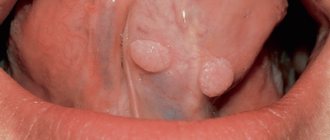
Papillomas are located in the mouth area, and they can be located in different areas. They have a pink color with a slight transparent tint. The size of the neoplasm does not exceed 10 mm. If treatment is not carried out, the so-called “leg” – the base – grows. Papillomas have uneven edges, their surface with slight bumps. Formations can be located in a single area, for example, only on the tongue, or they can be located in two or more areas. There are several types of localization:
- Throat papillomas - at the initial stage do not cause discomfort, but then lead to the development of diseases of the vocal cords and interfere with the swallowing of drinks and food. They have a light pinkish tint with white edges, and the surface is lumpy.
Throat papillomas
- Tongue - at the beginning they are felt as scratching and roughness on the tip of the tongue, and then cause pain; eating food leads to injury, as a result of which a person cannot speak and chew food normally.
Papilloma on the tongue
- Labial papillomas are visible to others from the very first day; when they grow, they affect the speech apparatus. Aesthetically it does not look attractive, similar to an advanced case of herpes.
Papilloma on the lip
- On the larynx - the most dangerous type of papilloma, since the process can lead to the development of hypoxia.
Papillomas on the larynx
- On the gum - has a reddish color, with pink edges. There is practically no pain, diagnosis is complicated, since the patient thinks that these are purely dental problems.
Papilloma on the gum
- On the mucous membrane, on the inside of the cheek - it has similar symptoms to all of the listed types.
Papilloma on the inside of the cheek
The appearance of the tumor depends on the area in which it is located. So, its color can be characterized from white to pink-red. Size varies by stage. The earlier a papilloma is detected, the easier it is to remove it, since its size is still small.
How to avoid relapse of the disease?
After the growth has been removed from the patient's tongue, the person should not relax. Typically, single papillomas do not recur, but with multiple formations, the reappearance of growths on the body occurs quite often.
Therefore, after surgical removal of papillomas from the surface of the tongue, you should:
- Take precautions and limit risk factors (see above).
- Take a course of antiviral treatment as recommended by your doctor.
- Seasonally take immunomodulatory drugs and vitamin complexes, which your doctor will also help you choose.
- Maintain oral hygiene and avoid tongue injuries.
One of the main conditions for achieving a favorable outcome in the fight against papillomas is choosing the right medical center, since much in the procedure for removing a tumor depends on the actions of the doctor. We hope that the recommendations of specialists from the LeaderStom network of dental clinics helped you clarify the picture of the growth that has developed on your tongue, and now you can solve this problem without unnecessary emotional distress.
Reasons for appearance
The manifestation of papilloma is associated with infection with a virus. The carrier does not always manifest the disease; the virus can remain latent for at least its entire life. Papillomas appear only if there are accompanying indicators. These include:
- getting injured;
- constant exposure to high temperatures (both climatic and related to production detail);
- genetic factors;
- weakening of the body's immune functions;
- hormonal imbalance that appeared in adolescence, during menopause, as a result of drug treatment.
There are more than a hundred viral agents that cause the disease. The level of danger, location and treatment rules for papilloma depend on the type of virus.
Risk factors
In addition to the obvious aesthetic component, papillomas cause discomfort in the mouth. Their presence interferes with chewing food and affects speech. Also, papilloma, if it is located on the root of the tongue or larynx, can lead to a more serious consequence - attacks of suffocation.
On the left is a normal larynx, on the right are advanced papillomas.
However, there are no less serious consequences that a neglected case can lead to. The main risk factor for a benign tumor is that it can develop into a malignant one. It is impossible to determine exactly at what stage this will happen and whether it will happen.
What is fibroma
This is a benign neoplasm in the form of a small node on a stalk or broad base, which consists of fragments of connective tissue. In most cases, it is localized on the mucous membranes of the gums, lips, palate, on the inside of the cheeks, and a little less often on the tongue. The pathological process is more often encountered by primary schoolchildren and adolescents aged 6-15 years.
The patient is not in pain; at a very early stage he does not notice the pathology. As the thickening grows, it is easily felt and accidentally injured.
Can fibroids in the mouth resolve on their own, without medical intervention? In most cases, surgical excision of the tumor is required. However, do not worry about the complexity and duration of the operation. In less than an hour, an experienced doctor will eliminate the defect without subsequent complications and a long rehabilitation period. Currently, clinics use laser and radio wave techniques.
Despite the relative harmlessness of fibrous growths, they still need to be treated even in the absence of discomfort. If the seals increase in size and are constantly exposed to traumatic effects, it is highly likely that an infection will enter the wound. In this case, treatment will be very long, difficult and will not always lead to positive dynamics. In addition, the lack of therapy sometimes leads to the degeneration of a neoplasm into a malignant one.
Kinds
| Type of neoplasm | Description of symptoms |
| epithelial hyperplasia | The virus is present in every person in a latent state; under certain conditions, the epithelium begins to divide and papillomas appear. There is little pain and removal is quite simple. |
| simple papillomas | Small growths that do not cause pain. Formed singly or in groups. |
| vulgar papillomas | They are similar to simple ones, with the difference that their formations are always group. |
| flat | They have practically no bulges, their color is darker. |
| threadlike | They are dangerous because they have a leg. |
| Pointed papillomas | They spread very quickly, scars after their removal are at risk of oncogenic risk. |
| fibropapillomas | A mushroom-shaped growth similar in color to the mucous membrane, easily injured. |
| Condylomas | Growths with a soft and thin stalk that are easily damaged. |
HPV symptoms
The papilloma virus infects the skin and mucous membranes, causing the proliferation of the epidermis and the appearance of pathological neoplasms:
- papillomas;
- warts;
- condylomas.
They look like small skin growths ranging in size from 1 to 5–8 mm, sometimes there are growths reaching 1–2 cm. Basically, such formations imitate healthy skin in color, but there are elements of dark brown and white shades.
The main places of distribution of papillomas:
- face and neck;
- external genitalia and groin area;
- the inner surface of the elbow and knee bends;
- soles of feet;
- fingers and skin around nails.
On the mucous membranes, elements can appear in the larynx, nasal passages; in girls, sometimes there are formations localized on the cervix.
Types of formations
There are several types of papillomas.
- Vulgar (common) warts. They are the most common type of neoplasm. They have a “leg”, rise noticeably above the surface of healthy skin, reaching a diameter of up to 1–2 cm. Sometimes hair grows from the central part of the wart. Such formations do not bother the child with pain or itching and appear on the arms, back or legs.
- Flat or juvenile papillomas. They look like small pigmented plaques of a fuzzy round shape, do not extend beyond the skin and form in groups. Such elements are characterized by a smooth surface and selectivity: they appear on the face, neck, legs, sometimes hands, but never in the armpits, genitals or skin folds. Occurs in children older than 5–6 years.
- Condylomas or genital papillomas. These elements resemble small papillae. They are pink in color and form in areas with thin skin: in the genital area, on the mucous membranes.
- Plantar warts. They affect the feet, occur under the big toes and visually resemble small round calluses.
Another variant of formations is filamentous warts or acrochords. They are similar to regular ones, but differ in elasticity and more compact sizes up to 5–6 mm. They prefer to appear under the mammary glands, in the groin, armpits, and are found on the neck and face.
The dangers of human papillomavirus
Some of the representatives of these microorganisms are harmless to humans, while others can provoke the growth of cancer cells. HPV is classified into two main types:
- Strains with high oncogenic risk. Such variants of the virus provoke the development of condylomas on the mucous membranes and in the genital area. Under unfavorable circumstances, they can cause an oncogenic mutation.
- Strains with low oncogenic risk. Viruses of this type cause warts, plantar lesions and juvenile papillomas. The risk of cell degeneration is minimal.
The greatest danger lies in girls: studies conducted in the USA have proven that 98% of cases of cervical cancer are associated with this virus. There is also a risk of developing cancer of the vagina, ovaries, anal canal, larynx, pharynx, and in boys, the genitals. Papillomas located in the anus and genital area require special attention.
Localization
Oral papillomas can be localized in one place or several. Areas of distribution include:
- language;
- sky;
- gums;
- lips;
- cheek;
- larynx.
The most painful sensations are caused by papillomas located on the tongue. At the same time , those located on the palate, root of the tongue, and larynx are considered the most dangerous , as they can lead to attacks of suffocation. New growths on the gums and cheeks are practically painless and can cause discomfort when eating. Papillomas on the lips are a serious aesthetic disadvantage.
Lip tumors
Of all the anatomical parts of the oral cavity, the red border of the lower lip is most often affected. Lower lip cancer accounts for approximately 12 - 25% of the total number of tumors in this location. For reasons that are not entirely clear, tumor lesions of the red border of the upper lip are an extremely rare occurrence. The disease most often occurs in people over 60 years of age. Men are more often affected, the male/female ratio approaches 13/1. Predisposing factors are solar radiation, smoking, and alcohol abuse.
Clinically, the disease manifests itself as an exophytic (i.e., protruding) tumor or, much more often, an ulcerative bleeding surface. Since it is very difficult not to notice this lesion, the vast majority of patients seek medical help at an early stage of the disease. This circumstance (treatment of the early stages of any malignant tumors always brings better results) allows us to count on high effectiveness of treatment. In addition, lower lip cancer rarely metastasizes to regional (cervical) lymph nodes. At the time of initial treatment, involvement of the cervical lymph nodes is diagnosed in only 2–12% of patients. Damage to regional lymph nodes significantly worsens the prognosis of the disease. Thus, the 5-year survival rate in the presence of this lesion drops from 90 to 50% and forces one to resort to more aggressive methods of treatment.
Treatment for lower lip cancer is determined by the stage of the disease. Early stages can be cured with surgery or radiation treatment. There are 2 types of radiation treatment: remote and contact therapy (so-called brachytherapy).
The lip has a rather complex anatomical structure, which includes muscles with different directions of muscle fibers, skin, salivary glands, and subcutaneous tissue. Naturally, the goal of surgical treatment is not only radical removal of the tumor, but also restoration of the structure and function of the lower lip. Modern methods and techniques of plastic surgery can achieve good cosmetic and functional results. This is illustrated by the photographs below of patients who underwent surgical treatment for cancer of the lower lip.
Develops against the background of obligate (warty precancer, limited dyskeratosis, Manganotti cheilitis, Bowen's disease) and facultative (leukoplakia verrucous, keratoacanthoma, cutaneous horn, papilloma with malignancy, erosive-ulcerative and hyperkeratotic form of lupus erythematosus and lichen planus, post-radiation cheilitis) precancerous processes .
Warty precancer is characterized by the appearance on the red border of the lip of a sharply demarcated hemispherical lesion with a diameter of 4 mm to 1 cm, which protrudes above the surrounding tissues and has a dense consistency. Its color varies from an almost normal red border to a stagnant red. The nodule may be covered with thin scales and resemble a wart or keratinizing papilloma. Malignancy can occur 1-2 months after the onset of the disease.
The productive form of focal dyskeratosis is characterized by excessive keratinization of the epithelium and looks like a white spot with flat protrusions on a red border or an area of hyperkeratosis with stylized horny protrusions. The destructive form (erythroplakia) is distinguished by the fact that limited defects of the epithelial cover appear on the red border of the lip - erosions, ulcers, cracks. Malignancy can occur 6 months after the onset of the disease.
Abrasive pre-cancer cheilitis Manga-Notty is characterized by the formation of an oval or irregularly shaped erosion on the lip, often with a smooth, polished surface of a deep red color. Crusts may form on the surface of the erosion, which may cause slight bleeding when removed. There is no tissue compaction at the base and around the erosion. Spontaneous epithelization with subsequent recurrence is possible. Malignancy occurs 3 months to 30 years after the onset of the disease.
Lip papilloma is a clearly circumscribed tumor from 1-2 mm to 2 cm in diameter on a wide base with a villous or rough surface. As keratinization increases, the tumor acquires a whitish tint. Lip papilloma is considered a progressive precancerous disease. Compaction of the base of the papilloma and the appearance of pain are signs of its degeneration.
Treatment of diffuse lesions of the lower lip (cheilitis, leukoplakia, etc.) is predominantly conservative. Focal lesions are treated surgically, laser and cryodestruction.
Lip cancer
The term “lip cancer” refers to malignant tumors that arise in the mucous membrane of the red border of the lip. Neoplasms that have developed on the skin near the lip or mucous membrane of the vestibule of the mouth are not included in this group of tumors.
In the structure of cancer diseases in Russia (1997), lower lip cancer accounted for 1.4% of the total number of malignant neoplasms with an incidence of 2.8 per 100,000 population. The disease is most often observed in men over 70 years of age. In rural areas, lip cancer occurs 1.5-2 times more often than in urban populations. In recent decades, there has been a gradual decline in incidence. Approximately 90% of tumors of the lower lip develop due to exposure to known carcinogens, the main of which are tobacco and tobacco-containing substances. Alcohol is comparable in action to tobacco and is part of a group of agents that can potentiate tobacco-induced carcinogenesis. Other important etiological factors are chronic injuries and inflammation of the lip, ultraviolet radiation, changes in weather conditions, and occupation (work in the oil refining, coal, textile industries).
International classification according to the TNM system
Anatomical areas and parts:
1.Upper lip, red border. 2.Lower lip, red border. 3. Angles of the lips (commissures). T - primary tumor: Tx - insufficient data to evaluate the primary tumor, T0 - primary tumor not determined, Tis - preinvasive carcinoma (carcinoma in situ), T1 - tumor up to 2 cm in greatest dimension, T2 - tumor up to 4 cm in greatest dimension , T3 - tumor more than 4 cm in greatest dimension, T4 - tumor spreads to adjacent structures - bone, tongue, facial skin.
N/pN - regional lymph nodes. Regional lymph nodes for all parts of the head and neck, with the exception of the nasopharynx and thyroid gland, are the lymph nodes of the neck, including: submental, submandibular, nodes at the base of the skull near the great vessels (deep cervical), in the bifurcation zone of the common carotid artery (deep cervical) , posterior cervical (superficial cervical), located along the accessory nerve, supraclavicular, preglottic and paratracheal, retropharyngeal, parotid, buccal, postauricular and occipital.
N/pNx - there is insufficient data to assess the condition of the regional lymph nodes, N/pN0 - there are no signs of metastatic lesions of the regional lymph nodes. pN0 - histological examination of material from a selected area of neck tissue includes 6 or more lymph nodes; histological examination of the material obtained using cervical lymphadenectomy includes 10 or more lymph nodes, N/pN1 - metastases in one lymph node on the affected side, up to 3 cm or less in the greatest dimension, N/pN2 - metastases in one or more lymph nodes on the affected side, up to 6 cm in greatest dimension; or metastases in the lymph nodes of the neck on both sides or the opposite side, up to 6 cm in greatest dimension, N/pN2a - metastases in one lymph node on the affected side, up to 6 cm in greatest dimension, N/pN2b - metastases in several lymph nodes on the affected side, up to 6 cm in the greatest dimension, N/pN2c - metastases in the lymph nodes on both sides or on the opposite side, up to 6 cm in the greatest dimension, N/pN3 - metastasis in the lymph node more than 6 cm in the greatest dimension.
Note. The lymph nodes on the affected side include the lymph nodes located in the midline.
M - distant metastases: Mx - the presence of distant metastases cannot be assessed, M0 - no distant metastases, M1 - distant metastases.
The requirements for determining the рТ category correspond to those for determining the T category.
Grouping by stages
Stage 0 TisN0М0 Stage I Т1N0М0 Stage II Т2 N0М0 Stage III Т3N0М0 Т1-3N1М0 Stage IVA Т4N0-1М0 Any ТN2М0 Stage IVB Any ТN3М0 Stage IVC Any Т Any NM1
Clinic . Cancer most often affects the lower lip midway between the midline and the commissure. The main morphological form is squamous cell carcinoma (80-95%). Metastasis later, mainly to regional lymph nodes (5-9.2%). In the clinical course of cancer of the lower lip, two groups are distinguished - exophytic and endophytic. There are transitional forms between them. Exophytic cancers of the lower lip include papillary and warty forms. The papillary form develops from papilloma or against the background of focal productive dyskeratosis. Exophytic cancer visually represents a tumor of irregular shape (round, lumpy, flat), rising above the surface of the red border. There is infiltration of the underlying tissues without clear boundaries. The warty (fungous) form develops against the background of productive dyskeratosis and is characterized by the fusion of multiple small growths on the lip (“cauliflower appearance”), a gradual increase in infiltration of the underlying tissues and tumor disintegration. Endophytic cancer of the lower lip is represented by ulcerative and ulcerative-infiltrative forms. The ulcerative form is characterized by the formation of an ulcerative defect on the lip with a gradual deepening of the ulcerative surface, its bottom becomes uneven, its shape is irregular, the edges rise above the level of the red border, everted. The ulcerative-infiltrative form is characterized by infiltrative growth, which makes it difficult to determine the boundaries of the tumor. Endophytic forms of cancer are more malignant.
Diagnosis is based on anamnesis, examination, palpation of regional lymph nodes (submental and submandibular lymph nodes are examined bimanually), cytological and/or histological examination data. When making a differential diagnosis, it is necessary to keep in mind tuberculosis and syphilitic lesions.
Treatment . In stages I and II, the standard treatment method is radiation therapy (short-focus radiotherapy, electron therapy, contact radiation, interstitial or combined radiation therapy) with a total focal dose of 60 Gy.
The treatment of choice for stage I lip cancer is cryodestruction. Block resection of the lip with simultaneous plastic surgery according to Bruns is rarely used due to the poor cosmetic effect. Surgery is more indicated in the presence of residual tumor after completion of a radical course of radiation therapy; it is performed within 3-6 weeks after the end of irradiation.
For stage II tumors, 2-3 weeks after completion of treatment of the primary lesion, prophylactic upper fascial-sheath excision of the neck tissue on both sides is performed. The block of tissue removed includes the tissue of the submental and submandibular region and the lymph nodes of the fork of the common carotid artery, the lower pole of the parotid salivary gland.
For stage III lower lip cancer, combined radiation therapy is used (external gamma or electron therapy + interstitial or short-focus radiotherapy). The presence of clinically detectable metastases in the lymph nodes is an indication for preoperative irradiation of the cervical lymphatic collector on the affected side. Preventive or therapeutic fascial-sheath removal of cervical tissue (on both sides) is carried out after complete subsidence of radiation reactions and completion of regression of the primary tumor focus. The method of choice for treatment of stage III lower lip cancer, especially when the tumor has spread to the corner of the mouth, is a combined one. In this case, lymphadenectomy is performed simultaneously with removal of the primary tumor. To eliminate lip tissue defects formed after treatment, primary reconstruction is performed using local or regional flaps.
Treatment for stage IV lower lip cancer is combined. For multiple metastases in the lymph nodes of the neck, treatment begins with preoperative external beam radiation therapy. 2-3 weeks after completion of the first stage of treatment, surgery is performed on the primary lesion with simultaneous cervical lymphadenectomy. Fascial-sheath excision of the cervical tissue is performed for single, dislocated metastatic lymph nodes that are not fused with adjacent anatomical structures of the neck. The presence of multiple displaced metastases in regional lymph nodes or single, but limitedly displaced, fused with the internal jugular vein and the sternocleidomastoid muscle is an indication for performing the Crail operation. In case of bilateral metastatic lesions of the lymph nodes, cervical lymphadenectomy is performed simultaneously on both sides. In elderly and weakened patients, this operation can be done first on one side, and after 2-3 weeks on the other.
In case of unresectable tumors and the presence of contraindications to combined treatment, a course of palliative remote gamma therapy is carried out according to a radical program (60-70 Gy). Chemotherapy is used as part of palliative chemoradiation treatment (see Cancer of the mucous membrane of the floor of the mouth).
Recurrent lower lip cancer is treated surgically and in combination. For late limited relapses (developing 1 year or more after completion of short-focus radiotherapy), a course of radiation therapy (60-70 Gy) can be repeated.
Forecast . According to the summary data of various authors, the five-year survival rate for stage I is 91.2-96.9%, for stage II - 82.8-83.6%, for stage III - 63.5-75.4%, for stage IV - no more thirty%.
Prevention of lip cancer consists of timely treatment of precancerous diseases, quitting smoking, eliminating unfavorable external factors (increased insolation, etc.), and using protective lip creams.
SIGN UP FOR A CONSULTATION by phone +7 (921) 951 - 7 - 951
Share link:
Diagnostics
Diagnosis should be carried out as soon as you detect formations in the oral cavity. Some types of papillomas grow very quickly and if treatment is not started immediately, this can lead to serious problems. Patients suspected of having papilloma are sent for histological examination. Violations of the basal layer are detected; if there is severe acanthosis, then this is evidence of the disease.
A specialist, when examining the oral cavity, by taking the necessary tests, finds out whether these are really papillomas and will separate them from condylomas, epitheliomas and other diseases whose symptoms are similar.
Treatment results
The photo below shows the results of removal of neck papillomas using radio wave surgery
Rarely occur again. Reappearance of papillomas is possible with high virus activity in the body. In this situation, additional treatment with immunomodulators or consultation with an immunologist may be required.
Treatment
Treatment of papilloma is complex; it includes not only drug therapy, but also vitamin supplementation. Traditional methods show effectiveness, but only if the inflammatory process has not yet started.
Sanitation of the oral cavity
Sanitation of the oral cavity is a mandatory stage in the treatment of papillomas.
Sanitation is a comprehensive measure aimed at removing concomitant diseases that affect the growth of the tumor or interfere with its treatment. It includes:
- treatment of inflammation;
- removal of plaque and tartar, treatment of caries and filling, installation of crowns, etc.;
- cleaning dentures;
- cleaning the braces (usually surgery, if necessary, is carried out after they are removed).
Sanitation of the oral cavity is necessary to reduce the risk of relapse. If they are not carried out, then the probability of re-growth increases by 2-3 times. After completing comprehensive oral therapy, the doctor prescribes special antibacterial tablets and rinsing with medical solutions for disinfection and healing.
Drug therapy
After sanitation, drug therapy is prescribed. Medications are used to boost weakened immunity. Panavir has proven itself, which must be used for two weeks. Also prescribed as therapy:
- Viferon (they need to lubricate the affected area, helps healing, has an antiseptic effect);
- Dimexide (gargle with it several times a day, avoid ingestion).
Likopid Isoprinosine tablets, Genferon suppositories, Allokin-Alpha solution are effective. The correct combination of medications is prescribed by a specialist after examining a specific case, identifying the cause and assessing the risks of relapse.
Traditional methods
You can alternate compresses with other treatment methods.
Alternative medicine methods have their place. They are not as effective as pharmaceutical drugs, but allergic reactions to them are extremely rare. There are such methods:
- crushed garlic mixed with Vaseline is applied to the affected area overnight;
- pickled lemon is fixed to the skin with a band-aid and waited for several hours;
- mashed potatoes with egg yolk are applied as a compress;
- lubricate the affected area with essential oils of juniper, orange, and tea tree.
Traditional methods are used for at least two weeks to achieve results. This makes it impossible to say that they are effective. To treat a viral disease, it is better to immediately consult a specialist.
Symptoms of the disease
Small tumors usually do not manifest themselves in any way. Medium and large papillomas cause discomfort. They interfere with normal air circulation, causing difficulty breathing. In the presence of such neoplasms, a person’s voice undergoes changes: it becomes nasal, hoarse, and speech is impaired. There are problems with swallowing food. The patient has a sensation of a foreign body in the throat. Large formations lead to a severe cough, and blood clots are observed in the sputum. In especially severe cases, the patient is diagnosed with airway obstruction and asthma attacks.
If the place of origin of neoplasms is the pharynx, they can most often be observed on the palatine tonsils and palatine arches. The most dangerous growths are in the larynx, since they are located next to the respiratory tract.
As we can see, the presence of such neoplasms causes severe discomfort to the patient, so if signs of the disease appear, you should contact an ENT doctor to diagnose the disease and have the papilloma professionally removed.
| Cost of admission | price, rub. |
| Initial visit (Payment is required upon initial visit) | 2000 |
| Repeated appointment | 1500 |
| Initial consultation with the head of the clinic (Payment is required upon initial visit) | 4000 |
| Repeated consultation with the head of the clinic | 2000 |
| Additional consultation during procedures | 500 |
| Adaptation of a child to the ENT office | 2000 |
| Adaptation of a child to the ENT office by the head of the clinic | 4000 |
| ENT diagnostics (carried out on the recommendation of an ENT doctor) | price, rub. |
| Endoscopy of the pharynx and larynx | 2500 |
| Videoendoscopy of the pharynx and larynx | 3000 |
| ENT manipulations | price, rub. |
| Application anesthesia | 500 |
| Infiltration anesthesia (local, injection, one injection) | 500 |
| Removal of pharyngeal papilloma | 5000 |
| Lubricating the back of the throat with medicinal solutions | 500 |
| Irrigation of the posterior pharyngeal wall using an ENT combine | 500 |
| ENT clinic discount | — 2000* |
| Cost of one day of treatment including discount | 5000 |
| Additional manipulations (carried out on the recommendation of an ENT doctor) | price, rub. |
| Session of infrared laser therapy of the posterior wall of the pharynx and palatine tonsils using the RIKTA device | 200 |
| Ultraviolet irradiation session of the posterior wall of the pharynx and palatine tonsils with the OUFd-01 Solnyshko irradiator | 200 |
| Ultrasonic medicinal irrigation session of the posterior pharyngeal wall and palatine tonsils using the TONSILLOR device | 1000 |
| Medicinal irrigation of the larynx using an ENT combine ATMOS 61 | 500 |
| Session of photodynamic therapy of the larynx and pharynx | 1000 |
| Percutaneous infrared laser treatment using the RVB device (Italy) | 200 |
* - discount is provided for complex treatment of the disease
Removal of papillomas
Removal of papillomas is a method used in 80% of cases. The surgical intervention is painless, because before it the oral cavity is anesthetized. High accuracy is achieved through the use of the latest surgical methods. It is not always possible to get rid of a tumor in one visit; in this case, complex therapy will be required.
Removal methods:
- Removal with a scalpel. This method is old and is used if the patient has a limited financial budget and there are no other options for removal. The formations are cut off and there is a possible risk of scarring. It is prohibited to use it to remove papillomas on the lips.
- Laser. The growth is burned out layer by layer, while the remaining tissue remains undamaged. The method is modern and effective, no scar remains. Its only disadvantage is the high cost of the procedure.
- Medical loop. The papilloma is captured and gradually removed from the tissue. The method avoids re-infection and is characterized by rapid healing.
- Ultrasound. High-intensity ultrasound destroys infected tissue while sparing others. The method is effective and safe, but it costs a lot.
- Electrocoagulation. Removal occurs by microwave or electric current. Used primarily to work with formations under the tongue.
- Conchotom. Extracting the affected areas of the mucosa using biopsy forceps. The method is effective and safe, indicated for growths in the oral cavity, and not outside it.
- Radio waves. The impact of the waves is painless and safe for the patient. Histology confirms the benign nature of the neoplasm.
Symptoms
The tumor grows and develops quite slowly, so for a long time the patient may not even be aware of its presence in the mouth. Fibroma of the oral mucosa looks like a hemispherical growth rising above the plane, covered with pinkish tissue. If you press it, pain or other discomfort does not appear. The surface is smooth, there are no irregularities or roughness on it.
The appearance of ulcers with such a diagnosis is very rare. In such cases, an infection is usually associated with the subsequent development of the inflammatory process. Swelling, redness, erosion occur, and pain is felt. The pain persists even if you do not touch the pathological area.
If you do not injure the formation, it may not change its size for quite a long time and remain in a stable state. If it is exposed to constant traumatic effects, there is a high risk of malignant degeneration, which is dangerous to the life and health of the patient.
Features of treatment and removal of papillomas in a child’s mouth
Laser removal of gum papilloma.
The disease can be congenital, in which case papillomas appear even in a newborn.
The virus is transmitted from mother to child, in utero, that is, a child may be born at a certain stage, and associated factors (virality, genetic predisposition, nutritional characteristics) will make it active.
The growths do not cause discomfort initially, but later cause itching. The child begins to scratch the wound, which leads to inflammatory processes. Drug treatment is used, among the permitted interventions are laser removal, cryodestruction, exposure to radio waves and electrocoagulation.
In a baby
There are several reasons for the development of the disease in newborns. Among them are transmission from a woman in labor to a baby and through contact-household transmission, when the virus was acquired through the use of toys, pacifiers, towels, mugs, etc.
Surgical intervention is unacceptable; ointments, gels and solutions are used at the beginning of therapy.
Treatment of oral fibroids
Surgery remains the most effective and most common therapeutic method. The seal is excised using a laser or radio waves. This procedure lasts about half an hour. If the tumor is very large, after its removal the wound is covered with a flap, which is formed by the doctor from the surrounding tissue.
When pathology is caused by taking certain medications, they should be completely eliminated and replaced with alternatives with similar properties. After discontinuation of drugs in such cases, the appearance of the mucous membranes is often restored without outside help, and the likelihood of relapse approaches zero. However, this does not apply to situations where the disease is advanced.
Surgery can also be avoided in case of traumatic effects of orthopedic structures. For example, when a crown, filling, or prosthesis puts pressure on the tissue. Elimination of the provoking factor often leads to a decrease or complete disappearance of a benign formation. Most likely, it will be necessary to dismantle the old structures and replace them with new ones.
On the Internet you can find stories of healing using home remedies. It is worth remembering that the disease cannot be treated with the help of folk recipes. Herbal decoctions and infusions, and other compositions are used only as an auxiliary element of complex therapy.


