Author
: Grachev Ilya Illarionovich
Editor
: Efremov Mikhail Mikhailovich
Date of publication: 06.11.2018 Date of update: 12.11.2020
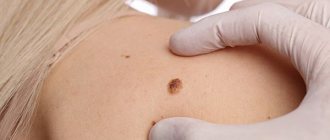
Nail psoriasis or onychia psoriasis is rarely primary. Typically, this form develops against the background of an existing disease. It affects approximately 10% of psoriasis patients. Interestingly, most often nail lesions accompany psoriatic joint lesions. Another feature is the predominant damage to the nail plates on the hands. Specialists from the Moscow Paramita clinic can effectively treat this disease.
Symptoms of onychodystrophy
Symptoms of onychodystrophy are directly dependent on the type of trophic disorder. General clinical signs characteristic of all congenital and acquired onychopathies include: changes in the strength, elasticity, thickness and color of the nail plate.
As a rule, dystrophic diseases are asymptomatic, but sometimes pain and inflammation may appear.
Visually uneven nails may have wavy, transverse or longitudinal striations, which deform the surface of the nail. Partial or complete loss of connection with the nail bed or matrix (plate growth zone) is possible. When pigmentation is disturbed, the plate loses its normal flesh color and acquires a color of varying intensity with a color change from whitish-yellow to almost black.
Treatment of onychodystrophy at the Podology Clinic
The Podology Clinic, which is one of the largest technically equipped specialized complexes in Russia, offers to take advantage of the priorities of innovative medical technologies aimed at solving a wide range of problems of the feet and nails. Experienced podologists who master all known techniques have at their disposal the most advanced expert-class diagnostic and treatment equipment, ensuring the most accurate and painless correction.
If you have problems with your fingernails and toenails, you shouldn’t leave them to chance. Only a timely visit to a professional involved in the treatment of various onychopathies will help to identify and carry out full correction at the earliest stages, without aggravating the development of dystrophic changes. And don’t forget that nails play not only an aesthetic role, but are also an indicator of your health.
Video review of the PinPointe FootLaser laser and interview with Dmitry Zakharchenko (CEO) on the latest device for the treatment of onychodystrophy and onychomycosis:
Ichthyosis
Ordinary or vulgar ichthyosis appears before the age of three years, but it is usually diagnosed before the third month of life. This is the most common form of ichthyosis, inherited in an autosomal dominant manner. First, the skin becomes dry and rough, then becomes covered with small whitish or gray-black scales tightly adjacent to each other. With ichthyosis, the area of the elbows, popliteal fossae, armpits and groin area are not affected.
Mucoid peeling appears on the palms, and the skin pattern becomes pronounced. The severity of ichthyosis depends on how deep the gene mutation is; an abortive course is possible, when the only manifestation of ichthyosis is dryness and slight flaking of the skin on the extensor surfaces.
With ichthyosis, hair, teeth and nails undergo dystrophic changes. Dry, brittle hair is typical, nails break off and split, and multiple caries develops. Quite often, ichthyosis is accompanied by eye damage - chronic conjunctivitis and retinitis. Patients with ichthyosis have a hereditary predisposition to myopia, which begins to manifest itself in childhood. Since immunity is reduced, allergic diseases and purulent infections are permanent. Later, disturbances in the functioning of internal organs occur, most often cardiovascular failure and liver disease.
Recessive ichthyosis occurs only in males, although it is inherited on the X chromosome and differs in that the cause of the disease is a defect in placental enzymes. Clinical manifestations appear in the second week of life, less often immediately after birth. The horny layers of the skin look like large dense scales of black-brown color and resemble scutes. The skin between the scales is covered with cracks, so it looks like the skin of a crocodile or snake. Children with recessive ichthyosis often have mental retardation, abnormalities in the skeletal structure, and epilepsy. Juvenile cataracts and hypogonadism occur in 10-12% of cases.
Congenital ichthyosis develops in utero at 4-5 months of pregnancy. At birth, the baby's skin is covered with thick horny scutes of gray-black color. With congenital ichthyosis, the scales can reach up to 1 cm in thickness, the scales have different shapes, smooth or jagged, the skin between them is covered with grooves and cracks. Due to the dense, well-cohesive scales, the baby's mouth is either stretched or sharply narrowed so that the feeding tube barely fits. The ear openings are deformed and filled with horny scales, the eyelids are everted due to stretching. Almost all infants have skeletal anomalies - clubfoot, clubhandedness; many children with a congenital form of ichthyosis have interdigital bridges on the feet and palms, and sometimes missing nails. Pregnancy is often premature, and the rate of stillbirth is quite high. Since there are anomalies incompatible with life, most children with the congenital form of ichthyosis die in the first days of life.
Epidermolytic ichthyosis is a form of congenital ichthyosis. The baby's skin is bright red, as if scalded by boiling water. Nikolsky's syndrome is positive, as with pemphigus of newborns - with a slight touch, rejection of the epidermal scales is observed. The skin on the palms and soles is white and significantly thickened. In some cases, with the epidermolytic form of ichthyosis, there may be hemorrhages in the skin and mucous membranes. This is an unfavorable sign; if hemorrhages occur, children most often die. With milder clinical manifestations of ichthyosis, the blisters become smaller over time, but throughout life the disease recurs in the form of outbreaks, and during a relapse of ichthyosis the temperature often rises to high levels. By the fourth year of life, horny layers appear in certain areas of the body in the form of thick dirty gray scales, which are localized mainly in places of natural skin folds.
There are often defects in the nervous, endocrine and other body systems; many children with congenital ichthyosis are later diagnosed with mental retardation and spastic paralysis, which is caused by the accumulation of phytanic acid in the tissues. Polyneuropathy, anemia, and infantilism complicate the course of ichthyosis. The mortality rate is very high due to associated complications and associated diseases.
Why does the disease develop?
The pathology is hereditary, and the main cause of ichthyosis is a mutation of certain groups of genes, which is transmitted to children from parents. In rare cases, the disease is acquired and becomes a consequence of a lack of vitamins, a manifestation of certain blood diseases, etc.
Patients have a disturbance in the metabolism of proteins and amino acids, the activity of oxidative enzymes in the epidermis increases, and the concentration of immunoglobulins decreases. The etiology and pathogenesis are still not well understood, but many researchers note the important role of vitamin A deficiency and the function of certain glands - the thyroid and adrenal glands.
In patients with a genetic form of ichthyosis, metabolic processes, as a rule, slow down, thermoregulation is disrupted, and sweat glands do not work well. High levels of enzymes lead to a significant increase in skin respiration. Cellular and humoral immunity is often reduced. Hyperkeratosis is often accompanied by endocrine diseases, diseases of the adrenal glands and reproductive system, and eye diseases. Patients often experience increased brittleness of hair and nails, and teeth are easily affected by caries.
The skin contains an excessive amount of structurally altered keratin. Due to the slowing of metabolic processes, old skin cells are not shed quickly enough, which leads to the formation of hard scales on the surface of the skin. Amino acid complexes accumulate between the layers of skin cells, accelerating the process of hardening and keratinization, as well as holding the scales together.
Causes of onychodystrophy
The causes of the occurrence can be either individual and very significant factors, or a combination of several factors that are of lesser importance for the human body as a whole. Here are some examples of various factors and causes of onychodystrophy:
- congenital anomalies;
- somatic and autoimmune pathologies;
- dermatoses;
- periungual and subungual neoplasms;
- deficiency of vitamins and minerals;
- traumatic injuries;
- stress;
- prolonged contact with water and chemical solutions;
- intoxication of the body, etc.
In other words, onychodystrophy can be an independent disease or an external manifestation of the internal processes of the body.
Causes of psoriasis on nails
Nail psoriasis is a chronic skin disease of an autoimmune (allergy to one's own tissues) nature. The disease has not been fully studied; there are many white spots left in it. But it is known for sure that heredity plays a large role in its development: in one family, several people often suffer from this non-contagious dermatosis. In recent years, assumptions about the leading importance of heredity have been confirmed by genetic research, that is, by the presence of special genes.
Psoriasis of any localization can spread throughout the body, so do not delay treatment.
See how easily the disease can be cured in 10-12 sessions.
But not everyone who has a family history gets sick. This also requires exposure to certain general and local factors. General (affecting the entire body) factors contributing to the development of nail psoriasis include:
- any chronic diseases and foci of infection;
- endocrine and allergic diseases;
- eating only high-calorie foods;
- regular drinking, smoking;
- constant emotional tension, stress, hard physical work;
- lack of physical activity, excess body weight;
- violation of the sleep-wake pattern;
- impaired peripheral circulation - with atherosclerosis of the extremities, obliterating endarteritis, etc.;
- anemia – the nutrition of the nail plates is impaired;
- existing psoriasis of any form, especially psoriatic arthritis.
Local factors (of no less importance):
- skin infections, especially fungal ones, affecting the nail plates;
- any mechanical and chemical effects on the nail plates.
When exposed to several causes, metabolism and immunity are disrupted. The body begins to reject its own cells, which leads to the appearance of a psoriatic inflammatory process in the nail area. It is important to notice the problem in time, consult a doctor and begin to treat the disease.
Can it be cured?
At the current stage of development, medicine is not yet able to cope with genetic diseases, so modern treatment of ichthyosis is aimed at reducing clinical manifestations and alleviating the patient’s condition. For this purpose they prescribe:
- vitamin complexes, iron-containing compounds, immunoglobulin;
- blood plasma transfusions;
- hormone therapy (in difficult cases);
- baths with the addition of various drugs, emollient ointments and creams;
- physiotherapy, spa treatment according to indications.
The choice of treatment methods depends on the degree of skin damage and the characteristics of the disease.
Types of pathology
More than 90% of patients develop ichthyosis vulgaris, a disease that first appears in the second or third month of life. Depending on the nature of the skin lesion, the following clinical forms are distinguished:
- xeroderma, or abortive form, is the mildest variant of the disease, in which the skin becomes dry and rough, but there are no scaly layers;
- a simple shape with small scales, thin at the edges and with a dense center, which cover the entire body up to the scalp;
- shiny shape with transparent scales located in the form of a mosaic mainly on the legs;
- white form with white or slightly yellowish scales, similar in appearance to asbestos fibers;
- serpentine in shape with rough keratinizations of brown, brown or gray color, reminiscent of snake scales.
Much less common is the lamellar form of the disease (collodion fetus), fetal (Harlequin fetus), linear, etc. Some of them are incompatible with life - for example, fetal ichthyosis develops in the second trimester of intrauterine development, and the child dies in the first days of life or is born already dead.
Where do nails grow from?
To understand the problem of nail psoriasis, it is important to understand how the so-called nail apparatus works.
The nail performs two functions: working and aesthetic. Firstly, the nail protects the fingertips from damage, increases accuracy and sensitivity when working with small objects, can be a weapon of attack or defense, and finally, we itch with the help of nails. Secondly, the aesthetic, or cosmetic, function of nails is also important, especially for women.
Nails are formed from the outer layer of skin - the epidermis. The nail apparatus includes:
- nail plate - the nail itself,
- matrix - it produces the nail plate, the nail socket, or lunula, is the only visible part of the matrix, this is a white moon-shaped area at the base of the nail plate,
Diagram of the nail apparatus
Appendix: list of references
Some of the scientific articles used:
- Rich P et al. Baseline nail disease in patients with moderate to severe psoriasis and response to treatment with infliximab during 1 year. J Am Acad Dermatol. 2008 Feb;58(2):224-31.
- Zaias N. Embryology of the human nail. Arch Dermatol. 1963 Jan;87:37-53.
- McGonagle D et al. The pathogenesis of psoriatic arthritis and associated nail disease: not autoimmune after all? Curr Opin Rheumatol. 2009 Jul;21(4):340-7.
- Cannavò SP et al. Treatment of psoriatic nails with topical cyclosporin: a prospective, randomized placebo-controlled study. Dermatology. 2003;206(2):153-6.
- Singh SK. Finger nail pitting in psoriasis and its relation with different variables. Indian J Dermatol. 2013 Jul;58(4):310-2. doi:10.4103/0019-5154.113955.
- van der Velden HM et al. Fingernail psoriasis reconsidered: a case-control study. J Am Acad Dermatol. 2013 Aug;69(2):245-52.
- Baran R. The burden of nail psoriasis: an introduction. Dermatology. 2010;221 Suppl 1:1-5.
- Gelfand JM et al. Prevalence and treatment of psoriasis in the United Kingdom: a population-based study. Arch Dermatol. 2005 Dec;141(12):1537-41.
- Gupta AK et al. A higher prevalence of onychomycosis in psoriatics compared with non-psoriatics: a multicentre study. Br J Dermatol. 1997 May;136(5):786-9.
Clinical manifestations
The main symptom of ichthyosis - characteristic hard, scaly formations on dry skin - appears already in the first year of life, much less often - over the next few years. The child's skin becomes rough, and transparent, whitish or grayish scales appear on it, adjacent to each other. Only the elbow bends, the hollows under the knees and axillary areas, as well as the groin area remain clean. The palms and feet are peeling, and the skin pattern is clearly visible on them. In the mildest cases, the disease manifests itself only as slight peeling and dry skin.
Currently, there are more than fifty clinical and morphological forms of the disease, but in all cases the following manifestations are present:
- the stratum corneum of the epidermis is thickened, scaly formations and keratinization zones form on it;
- the skin is dry and flaky, the sebaceous glands practically do not work;
- the nail plates peel or break, in some cases they are deformed and resemble bird claws, peeling off from the soft tissues;
- the skin pattern becomes deeper and more expressive;
- There are rashes of different shapes and colors.
Skin ichthyosis usually worsens with the onset of cold weather. In warm weather, remission usually occurs. During puberty in adolescents, painful symptoms decrease, only to return in adulthood.