Lichen planus (LP) of the vulva, as an isolated lesion or a manifestation of widespread LP, can be characterized by polygonal papules typical for this dermatosis or atypical: pigmented, erosive, hypertrophic elements [1-3].
Among the atypical forms of vulvar LP, the most rare is hypertrophic. Typically, the hypertrophic form of LP manifests itself as itchy purple papules and polygonal plaques with a Wackham mesh on the surface, affecting mainly the lower extremities - the legs, as well as the ankle and interphalangeal joints. In this case, histological signs characteristic of LP, such as hypergranulosis and basal cell vacuolar degeneration, may be absent [4]. This circumstance should be taken into account when performing histological confirmation of this form of LP. The histological diagnosis of hypertrophic LP is complicated by the presence of pseudocarcinomatous hyperplasia (PCH) of the epidermis in long-term lesions that are constantly injured by scratching, which can be difficult to distinguish from squamous cell carcinoma (SCC) of the skin [5].
Clinical observation
Patient B
., 68 years old, was admitted to the department of dermatovenerology and dermato-oncology of MONIKI with complaints of intensely itchy rashes on the skin of the torso, limbs and genitals. She believes that she fell ill 15 years ago, when, against the background of menopause, she noted the appearance of intense itching of the genitals, and therefore the gynecologist recommended the local use of baby cream, pumpkin and sea buckthorn oils. Despite the increased itching, she did not consult a doctor. After 10 years, an itchy light pink spot appeared in the area of the right ankle joint. A local dermatologist diagnosed eczema and prescribed local treatment with corticosteroid ointments (without effect). Over the next 4 years, the lesion continued to increase in size. A year ago, after the death of her husband, the itching sharply intensified, new rashes appeared on the skin of the torso and upper extremities, and therefore she was hospitalized in the department of MONIKI.
On examination: the pathological process is widespread, symmetrical, localized on the skin of the limbs, torso and genitals. On the skin of the lower extremities, mainly the legs and ankle joints, there are hypertrophic papules and plaques of a violet-brown color with a cellular surface, clear boundaries and mealy peeling on the surface, as well as atrophic pale pink plaques with a purple rim along the periphery (Fig. 1, 2 ).
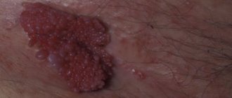
Rice. 1. Patient B. Hypertrophic plaque of purple-brown color in the ankle joint.
Rice. 2. The same patient. An atrophic pale pink lesion with a violet rim along the periphery in the area of the ankle joint and lower leg. When illuminated from the side and the elements are lubricated with oil, a network of whitish lines is visualized on the surface. On the skin of the left forearm there is a longitudinal lilac plaque with a honeycomb surface, 5x10 cm in size. On the skin of the chest there are multiple polygonal pink-violet shiny papules with a central umbilical depression. The mucous membrane of the labia minora is whitish; in the area of the left labia minora a dense hypertrophic whitish plaque with a spongy surface was detected. Multiple small erosions and excoriations were noted on the mucosa of the labia majora and minora (Fig. 3).
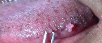
Rice. 3. The same patient. A dense hypertrophic whitish plaque with a spongy surface on the mucous membrane of the labia minora, against this background there are small erosions. The nails on the upper extremities have longitudinal striations.
Laboratory examination data: results of a general urine test, blood coagulogram, blood test for thyroid hormones, glycemic profile within normal limits.
Histological examination of the lesion on the mucous membrane of the genital organs: epidermis with compact orthohyperkeratosis, focal hypergranulosis, uneven acanthosis, apoptosis of single keratinocytes; in the upper parts of the dermis there is fibrosis, angiomatosis, perivascular lymphohistiocytic infiltrates. Conclusion: histological changes may correspond to the hypertrophic form of LP.
The patient received treatment: pentoxifylline 5.0 ml per 100.0 ml of saline intravenous drip every other day; chloropyramine intramuscularly at night 2.0 ml; vitamins B1, B6 1.0 ml intramuscularly every other day, vitamin B12 500 mcg once a day intramuscularly, tetracycline 0.1 g orally 2 times a day for 10 days, delagil 0.25 mg orally 2 times a day course for 20 days, locally on lesions in the area of the trunk and limbs, applications of Akriderm ointment 2 times a day, on the genitals - applications of 0.1% Protopic cream 2 times a day.
A week after the start of treatment, there was a significant reduction in itching and flattening of the lesions in the torso, limbs and genitals. 20 days after the start of treatment, the itching disappeared, the rash almost completely regressed with the formation of pigmentation and congestive hyperemia, and on the genitals - depigmentation and atrophy (Fig. 4).
Rice. 4. The same patient after treatment. a — significant flattening of the lesions in the area of the right shin; b - the plaque in the area of the left shin has softened and turned pale; c — depigmentation and atrophy at the site of former lesions in the area of the external genitalia.
Discussion
Due to the anatomical and physiological characteristics of the vulva (high humidity, the influence of friction, occlusion, high sensitivity to secondary infection), dermatoses in this area have a clinical uniqueness and are often characterized by atypical forms [6]. This is important to take into account in the differential diagnosis of such a precancerous form of LP as hypertrophic.
First of all, hypertrophic LP of the vulva should be differentiated from verrucous carcinoma of the vulva, characterized by asymptomatic, slowly growing exophytic lesions, rarely accompanied by ulceration. Histologically, it manifests itself as acanthotic cords with minimal nuclear atypia, areas of hyperkeratosis on the surface of the tumor with minimal keratin formation within it, as well as diffuse chronic inflammation of the stroma [7].
In such cases, the presence of Wackham's mesh lesions on the surface, as well as the presence of lesions of the oral mucosa and nails characteristic of LLP, may indicate in favor of LP.
Like other chronic inflammatory diseases characterized by scratching and inflammation, hypertrophic vulvar SCC may be accompanied by signs of epidermal SCC, which is usually difficult to distinguish from cutaneous SCC [4, 5].
The difficulty of differential diagnosis of hypertrophic LP of the vulva with SCC of the skin is also associated with the possibility of malignant transformation of the former [8]. The importance of distinguishing these processes is evidenced by four cases of erroneous diagnosis of RCC in hypertrophic LP described in the literature [4, 9].
SCC of the skin is characterized by hyperplasia of the epidermis and adnexal epithelium, severe irregular acanthosis with the formation of horny cysts, infiltration of the reticular layer of the dermis, vascular and perineural invasion. Unlike SCC, SCC of the epidermis is not accompanied by infiltration of the reticular layer of the dermis, as well as vascular and perineural invasion [4, 9].
Therefore, when performing histological verification of hypertrophic LP, a deep biopsy of the lesion should be performed, sufficient for a differential diagnosis with RCC [5].
Histological features of hypertrophic LP include orthohyperkeratosis, wedge-shaped hypergranulosis and psoriasiform epidermal hyperplasia [10], as well as lichenoid borderline dermatitis with the presence of eosinophils [11]. In this case, the presence of typical signs of PCD of the epidermis is possible, but there is no cytological atypia, pronounced solar elastosis, deep spread of acanthotic cords into the dermis, vascular and perineural invasion [4, 9].
Staining for elastin to identify perforated elastic fibers can help distinguish hypertrophic LP from SCC, which is rarely observed in hypertrophic LP and often observed in skin SCC [12, 13].
Vulvar LP is usually treated with applications of corticosteroids or angioneurin inhibitors (tacrolimus and pimecrolimus ointments). In severe cases resistant to therapy, methotrexate and cyclosporine are used [2].
Experience in the treatment of vulvar hypertrophic LP is limited, which is due to the rarity of the pathological process. Only one report indicated the effectiveness of intralesional injections of corticosteroid hormones in this disease [14].
Lichen planus - causes
Although the reasons for the appearance of lichen on the skin or mucous membranes remain a mystery, this disease is often associated with autoimmune diseases. The immunological basis of the disease is indicated by the frequent coexistence of lichens with autoimmune diseases, that is, those in which cells of the immune system attack the body for unknown reasons, destroying “healthy” cells.
Lichen planus may appear in people diagnosed with diabetes, vitiligo, alopecia areata, certain liver diseases (including hepatitis and cirrhosis), and ulcerative enteritis (Crohn's disease). Likewise, lichen planus is more common in people who have had a transplant (especially graft-versus-host disease - GVHD) or are taking certain medications, such as penicillamine, methyldopt.
Types of lichen planus
There are many varieties of lichen planus, including: lichen planus, pigmented, nodular, pemphigoid, linear, follicular, erosive.
The type of lichen can also be determined by the place of its appearance: lichen planus in the mouth, lichen on the nails, lichen of the perianal area, lichen on the genitals: in men, lichen planus is found on the foreskin and on the head, in women, lichen can be found on the genitals lips - and resemble leukoplakia.
Diagnostics
If a rash appears on the pubis or genitals, you should contact a dermatovenerologist. The diagnosis of lichen can be made based on the following studies:
- initial examination, history taking;
- microscopic examination of a skin sample from the site of the lesion (for this, a scraping is taken);
- irradiation with a special lamp;
- clinical blood test.
Research helps to determine the nature of lichen - infectious, neurological or allergic, and to choose adequate treatment. Additionally, a test is carried out for the presence of sexually transmitted infections.
A blood test may be needed to make a diagnosis
Help from a dermatologist in Moscow
Diagnosis and treatment of pityriasis rosea is the field of activity of a dermatologist. Depending on the form of the disease and the extent of skin damage, the patient is provided with recommendations. With lichen, the emotional and psychological background of a person is important, so promptly making a correct diagnosis allows you to quickly overcome the disease.
You can make an appointment with a dermatologist at Meditsina JSC (academician Roitberg’s clinic) by phone. You should not self-medicate. The reason is that, according to external signs, skin rashes may turn out to be differential diseases, for example, toxicerma, psoriasis, mycosis, secondary syphilis. In these cases, it is necessary to prescribe another therapy, taking into account the specific clinical picture and medical history of the patient.
The danger of skin pathologies on the face
Shingles becomes the most dangerous when it appears on the face. It can have an ocular form - spread to the orbital nerve, causing inflammation of both eyelids, the skin on the forehead, nose, and crown area. Ringworm on the eye also appears in case of damage to the maxillary nerve. Infection on the cornea can lead to blindness. If scaly lichen appears on the eyelid, this is also dangerous due to loss of clarity of vision and is fraught with pain.
If fungal lichen is not treated properly, a number of consequences are possible:
- development of a chronic form of the disease;
- inflammation on the skin will spread to the eyes and mouth;
- spread to other areas of the body;
- relapses;
- scars.
Lichen planus - symptoms
Symptoms of the disease are well expressed:
- red or purple papules;
- skin lesions are usually shiny;
- formations that tend to stick together and appear in groups;
- whitish lines on the surface of the skin (the so-called Wickham grid);
- itchy skin.
Itchy skin
Symptoms of lichen planus usually appear suddenly - skin lesions during lichen planus are usually limited, the rash does not increase after the first symptoms appear.
Diagnostics
In advanced stages, it is difficult to confuse lichen sclerosus with any other disease.
At the same time, in the initial stages, the pathology has common symptoms with diabetes mellitus, vitiligo or vulvovaginitis. This may cause confusion. Diagnosis of pathology always begins with a questioning and examination of the woman. Already at this time, an experienced physician will be able to suspect the presence of lichen and make a preliminary diagnosis.
To confirm it, he may prescribe a regular or extended vulvoscopy (a procedure during which the tissues of the vulva are examined using a colposcope).
As for laboratory tests, they may include:
- blood test for glucose;
- PCR (to detect HPV);
- cytological examination of material taken from the tissues of the external genitalia (necessary to exclude cancer);
- immunogram (to understand whether the patient has immune disorders).
Information about pityriasis rosea: causes, symptoms, manifestation
Medicine does not know the exact reasons for the development of lichen deprivation in men and women. Research is being conducted in the field of the influence of herpes viruses types 6 and 7, but a clear etiological agent has not yet been identified. The causes of pityriasis rosea continue to be studied. We only know that a weakened immune system can become a trigger at any time. The causes of pityriasis rosea lie precisely in a decrease in the body's defenses due to bacterial and infectious diseases.
Symptoms and clinical picture of the disease
Due to colds, hypothermia, severe emotional states, and stress, a rash may appear on the skin. Symptoms of pityriasis rosea begin to appear from this point. The classic clinical picture is the formation of the main lesion in the form of a medallion with a diameter of 2 to 10 cm. Within 7-14 days after its appearance, the rash spreads in the form of plaques and papules of pink and yellow-brown color. They are smaller than the main lesion - their diameter can be from 0.5 to 2 cm. In appearance, the rashes can be confused with ringworm due to the scaly edge of the rashes. A few days after the rash, the spots turn pale, wrinkle and the stratum corneum cracks. The central part of the plaques remains smooth. Symptoms of pityriasis rosea may be accompanied by itching, fatigue, fever, general intoxication, and enlarged lymph nodes in the neck and chin.
Are you experiencing symptoms of pityriasis rosea?
Only a doctor can accurately diagnose the disease. Don't delay your consultation - call
Types of disease
Pityriasis rosea can have a classic appearance, when the clinical picture develops in stages in accordance with the generally accepted system - from the appearance of a “maternal” plaque to smaller rashes in the chest, back, abdomen, thighs, and on the flexor surfaces of the limbs. In the medical classification, several more forms of the disease are distinguished. Zhiber's lichen occurs:
- urticarial - characterized by the presence not of plaques, but of a rash in the form of blisters. There is severe itching. In appearance it resembles urticaria;
- vesicular - manifested by generalized rashes of vesicles with severe itching. The diameter of bubbles with clear or cloudy liquid is from 2 to 6 mm. They often form “rosettes”;
- papular - rare. It is characterized by the appearance of cavity-free formations above the surface of the skin. Small papules with a diameter of 1-2 mm;
- hemorrhagic – point hemorrhages (bleedings) occur, so the color of the plaques is darker than usual;
- follicular - rashes are grouped into round plaques of follicular papules, which can occur in parallel with classic plaques;
- unilateral;
- hypopigmented - most often occurs in people with dark or dark skin. Lichen inversus is characterized by rashes in the axillary and groin areas and in the popliteal fossae;
- asbestos-like - is extremely rare and appears on the scalp in the form of gray plaques;
- Darier's giant pityriasis rosea - the formation of large diameter plaques from 5-7 cm. In severe cases, they reach the size of the patient's palm;
- ring-shaped ring-shaped pink lichen of Vidal has an atypical location - mainly in the groin or axillary region. The rash appears ring-shaped.
Gender and age characteristics
Women, adolescents and children are susceptible to the occurrence of pityriasis rosea. Different forms of the disease affect specific groups of people. For example, the vesicular form is diagnosed more often in children and adolescents. The papular form is diagnosed in most cases in pregnant women and young children. Unilateral – occurs equally in both adults and children.
How does disease transmission occur?
Research has not given a clear answer to the question of infection. Theoretically, pityriasis rosea is transmitted by tactile contact, but this occurs extremely rarely. There must be provoking factors for infection to occur. We are talking about low immunity, past viral and infectious diseases, and colds. Relapse is possible in people with HIV, oncology and blood diseases.
What are the risks for the sick person and his partner?
Whether it is possible to receive lichen as a “gift” through sexual contact with an infected person depends on the state of the person’s immunity, as well as on the type of disease. That is why such dermatoses are called conditionally infectious.
The most unpleasant ones on the list are the girdling and pink subspecies:
- Shingles cannot be contracted through airborne droplets; close contact is required, especially sexual contact. Using the patient's personal belongings also threatens with bad consequences. But these warnings apply only to the stage of the disease when a new rash appears. If the blisters are covered with scabs, the disease automatically becomes non-contagious.
- Due to little knowledge of the mechanism of activation of pityriasis rosea, it is not possible to accurately answer the question of its contagiousness. Therefore, it is assumed that this dermatosis is transmitted infrequently by contact. Doctors still recommend abstaining from sexual activity during illness, because sweat and water procedures are not the best companions for this dermatosis.