Scraping for pathogenic skin fungi is a microscopic analysis that is performed to identify fungal infections of the epidermis (mycosis). Using this technique, it is possible to determine the fact of infection with a fungus (preliminary analysis) and determine the type of pathogen (final analysis). The causative agents of mycoses can be pathogenic and opportunistic fungi. In order for the dermatologist to prescribe an effective course of therapy, the patient is sent for mycological examination.
Classification of pathogenic skin fungi
According to the classification, there are 5 main types of fungal diseases: erythrasma (pseudomycosis); deep mycoses; candidiasis; ringworm; keratomycosis. Erythrasma is a pathological fungal infection that is quite difficult to diagnose. Erythrasma can be detected only by scraping for fungi on the skin.
Content:
- Classification of pathogenic skin fungi
- Symptoms of fungal skin diseases
- Methods for diagnosing fungal diseases
- Indications and contraindications for taking scrapings for pathogenic skin fungi
- Preparation and collection of biomaterial for analysis
- Decoding the analysis results
Deep mycoses affect the larynx, bronchi and lungs. The disease occurs in people with weakened immune systems and long-term illnesses. Deep mycoses include: mucorosis, aspergillosis and penicilliosis. Fungi often affect the nasal cavity and cause diseases of the lungs, ears and the formation of abscesses.
Candida fungi look like yeast under a microscope. They are present in small quantities on the body, but when the immune system is suppressed, the fungi begin to multiply intensively, as a result of which a person becomes ill with candidiasis. Candidiasis (thrush) can affect not only the skin, but also the intestines, oral cavity, and intimate organs. The proliferation of fungi and an increase in their number can be confirmed by passing a general blood test and scraping for pathogenic mycoses.
Microsporia, rubromycosis, trichophytosis, epidermomycosis, epidermophytosis are ringworms that cause inflammation of the skin, hair and nail plates. You can get the infection from pets or sick people. Heterotrophic microorganisms love moisture and begin to multiply in a damp environment, so you can “pick up” an infection in the shower, swimming pool, sauna, and even in the gym - in familiar common areas.
Keratomycosis occurs as follows: the stratum corneum of the epidermis is damaged, but there are no inflammatory processes. Keratomycosis is fraught with the fact that because of them two serious illnesses can develop - pityriasis versicolor and trichosporia nodosum. Lichen is diagnosed in 15-20% of the world's population, but people diagnosed with AIDS and tuberculosis are more susceptible to the disease.
Why do analysis?
Treatment of fungal and bacterial diseases differs radically. It is important to establish the cause of skin problems in order to prescribe adequate therapy.
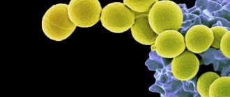
Fungi are one of the most resistant and contagious microorganisms. Mycoses occupy first place among all infectious skin diseases. Fungi do not threaten the patient’s life, but significantly reduce its quality. The situation is complicated by the fact that mycoses are very resistant to treatment. The earlier the disease is detected and the smaller the area it affects, the faster the recovery will occur.
Symptoms of fungal skin diseases
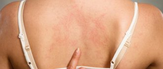
It is difficult to determine the causative agent of infection in the early stages, since there are no external manifestations of the disease. With a large number of pathogenic microorganisms, the first symptoms appear: the skin becomes pale and looks unhealthy; peeling of the skin is observed; hair becomes brittle and begins to fall out; spots form on the skin with an unpleasant odor; the structure of the nails is destroyed; discomfort, itching and burning are felt.
Pathogenic fungi reproduce in favorable conditions (humid environment) and can live outside the human body for a long period of time. It is possible to become infected through direct contact, when the pathogen comes into contact with healthy areas of the body. You can “get” an infection through someone else’s shoes or clothes; this often happens on the beach, in a bathhouse or in a swimming pool.
The incubation period lasts from 2-3 days to 3-4 months. The first symptoms of the disease are cracks in the skin, itching, rashes, and exfoliation of the upper layer of the epidermis.
What the analysis shows
Skin scraping shows the presence or absence of: fungi and subcutaneous parasites.
There are many types of fungal infections. It should be remembered that the fungus is contagious, and the carrier can be not only an infected person, but also an animal. Scraping the skin affected by the fungus will help identify the type of fungus and, accordingly, make it possible to choose the appropriate treatment.
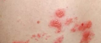
Signs of skin damage by fungus:
- The affected areas differ in color, bumpiness, and peeling of the skin. In case of injury, the area may become larger in size and ulcers may appear. Itching bothers me;
- If the hands and feet are affected, severe itching and peeling appear; the skin becomes thinner and becomes covered with erosions. The nail plate may thicken in size and turn yellow;
- Fungus on the scalp makes hair dull and unhealthy. Peeling, itching, and plaque appear on the hair. Hair may fall out.
Demodex is a microscopic mite that parasitizes the sebaceous glands of the skin, hair follicles of humans and animals.
Signs of demodicosis can be confused with allergies and dermatitis. But there are such differences:
- The rash is accompanied by itching, burning, and discomfort. When the eyelids are affected, itching appears, the eyes become inflamed and swollen. Eyelashes become coated and fall out;
- There is an oily sheen on the face. The affected areas of the skin turn red, swell, and peeling and roughening may begin.
- Over time, scars appear. There is a feeling that something is moving under the skin.
It is impossible to see this mite without a microscope. Therefore, the dermatologist scrapes the skin, then evaluates the results and prescribes treatment.
Skin scraping will help identify scabies. Scabies is an infectious skin disease caused by the scabies mite. The tick can be seen with the naked eye (a white or yellow, small dot).
The tick can penetrate human skin very quickly - within 15 minutes. Scabies is highly contagious, with an incubation period of up to 14 days. The main route of infection is through touch.
Symptoms of scabies:
- Itching, especially intense in the evening and at night;
- Rashes;
- General malaise, fever in children.
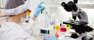
Methods for diagnosing fungal diseases
Scrapings are taken exclusively from smooth skin.
A dermatologist most often prescribes this test to patients with lichen that affects the shoulders, back, chest and neck. The doctor scrapes off the top layer of dead skin and obtains a sample containing mycelium and fungal spores.
Tests for heterotrophic microorganisms include:
- scraping for pathogenic skin fungi;
- microscopic diagnostics;
- cultural research.
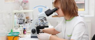
Using microscopic diagnostics, clear contours of the microorganism are revealed. To do this, the biomaterial is clarified in an alkaline environment, which dissolves organic matter and only fungi remain for further diagnosis.
A culture test will help identify the causative agent of the infection. The analysis is performed within 7-10 days, and the doctor can make the final diagnosis based on the results of cultural diagnostics on the 13th day, since primary fungi grow over a long period of time. Other methods for diagnosing skin diseases caused by microorganisms include:
- enzyme immunoassay blood test;
- PCR research;
- sowing.
Get tested for skin fungus in Moscow
Our clinic works with one of the largest laboratory complexes in Moscow, which allows us to perform tests quickly, efficiently and at an affordable cost. The price of scraping for skin fungus does not include a consultation with a dermatologist on the results of the study and treatment of mycosis, but you can sign up for it separately.
Reception is carried out at a time convenient for the client. You don't have to wait in line or adjust to your local doctor's schedule.
Are you looking for where to have your skin scraped tested for fungus in Moscow quickly and efficiently? Come to the Kutuzovsky Children's Center.
Leave feedback
Indications and contraindications for taking scrapings for pathogenic skin fungi
The procedure is indicated for the purpose of preventive diagnostics for visiting a swimming pool, bathhouse, sauna and other public places. The analysis begins when mycoses are suspected, in particular due to the patient’s complaints of cracks, rashes, itching and peeling of the epidermis. Using the study, you can detect microorganisms, determine their type and make an accurate diagnosis.
There are no contraindications to the manipulation, since it is painless and is carried out within a few minutes. Taking a scraping does not cause discomfort or other unpleasant sensations.
References
- Basal cell skin cancer. Clinical recommendations. Association of Oncologists of Russia, 2022. - 85 p.
- Squamous cell skin cancer. Clinical recommendations. Association of Oncologists of Russia, 2022. - 31 p.
- Kumar, V., Abbas, A., Fausto, N. et al. Robbins and Cotran Pathologic Basis of Disease, 2014. - Vol. 1(7), 3(25) — 1464 p.
- Ferrante di Ruffano, L., Dinnes, J., Chuchu, N. et al. Exfoliative cytology for diagnosing basal cell carcinoma and other skin cancers in adults. Cochrane Database of Systematic Reviews, 2018 - Vol 12.
Preparation and collection of biomaterial for analysis
The patient should prepare in advance for the upcoming manipulation: a few days before the date of the study, hygiene procedures should not be carried out; It is prohibited to use cosmetics and apply them to the places from which the biomaterial will be taken; for a sample near the nail plates, you cannot first apply coloring varnishes and cosmetics to them; you should stop using certain medications (it is advisable to consult a dermatologist about the use of medications).
Biomaterial from the affected area of the epidermis is taken with a sterile scalpel or special instruments. It is necessary to separate the damaged skin near the inflammatory and affected area, since in this place the concentration of microorganisms will be maximum; to scrape the area between the fingers, it is necessary to take the stratum corneum.
After taking a scraping, a special solution is added to the biomaterial to create an alkaline environment and destroy the keratin. A day later, the sediment in a container or test tube is examined using various methods.
Best materials of the month
- Coronaviruses: SARS-CoV-2 (COVID-19)
- Antibiotics for the prevention and treatment of COVID-19: how effective are they?
- The most common "office" diseases
- Does vodka kill coronavirus?
- How to stay alive on our roads?
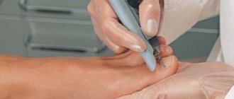
How to do scraping
First of all, the doctor conducts a survey, identifies complaints and medical history.
Before taking a scraping, the dermatologist cleans the surface of the skin from dust and sebum. To carry out scraping, a scalpel, a special spatula, and scissors (to take a nail sample) are usually used. If a scraping needs to be taken from the scalp, the doctor will trim the affected area.
The procedure is absolutely painless. The material obtained during the analysis is delivered to the laboratory. Based on the results, the dermatologist selects a course of treatment.
How much does the demodex detection procedure cost?
Depending on the place where the sample is taken for demodex scraping, the cost of the procedure varies. Moreover, the price of the study depends on the region of residence, the type of medical institution and the speed of obtaining the result.
Attention!
This article is posted for informational purposes only and under no circumstances constitutes scientific material or medical advice and should not serve as a substitute for an in-person consultation with a professional physician.
For diagnostics, diagnosis and treatment, contact qualified doctors! Number of reads: 23807 Date of publication: 09.28.2018
Dermatologists - search service and appointment with dermatologists in Moscow
How do you know when it's time to sound the alarm?
Every person thinks that bad things will happen to anyone, but not to him. That is why in most cases people turn to a mycologist at an advanced stage of the disease. If you suspect that you have nail fungus, our advice is to immediately get tested and begin treatment.
Look at what changes are tested for nail fungus:
- Change in nail color. The nail has become opaque and it is impossible to see the nail bed. The surface of the nail has a yellowish tint.
- The appearance of a gap between the nail bed and the nail. In this interval, a focus of infection is formed.
- Thickening (keratinization) of the nail plate. The thicker the nail, the more deeply the fungus has taken root.
- The appearance of white dots. The surface of the nail becomes loose and rough.
- Swelling and redness of the skin around the nail. As a result, after some time the nail plate begins to deform.
- Painting nails grayish-yellow or brown. This sign is an indicator of the advanced stage of the disease. At this stage, the nails begin to peel off.
How to prepare for analysis
To obtain reliable information about the presence of a parasitic mite on the eyelashes or facial skin, the following rules must be followed before taking the test:
- Do not wash your face with products containing a high alkaline content for 24 hours before the test;
- stop using decorative and medicinal cosmetics, ointments and creams two days before visiting a doctor;
- Avoid contact with eyelashes with shampoo two days before the test;
- You cannot use eye drops the day before the procedure, with the exception of drops that are prescribed for serious eye diseases.
The parasitic mite is most active in the evening hours, as it does not tolerate ultraviolet light. Therefore, the most favorable time to submit a scraping for demodex is the evening hours. But in medical laboratories, material is collected for demodex in the morning, which can make it difficult to identify the causative agent of the disease, provoke its active reproduction and aggravate the patient’s condition.
When a positive result is given
When testing for demodicosis in a private laboratory, the patient receives the test result within an hour. State clinics provide results within 24 hours.
A positive diagnosis is made when a tick, its eggs or larvae are identified. If an empty shell of a parasitic mite is found on the skin, the demodex test is re-administered. If several individuals are detected within one cell, the result is considered conditionally negative and treatment is not prescribed, since this number of mites is not capable of harming human health (changes in appearance or hair).
When a parasite is detected, it is necessary to determine the type of tick (long or short), since this fact affects the duration of treatment and medication prescription. The shortest type is the most difficult to treat.