Scraping for pathogenic skin fungi is a microscopic analysis that is performed to identify fungal infections of the epidermis (mycosis). Using this technique, it is possible to determine the fact of infection with a fungus (preliminary analysis) and determine the type of pathogen (final analysis). The causative agents of mycoses can be pathogenic and opportunistic fungi. In order for the dermatologist to prescribe an effective course of therapy, the patient is sent for mycological examination.
general characteristics
Fungal skin diseases (dermatomycosis), caused by pathogenic dermatomycete fungi (dermatophytes), are divided into 4 groups. The first group is keratomycosis. Pathogens parasitize in the most superficial parts of the stratum corneum of the skin or on the cuticle of the hair. This group includes pityriasis versicolor, erythrasma, nodular trichosporia and axillary trichomycosis. The second group is epidermomycosis. The pathogens also parasitize in the stratum corneum, often affecting the nails, but, unlike the pathogens of keratomycosis, they cause an inflammatory reaction from the underlying layers of the skin. This group includes: athlete's foot, athlete's foot and superficial yeast lesions of the skin and mucous membranes (candidiasis). The third group is trichomycosis. They differ from the causative agents of epidermomycosis in their ability to infect not only nails, but also hair, growing into their cuticle and penetrating into the cortex of the hair. This group consists of: trichophytosis, microsporia and scab. The fourth group is deep dermatomycosis: actinomycosis, deep blastomycosis, chromomycosis, sporotrichosis, as well as visceral forms of candidiasis and mycosis caused by certain molds. This study determines only the presence of a dermatophyte, without determining the species.
Methods for diagnosing fungal diseases
Scrapings are taken exclusively from smooth skin.
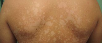
A dermatologist most often prescribes this test to patients with lichen that affects the shoulders, back, chest and neck. The doctor scrapes off the top layer of dead skin and obtains a sample containing mycelium and fungal spores.
Tests for heterotrophic microorganisms include:
- scraping for pathogenic skin fungi;
- microscopic diagnostics;
- cultural research.
Using microscopic diagnostics, clear contours of the microorganism are revealed. To do this, the biomaterial is clarified in an alkaline environment, which dissolves organic matter and only fungi remain for further diagnosis.
A culture test will help identify the causative agent of the infection. The analysis is performed within 7-10 days, and the doctor can make the final diagnosis based on the results of cultural diagnostics on the 13th day, since primary fungi grow over a long period of time. Other methods for diagnosing skin diseases caused by microorganisms include:
- enzyme immunoassay blood test;
- PCR research;
- sowing.
Patient preparation rules
scraping the epidermis
Standard preparation conditions (unless otherwise determined by the doctor):
3 days in advance Stop taking any cosmetic products for facial skin care (creams, soaps, lotions, etc.), local medications.
Do not wash your face on the day of delivery. hair Standard preparation conditions (unless otherwise determined by the doctor):
3 days in advance Stop taking any hair care products, topical medications.
Notes:
To examine hair from the affected part of the head, cut it into a clean container at a distance of 0.1 mm from the roots or pull it out with hair follicles; do not wash your hair on the day of submitting the material.
nails Standard preparation conditions (unless otherwise determined by the doctor):
3 days in advance Stop taking any cosmetics (varnishes, nail strengtheners, etc.), local medications.
Notes:
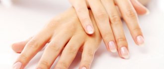
Nails must be clean and dry. You should also not treat your nails with a metal file before the procedure. Trim the nails of both hands (the length of the cut nails should be at least 2 mm). BM is delivered in a sterile plastic container with a screw cap or in a paper envelope.
You can add this study to your cart on this page
Symptoms of fungal skin diseases
It is difficult to determine the causative agent of infection in the early stages, since there are no external manifestations of the disease. With a large number of pathogenic microorganisms, the first symptoms appear: the skin becomes pale and looks unhealthy; peeling of the skin is observed; hair becomes brittle and begins to fall out; spots form on the skin with an unpleasant odor; the structure of the nails is destroyed; discomfort, itching and burning are felt.
Pathogenic fungi reproduce in favorable conditions (humid environment) and can live outside the human body for a long period of time. It is possible to become infected through direct contact, when the pathogen comes into contact with healthy areas of the body. You can “get” an infection through someone else’s shoes or clothes; this often happens on the beach, in a bathhouse or in a swimming pool.
The incubation period lasts from 2-3 days to 3-4 months. The first symptoms of the disease are cracks in the skin, itching, rashes, and exfoliation of the upper layer of the epidermis.
Recovery time
In the most severe cases of long-term, advanced mycosis, the average duration of therapy will take 6-7 months. This period of time is determined by the rate of growth of toenails - the nail plate completely changes just during this time. This period does not mean that the patient will need to regularly visit the doctor for the entire six months. Scheduled follow-up visits to the doctor are necessary only 2-3 times during therapy, including the initial appointment (now we are not talking about visits for a hardware pedicure procedure).
Decoding the research results
The result is considered positive when mycelium, pseudomycelium and fungal spores are detected in the scrapings, as well as in the presence of symptoms of mycotic skin lesions. It should be understood that mushrooms are a component of normal human microflora. The absence of symptoms with a positive fungal test result indicates a healthy carrier state. In addition, this diagnostic method does not allow identifying the type of pathogen. This requires additional research, including inoculation of skin scrapings on nutrient media.
Do you need to do a skin scraping test for fungi?
The Med-Yutas Medical Center conducts microscopic examinations for fungi in Moscow clinics located at st.
Grimau, 10A, building 2 (metro station Akademicheskaya) and Michurinsky Avenue, 21B (metro station Ramenki). To obtain a consultation and make an appointment, use the online registration form or contact the clinic reception. The procedure is performed by a laboratory microbiologist, duration – 10 minutes. The cost of examining skin and nails for fungus is from 790 rubles.
INITIAL CONSULTATION
from 2,500 rub.
Is it really being treated?
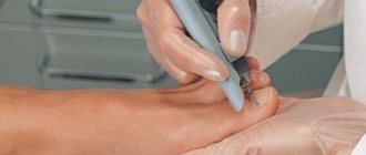
Today, the problem of successful therapy of mycoses can be solved. But it must be approached professionally. One of the important points: in addition to drug treatment, hardware treatment with diamond cutters is strongly recommended for patients with nail mycoses. Its essence is to remove as much as possible the part of the nail affected by the mycelium of the fungus, for better penetration of external therapy drugs and speed up recovery. It should be borne in mind that, unfortunately, neither ointments nor solutions penetrate the nail. If you do not remove several layers of the nail plate affected by mycelium, then during the growth of a healthy nail, it will be re-affected by screening from the infected part of the nail plate.
Preparation and collection of biomaterial for analysis
The patient should prepare in advance for the upcoming manipulation: a few days before the date of the study, hygiene procedures should not be carried out; It is prohibited to use cosmetics and apply them to the places from which the biomaterial will be taken; for a sample near the nail plates, you cannot first apply coloring varnishes and cosmetics to them; you should stop using certain medications (it is advisable to consult a dermatologist about the use of medications).
Biomaterial from the affected area of the epidermis is taken with a sterile scalpel or special instruments. It is necessary to separate the damaged skin near the inflammatory and affected area, since in this place the concentration of microorganisms will be maximum; to scrape the area between the fingers, it is necessary to take the stratum corneum.
After taking a scraping, a special solution is added to the biomaterial to create an alkaline environment and destroy the keratin. A day later, the sediment in a container or test tube is examined using various methods.
Best materials of the month
- Coronaviruses: SARS-CoV-2 (COVID-19)
- Antibiotics for the prevention and treatment of COVID-19: how effective are they?
- The most common "office" diseases
- Does vodka kill coronavirus?
- How to stay alive on our roads?
Why do analysis?
Treatment of fungal and bacterial diseases differs radically. It is important to establish the cause of skin problems in order to prescribe adequate therapy.
Fungi are one of the most resistant and contagious microorganisms. Mycoses occupy first place among all infectious skin diseases. Fungi do not threaten the patient’s life, but significantly reduce its quality. The situation is complicated by the fact that mycoses are very resistant to treatment. The earlier the disease is detected and the smaller the area it affects, the faster the recovery will occur.
Case study
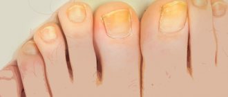
Patient A. came for an initial appointment in November 2009 with complaints of changes in the structure and color of nails, itching in the groin area. He has been ill, according to him, for about six years. Treatment with Orungal, Lamisil and Batrafen Lac did not bring relief. The patient experienced great anxiety and even despair when the process moved to his fingernails, which became the decisive impetus for visiting the mycologist. Upon examination, the nails of A.'s big toes are completely affected: thickened, crumbling, yellow in color. Detachment of the epidermis, itching, and cracks in the skin between the fingers were also observed. On the first and fifth fingers of the right hand there are yellow spots, thickening and detachment from the nail bed. Inguinal region: large, clearly defined scaly plaques, excoriations.
At the beginning of December, the results of A.'s tests were completely ready. Using an accurate history, physical examination and laboratory methods, eczema, onychodystrophy, psoriasis of skin folds, erythrasma and candidiasis of skin folds - severe skin diseases with identical external signs - were excluded. To confirm the fungal etiology of this process, material was taken from the nail plates and the skin of the groin area. In both cases, microscopy revealed fungal mycelium, and the exact pathogen was identified in culture - Trichophyton rubrum.
Already at the first examination, it became obvious that for a successful recovery the patient needed a therapeutic spectrum, including broad external therapy, therapy with systemic antimycotics, and therapy for dystrophic processes in the nail bed that arose as a result of the duration of the disease. In addition, recommendations were given aimed at improving microcirculation and improving blood flow to the nail plates, which should ensure the delivery of the necessary nutrients, also prescribed to accelerate nail growth and reduce treatment time.
The third visit was scheduled for A. at the end of February. Upon examination, a significant improvement in condition was observed. So, in particular, in the groin area the skin is absolutely healthy, blood biochemistry tests are normal. The treatment regimen has been adjusted (in the process this must be done once every two months).
The last visit took place at the end of April 2010. Based on the results of the examination and repeated tests (blood biochemistry and microscopy), patient A. is absolutely healthy.
Examination of the skin and nail plates for superficial mycoses
Microscopic examination of scrapings from smooth skin and nail plates, used for the diagnosis of superficial mycoses of the skin and its appendages.
Synonyms Russian
Microscopy of skin scrapings and nail plates for fungi.
English synonyms
- Direct microscopy, Superficial mycoses
- KOH-test
Research method
Microscopy.
What biomaterial can be used for research?
Nails, scraping.
How to properly prepare for research?
No preparation required.
General information about the study
Superficial mycoses of the skin are a group of diseases of the skin and its appendages (nails and hair), the causative agents of which are fungi that are capable of superficial invasion of the epidermis, hair and hair follicles and the nail apparatus. Most often, superficial mycoses are caused by the presence of fungi of the genus Dermatophytes (in this case we speak of dermatophytosis), but the disease can also be caused by fungi of the genus Candida, Malassezia, Trichosporon and Hortaea.
Clinical examination may suggest the presence of smooth skin mycosis or nail mycosis (onychomycosis), but is not used as a definitive diagnostic method, since many other diseases may have a similar clinical picture (for example, nail psoriasis may resemble onychomycosis). The main method for diagnosing this group of diseases is direct microscopy, in which the material under study (skin flakes obtained by scraping at the lesion, or fragments of the affected nail plate) is examined under a microscope after treatment with a solution of potassium hydroxide (KOH). Microscopy makes it possible to identify hyphae/pseudohyphae and yeast cells of the fungus and characterize their morphology, on the basis of which the diagnosis of “superficial mycosis with clarification of its nature” can be confirmed. For example, dermatophyte fungi (Trichophyton, Microsporum and Epidermophyton) are represented by multiple tube-like structures with true septa and spore formation. Fungi of the genus Candida, on the contrary, are represented by elongated yeast cells (pseudohyphae), and do not form true spores. Fungi of the genus Malassezia produce round yeast cells and elongated pseudohyphae.
The success of a microscopic examination in cases of suspected mycosis largely depends on the quality of the biomaterial taken. To diagnose superficial skin mycoses, skin scrapings are performed from the affected area. Dermatophyte fungi persist in skin flakes for several months, and Candida fungi for several weeks.
The pathogens of superficial mycoses are in fact often part of the normal skin microbiota or temporarily colonize it (“healthy carriage”) and can be identified by microscopy of scrapings from normal skin or scrapings from the distal part of the nail plates in many healthy people. For this reason, a positive test result in the absence of any clinical signs of mycosis has no clinical significance. This statement, however, does not apply to the analysis of the subungual contents of the proximal nail plates, which normally do not contain any microorganisms, including fungi.
It should be noted that microscopy does not allow specifying the type of fungus: for these purposes, inoculation on a nutrient medium is used (microbiological method). A negative test result does not completely exclude superficial mycosis.
What is the research used for?
- For the diagnosis of superficial skin mycoses (dermatophytosis, candidiasis, pityriasis versicolor and others) and onychomycosis.
When is the study scheduled?
- With symptoms of superficial mycosis of the skin (single or multiple foci of peeling and hyperemia with a clear edge, accompanied by itching);
- in the presence of symptoms of onychomycosis: changes in color (yellow, whitish), thickness (subungual hyperkeratosis) and shape of the nail (cracks, onychogryphosis).
What do the results mean?
Reference values: negative.
Positive result:
- dermatophytosis, candidiasis or other superficial mycosis of the skin or nails;
- "healthy carrier"
Negative result:
- norm;
- incorrect collection of biomaterial for research.
Important Notes
- Microscopy does not allow us to determine the specific type of fungus.
Also recommended
- Culture of Candida spp./yeast-like fungi with selection of antimycotic drugs
Who orders the study?
Dermatovenerologist, mycologist, general practitioner.
Literature
- Burns T., Breathnach S., Cox N., Griffiths C. Rook's Texbook of Dermatology / T. Burns, S. Breathnach, N. Cox, C. Griffiths; 8th ed. – Wiley-Blackwell, 2010.
- Wolff K., Johnson RA Fitzpatrick's Color Atlas and Synopsis of Clinical Dermatology / K. Wolff, RA Johnson; 6th ed. – McGrawHill, 2009.
What is the danger?
Without treatment, a fungal infection can serve as an “entry gate” for infectious diseases, such as erysipelas. The worst thing is that in many patients this process sooner or later moves from the feet to the hands. This is a real tragedy for women. If your toenails can be hidden in shoes or socks, then your hands are always visible. Extending nails, using polish, etc. only worsens the process. Such tricks form the so-called “greenhouse effect” and the fungus begins to feel just great, multiplying and destroying your nails, health and peace of mind.
Unfortunately, once the fungus has taken root, it will never leave its host without treatment. The process can only progress and spread to other parts of the body. So, itchy, scaly spots may appear in the groin area, head, thighs and other parts of the body.
We should not forget about the constant allergization of the body, weakening of the immune system, exacerbation and worsening of chronic diseases such as diabetes and bronchial asthma.