Otitis externa is an inflammation of the outer ear, which includes the pinna and external auditory canal (EA).
Author:
- Filatova Evgenia Vladimirovna
otolaryngologist, rhinosurgeon
3.80 (Votes: 10)
- Causes of a boil in the external auditory canal
- Causes of development of bullous otitis media
- Treatment
Otitis externa is an inflammation of the outer ear, which includes the pinna and external auditory canal (EA). Like most inflammatory diseases of the ENT organs, otitis externa can be acute or chronic.
Features of the structure of the outer ear
The outer ear consists of the following components:
- The outer funnel-shaped structure is the auricle.
- The S-shaped tube is the auditory canal that passes through the temporal bone.
- Eardrum.
The pinna helps to capture sound waves and transmit them through the ear canal to the eardrum. The latter is a translucent membrane covered with a thin layer of skin. The attachment of one of the auditory ossicles, the malleus, maintains the cone shape of the eardrum. Sound waves entering the external acoustic passage change the pressure on the membrane, which, vibrating in response, reproduces the vibrations of the source of the sound waves.
Diagnostics
The diagnosis is made based on the patient's complaints, medical history and characteristic clinical picture.
This disease should be differentiated from mastoiditis (inflammation of the mastoid process). With the development of a boil, the pain is most pronounced around the auricle, especially in the area of its attachment; no changes are observed in the eardrum, but with mastoiditis, the eardrum is infiltrated and hyperemic. Make an appointment right now!
Call us by phone or use the feedback form
Sign up
Etiology and pathogenesis
The causes of external otitis are divided into infectious and non-infectious.
Infectious causes
Excessive growth of bacteria in the ear canal occurs when there is increased humidity in the ear canal (sweating, swimming) or local trauma that allows bacteria to penetrate through microcracks in the skin. Staphylococcus aureus is a common pathogen of otitis externa.
Often the cause of the disease is candidiasis due to long-term use of local antibacterial drugs with glucocorticosteroids.
Non-infectious etiology
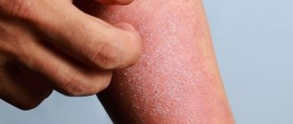
In the absence of bacterial flora, skin diseases lead to external otitis: atopic dermatitis, psoriasis and acne. The consequence of the disease is microcracks in the skin and the development of a secondary bacterial infection.
Predisposing factors for external otitis:
- hot and humid climate;
- swimming lessons;
- elderly age;
- dermatological diseases (eczema, psoriasis);
- narrow ear canals (with Down syndrome);
- ear surgery;
- polyps in the area of the external auditory canal;
- radiation therapy to the head and neck;
- diabetes;
- uncontrolled use of local medications;
- improper use of ear sticks, earplugs;
- lack of hygiene when using a hearing aid.
What underlies the pathogenesis of otitis externa? Any disturbance in wax formation (moisture), canal injury or blockage disrupts the ear canal's defense mechanisms and stimulates bacterial growth.
The skin turns red, swells, and the local temperature rises. This, in turn, leads to the formation of crusts and mucopurulent discharge. The narrowing of the canal, combined with blockage, promotes even greater bacterial growth and the spread of the infectious process.
Epidemiology
The prevalence of inflammatory diseases of the external ear ranges from 17 to 30% among all otiatric pathologies. The growth of this pathology is facilitated by deteriorating environmental conditions, an increase in the level of resistance of the flora, an increase in the number of people with metabolic disorders, immune status, including allergopathology, irrational treatment of acute inflammatory pathology, untimely contact with an otorhinolaryngologist (ENT doctor) and a number of others moments.
Otitis externa is a fairly common disease, but the epidemiology has not yet been sufficiently studied, including due to the different designations of the same type of pathological process. Inflammatory diseases of the outer ear occur in all countries and regions of the globe, but are most often observed in hot and humid climatic regions; an increase in incidence is noted in the warm season. On average, every 10th person experiences this disease at least once during their lifetime, and 3–5% of the population suffers from a chronic form of otitis externa. On average, 0.4% of the population suffers from acute external otitis every year. The disease is most common among people exposed to high humidity for a long time.
Otitis externa occurs in all age groups, the highest prevalence is observed in older children and young adults, then increases slightly after 65 years. The incidence of inflammatory diseases of the outer and middle ear in men and women is approximately the same. No racial differences have been identified in the epidemiology of otitis externa.
Classification
Depending on the clinical manifestations in the pathogenesis of external otitis, three forms are distinguished:
- Acute (up to 1 month).
- Chronic (3 months).
- Recurrent (more than 3 relapses per year).
Based on the severity of the lesion, otitis externa is divided into:
- for mild otitis media (inflammation without signs of suppuration);
- otitis media of moderate severity (severe tissue swelling, increasing pain, symptoms of general intoxication);
- Severe otitis (suppuration of the lesion, unbearable pain, symptoms of general intoxication).
Acute diffuse otitis media
Causes:
- excessive hygiene of the ear canal;
- excess moisture in the ear (water ingress);
- ear skin injuries;
- hypothermia.
The causative agent of diffuse otitis externa is Staphylococcus aureus.
Symptoms of acute diffuse otitis:
- itching and hyperthermia in the area of inflammation;
- bursting pain in the ear;
- swelling and narrowing of the auditory canal;
- hearing impairment;
- serous-purulent discharge from the ear;
- lymphadenitis;
- symptoms of general intoxication, fever;
- insomnia, weight loss (due to pain when chewing).
Manifestations of acute diffuse otitis are short-lived and subside by the end of 2 weeks of the disease. If left untreated, the acute stage of otitis externa becomes chronic, complicated by inflammation of the tissues of the parotid region.
Diagnostic criteria for acute diffuse otitis:
- Examination: pain on palpation of the ear, thickening of the shell skin.
- Otoscopy: hyperemia and swelling of the tissues of the ear canal, erosion with purulent discharge, desquamation of the epithelium of the ear canal, identification of the eardrum is difficult.
Advanced stages of diffuse otitis during otoscopy are characterized by cracks, swelling and suppuration of the tissue. In the resolution stage, the patient is recommended physical procedures: UHF, laser therapy.
Differential diagnosis is carried out with erysipelas and eczema.
Treatment of acute diffuse external otitis includes:
- Taking anti-inflammatory, antihistamine, immunostimulating drugs, antibiotics.
- Toilet the outer ear and introduce turunda with antibacterial, hormonal ointments into the ear canal.
- Instillation of antibacterial ear drops.
If the causative agent of diffuse otitis is a fungus, then antifungal drugs are prescribed.
Publications in the media
Otitis externa - inflammation of the external auditory canal; the incidence is higher in the summer months. In pathogenesis, great importance belongs to the state of the body's immune system. There are limited and diffuse external otitis • Acute limited external otitis (furuncle of the auditory canal) occurs as a result of infection (usually by staphylococcus) of the hair follicles and sebaceous glands of the membranous cartilaginous part of the external auditory canal during microtrauma due to manipulation in the external auditory canal with sharp objects (hairpin, match, etc.). More often occurs in people suffering from diabetes, hypovitaminosis (A, C, group B) • Acute diffuse external otitis is a common form that occurs mainly in chronic purulent otitis media due to infection of the skin and subcutaneous tissue of the external auditory canal by various microorganisms •
Chronic external otitis - diffuse external otitis lasting more than 6 weeks • Eczematous external otitis often accompanies ordinary atopic eczema or other primary diseases of the skin • Malignant (necrotizing) otitis - a rare, most severe form of the disease, characterized by high activity, rapid spread of the infectious process in depth tissues, rapid growth of granulations and sequestration of bone tissue. It occurs either as osteomyelitis or as cellulitis in elderly people suffering from type 1 diabetes, with infection of the external auditory canal by Pseudomonas aeruginosa. Occurs at a young age in patients with immunodeficiency conditions; it is rarely reported in children.
Etiology • Acute and chronic external otitis •• Injuries of the external auditory canal •• Bacterial infection (Pseudomonas aeruginosa, staphylococci, streptococci, gram-negative bacilli) •• Fungal infection (Candida fungi, molds) •• Viral infection (influenza virus, HSV) ••
Previous antibiotic therapy • Eczematous external otitis (associated with primary diseases) •• Eczema •• Seborrhea •• Contact dermatitis •• Hypersensitivity to drugs prescribed locally • Malignant external otitis - Pseudomonas aeruginosa is considered the main causative agent in patients suffering from type 1 diabetes.
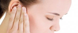
Clinical picture • Pain in the ear, radiating to the eye, teeth, neck. The pain intensifies when chewing, talking, pressing on the tragus, retracting the auricle • Ear congestion • Itching (with fungal infection) • Regional lymphadenitis • Putrefactive discharge from the external auditory canal • Damage to the cranial nerves (VII, IX–XII pairs).
Otoscopy • Furuncle of the external auditory canal - limited infiltrate, narrowing the lumen of the auditory canal • Diffuse external otitis •• Acute form. Hyperemia, infiltration of the skin of the membranous-cartilaginous part of the ear canal, narrowing of the lumen of the ear canal to varying degrees. In its depths you can see a mushy mass consisting of desquamated epidermis and pus with a pungent putrefactive odor. The eardrum is moderately hyperemic and covered with desquamated epidermis •• Chronic form. Thickening of the skin of the ear canal and eardrum •• Fungal infection (otomycosis). The auditory canal is narrowed along its entire length, both in the bone and in the membranous-cartilaginous sections due to skin infiltration. The eardrum is accessible for inspection ••• When affected by mold fungi of the genus Aspergillus, the pathological discharge in the external auditory canal resembles blotting paper. The color of the discharge is black-brown when infected with fungi Aspergillus niger, yellowish or green - Aspergillus flavus, Aspergillus graneus, gray-black - Aspergillus fumigatus ••• With the etiological role of yeast-like fungi of the genus Candida, the otoscopic picture resembles weeping eczema of the outer ear. Pathological discharge in the ear canal looks like whitish or yellowish crusts, pityriasis scales or caseous masses that cover the lumen of the ear canal. The process often extends to the auricle and postauricular area.
Special studies • Microscopy of a scraping of the external auditory canal, culture on nutrient media to identify the type of fungus • Bacteriological examination - Gram staining and culture of the discharge of the external auditory canal on nutrient media • X-ray examination for malignant otitis externa.
Differential diagnosis • Otitis media • Palsy of cranial nerves (VII, IX–XII pairs) • Eruption of wisdom teeth • Arthrosis of the mandibular joint.
TREATMENT is often outpatient; for malignant external otitis, hospitalization is necessary • Etiotropic therapy •• For bacterial otitis - antibiotics depending on the type of pathogen •• For infection with yeast-like fungi of the genus Candida - nystatin, levorin •• For infection with mold fungi - neomycin + polymyxin B + lidocaine , amphotericin B, amphotericin B+methylglucamine, mycoheptin • Antihistamines • Analgesics - according to indications • Autohemotherapy, immunostimulants • Correction of carbohydrate metabolism (especially in malignant external otitis) • Local treatment for limited and diffuse bacterial external otitis •• Careful toilet of the external auditory canal 2 times a day, washing it with a warm solution of boric acid or nitrofural (1:5000); for itching - instillation of 1% menthol solution in peach oil into the outer ear, administration of 2% sulfathiazole ointment or 1–2% yellow mercury ointment •• Introduction of turundum with 3% alcohol solution of boric acid into the external auditory canal, for more in the later stages - streptocidal or 1% chloramphenicol emulsion •• Lubricating the ear canal with 2–3% silver nitrate solution or 1–2% brilliant green alcohol solution •• Prednisolone ointment, 1% hydrocortisone emulsion, 5 drops in the ear 3 times /day for 7 days •• UHF therapy, ultraviolet irradiation, helium-neon laser therapy • Local treatment of otomycosis •• Instillation of 2% nitrofungin solution into the external auditory canal •• Introduction of nystatin ointment, levorin into the external auditory canal •• 1 % clotrimazole cream •• 2% alcoholic solution of flavofungin • Treatment of malignant external otitis •• Instillation of a solution of a specific bacteriophage against Pseudomonas aeruginosa •• Instillation of 2% solution of boric acid •• Hyperbaric oxygenation •• Antibiotics (carbenicillin, piperacillin, azlocillin, ceftazidime, imepenem + cilastatin and ciprofloxacin).
Surgical treatment is indicated only for malignant otitis externa - necrotic tissue is removed.
Observation • Acute otitis externa - improvement usually occurs within 48 hours after the start of treatment • Chronic otitis externa •• Re-sanitation of the external auditory canal is necessary every 2-3 weeks •• It may be necessary to change local medications • Malignant otitis externa •• Daily observation in an inpatient setting •• Carrying out basic auditory and vestibular tests at the beginning and end of treatment.
Complications • Malignant otitis externa - infection may spread to adjacent bone structures and brain • Acoustic chondritis in acute external otitis. Course and prognosis • Acute external otitis - rapid improvement in response to treatment • Chronic external otitis - with repeated sanitation and adequate antibiotic therapy in most cases responds well to treatment • Eczematous external otitis - recovery occurs when the primary skin disease is cured • Malignant external otitis - usually resolves successfully with repeated sanitation and long-term parenteral antibiotic therapy. The relatively high mortality rate may be due to concomitant diseases. Age-related characteristics - malignant external otitis often occurs in the elderly.
Prevention • Refusal of prolonged stay in conditions of high humidity or dampness • Prevention of traumatic effects on the ear canal • Diagnosis and treatment of concomitant systemic diseases.
ICD-10 • H60 Otitis externa
Note. Cholesteatoma is a dense layer of epidermal masses and their breakdown products, mainly cholesterol, a white formation that usually has a connective tissue membrane covered with stratified squamous epithelium, which fits tightly to the bone and often grows into it. Under the influence of decay products and chemical components of cholesteatoma, in particular collagenase, bone destruction occurs. Growing cholesteatoma can cause extensive destruction in the temporal bone, which often leads to the so-called natural radical surgery, as well as various intracranial complications.
Limited (local) otitis externa or boil of the external auditory canal
Causes:
- Excessive hygiene of the external auditory canal.
- Excessive moisture in the ear (water ingress).
- Injuries to the skin of the outer ear.
The causative agent of the disease is streptococcus.
Local external otitis is characterized by the formation of a boil and begins with local symptoms: severe itching in the area of the outer ear. As the boil enlarges, compression of the nerve receptors occurs and the itching turns into unbearable pain. The pain is localized only on the affected side. These manifestations enter the infiltration stage of otitis externa.
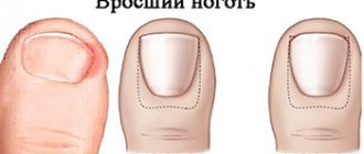
At the abscess formation stage, the forming boil completely closes the lumen of the external auditory canal, which significantly reduces hearing. In the center of the boil, a purulent core with a hair inside is determined.
After opening the boil and expiration of the purulent contents, at the resolution stage, the pain decreases.
Diagnostic criteria for limited external otitis:
- Examination and palpation: pain when pulling the ear and pressing on the tragus, thickening of the auricle.
- Otoscopy: boil in the initial or maturing stage.
- Audiometry and hearing testing using a tuning fork: conductive hearing loss.
- Bacteriological culture of pus: detection of Staphylococcus aureus or its absence.
Treatment of external limited otitis includes:
- Toilet of the external auditory canal.
- Introduction of turunda with antibacterial ointment into the ear canal.
- Instillation of antibacterial ear drops.
- Taking analgesic and anti-inflammatory drugs.
- Antibiotic therapy (for furunculosis).
- Immunostimulating therapy.
If the boil completely blocks the auditory canal, it is opened and drained. In the resolution stage, patients are prescribed UHF therapy.
Risk factors
- Swimming or other exposure to water. Excess moisture leads to skin irritation and destruction of the skin-sulfur barrier.
- Injury. This includes excessive or aggressive ear cleaning. Not only leads to a decrease in sulfur, but can also lead to injury to the skin, allowing microorganisms to gain access to its deeper layers.
- Obstruction of the NSP. Earwax, foreign body, less often - devices that close the ear canal: hearing aids, in-ear headphones.
- Allergic contact dermatitis. Exposure to shampoo, cosmetics, allergic reaction to metal earrings.
- Dermatological diseases. Psoriasis, atopic dermatitis.
- Radiation therapy. It can lead to ischemia (local decrease in blood supply) of the NSP, alter the production of sulfur and its normal excretion.
Normally, NSP is populated by aerobic and anaerobic bacteria. Staphylococci are the most common bacteria (63%) found in the ear canal, most commonly Staphylococcus auricularis and Staphylococcus epidermidis.
The most common pathogenic organisms causing inflammation of the outer ear are Pseudomonas aeruginosa (38%). Less commonly - Staphylococcus epidermidis (9%) and Staphylococcus aureus (8%). Anaerobic bacteria are detected in 4-25% of cases. Fungal infections account for 2-10% of cases of ANO.
Acute bullous or hemorrhagic otitis externa
Causes:
- bacteria;
- long-term acute respiratory viral infection;
- flu.
In acute bullous external otitis, bullae (blisters) filled with blood are noted on the eardrum.
Treatment of acute bullous external otitis includes:
- Toilet of the external auditory canal.
- Introduction of turunda with antibacterial ointment into the ear canal.
- Instillation of antibacterial ear drops.
- Taking analgesic and anti-inflammatory drugs.
- Antibiotic therapy.
- Immunostimulating therapy.
- Prescription of agents that reduce the permeability of the vascular wall.
4.Treatment
The basis of etiopathogenetic therapy is the prescription of antibiotics and strengthening the immune system. Alcohol turunds, bactericidal ointments, UV irradiation, UHF are used locally. In case of rapid painful development, the abscess is opened surgically, observing all antiseptic precautions; The rod is removed, rubber drainage is used according to indications, then treatment is carried out according to the above scheme. The prognosis is favorable in case of timely adequate intervention, sanitation of existing foci of chronic infection (teeth, tonsils, etc.) and effective correction of endocrine and metabolic processes, if necessary.
Myringitis
Myringitis is an inflammation of the eardrum.
Most often, myringitis develops against the background of existing ear diseases. Predisposing factors of inflammation are:
- mechanical, chemical, thermal injuries to the ear;
- hypothermia;
- water getting into the ear;
- uncontrolled use of antibacterial and hormonal drugs.
The first signs of myringitis are difficult to distinguish due to the predominance of the clinical cause of the disease. In the midst of inflammation, the leading symptoms of myringitis are:
- Bursting pain in the ear, often unilateral, aggravated by chewing and talking.
- Itching, feeling of heaviness in the ear.
- Hearing loss.
- Noise in ears.
If left untreated, the patient develops symptoms of general intoxication and serous-purulent exudate is released from the ear canal. Often the discharge contains streaks of blood.
Diagnostic criteria for myringitis:
- Examination and palpation: pain when pulling the ear and pressing on the tragus, discharge of pus from the ear canal.
- Otoscopy: swelling and hyperemia of the eardrum, the presence of pus in the ear canal.
- Audiometry and hearing testing using a tuning fork: conductive hearing loss.
- Bacteriological culture of pus: detection of Staphylococcus aureus or its absence.
- Complete blood count: leukocytosis, increased ESR.
Differential diagnosis of myringitis is carried out with otomycosis, purulent otitis media.
Therapy for acute myringitis:
- Antibiotic therapy.
- Taking analgesic and anti-inflammatory drugs.
- Instillation of antibacterial ear drops.
- Detoxification therapy.
- Rinsing the ear canal with antiseptic solutions.
If the etiology of myringitis is viral in nature, then antiviral drugs are prescribed.
Dermatitis of the ear
Dermatitis is classified as a separate group of diseases, since inflammation of the skin of the ear occurs more often without affecting the external auditory canal. The causes of dermatitis are nickel-containing jewelry, hair sprays and gels, and existing allergic diseases. The clinic is characterized by peeling, redness and thickening of the skin with the formation of small blisters.
Diagnosis of auricular dermatitis is based on an examination by an ENT doctor and an allergist. Treatment includes taking antihistamines, anti-inflammatory and immunostimulating drugs.
Erysipelas of the auricle
Erysipelas is an infection of the ear caused by streptococcus. Predisposing factors for erysipelas: hypothermia, decreased immunity, microtrauma of the skin, contact with a carrier of streptococcus.
Forms of erysipelas
The disease begins with symptoms of general intoxication. The severity of the infection depends on the degree of damage, and therefore three forms of the disease are distinguished:
- Mild degree - erythematous. Characterized by pain and hyperemia. The affected area rises slightly above healthy skin and has the appearance of a “varnished” film.
- The average degree is bullous. Signs of inflammation include blisters filled with purulent contents.
- Severe degree – necrotic. At this stage, the lesion affects the deep layers of the skin and is accompanied by the formation of ulcers covered with purulent plaque.
Treatment of the necrotic form consists of surgical debridement and excision of necrotic tissue. Conservative therapy includes the prescription of antibacterial, detoxification and immunostimulating drugs. In severe cases, administration of antistreptococcal gamma globulin is indicated.
Prevention
Prevention of ONO is carried out in case of frequently recurring episodes of the disease, concomitant dermatological diseases, as well as with regular contact with water - in athletes.
When engaging in water sports or frequently visiting the pool, it is recommended to use swimming caps or special earplugs, and dry your ears after swimming. The outer ear can be gently wiped with a towel. To remove residual water, shake your head vigorously in different directions.
Author:
Chekaldina Elena Vladimirovna otorhinolaryngologist, Ph.D.
Malignant otitis externa (necrotizing otitis externa)
Necrotizing external otitis is a complication of diffuse external otitis. In this case, the pathological process spreads to the temporal bone, affecting the temporomandibular joint and cranial nerves.
More often, the malignant form of otitis externa is determined in people with AIDS, diabetes mellitus, and in people after chemotherapy. The causative agent of the disease is Pseudomonas aeruginosa.
The clinical picture of necrotizing external otitis is characterized by pain, discharge of pus from the ear canal, and fulminant hearing loss. After damage to the cranial nerves, difficulty swallowing, dizziness and paralysis of the facial muscles appear.
Bacteriological culture of purulent discharge and CT scan of the temporal bones are of leading diagnostic importance. Treatment of malignant external otitis includes antibacterial, detoxification, immunostimulating therapy, administration of gamma globulin and surgery (excision of necrotic tissue and skin plastic surgery).
Chondroperichondritis of the auricle
Chondroperichondritis is an inflammation of the cartilage tissue and perichondrium of the auricle. The causes of inflammation are thermal, chemical and mechanical injuries to the ears, skin cracks, erysipelas, furunculosis with otitis externa. The clinical picture is dominated by swelling and hyperemia of the cartilage, and an increase in general temperature. Treatment includes anti-inflammatory and antibacterial therapy.
Question answer
What should you be wary of when treating otitis externa?
If severe itching and burning occurs after instilling drops or applying ointment, then you should stop treatment and consult a doctor. Symptoms may indicate a ruptured eardrum, which in most cases threatens permanent hearing loss.
What not to do if you have ear pain?
If you experience the slightest discomfort, you should immediately contact an ENT doctor. Self-medication is fraught with deterioration of the condition and the development of deep ear lesions.