Symptoms of melanoma are a set of indicators according to which ordinary moles on the body, which everyone has, are beginning to degenerate or have already degenerated into a malignant tumor. The appearance of melanoma depends on its type and stage; superficial spreading, acral lentiginous, nodular melanomas and lentigo are known.
More than 20% of melanomas belong to the superficial spreading variety. However, patients rarely live more than 5 years. However, with timely and high-quality treatment, life expectancy can be significantly increased, and for this it is important to know what melanoma itself looks like in order to recognize it in time.
Manifestations in the early stages
Content:
- Manifestations in the early stages
- How does amelanotic melanoma manifest?
- Signs of metastasis in the final stages
- Well-being when sick
- Signs of a mole degenerating into melanoma
- Appearance of melanoma and how to distinguish it from a mole
Melanomas can appear on any part of the body, but most often the tumors are located on the legs or back. Melanoma can also appear on the palms, mucous membranes of the mouth, vagina, rectum, and scalp. In aging skin, melanomas often manifest in the facial area.
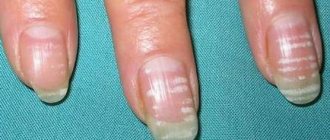
Subungual or acrolentiginous variety
In 3% of all diagnosed cases of melanoma, specialists identify cancer of the nail plates. This disease has its own specific symptoms, which include the appearance of a tumor on the finger, darkening of the nail or the appearance of a clear dark stripe on it, cracking of the nail plate over time with the release of sanguineous fluid or pus. It is important to know that nail melanoma most often occurs in the area of the thumb. As it grows, the tumor changes color to bluish-red or black and its shape to mushroom-shaped.
Cutaneous melanoma
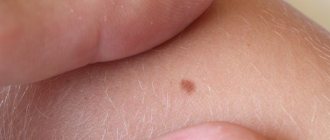
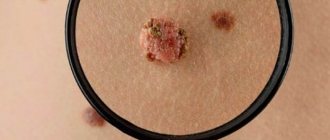
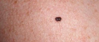
The symptoms of cutaneous melanoma are determined by the naked eye, so you should always pay attention to your own health so as not to miss changes in the development of a mole and seek medical help in a timely manner. The main symptom of melanoma manifestation is the degeneration of a mole (nevus). This degeneration can be expressed in different forms, for example, the mole may begin to enlarge, its contour becomes jagged and asymmetrical, the pigmentation color darkens or acquires blue or reddish shades, itching occurs in the area of the formation, as well as swelling or wet discharge. Against the background of the above symptoms, regional lymph nodes may enlarge.
A mole can begin to degenerate after an injury, or for no apparent reason. Most often, hair falls out in the pigmentation zone, the skin begins to peel off and form crusts, and the skin pattern on the surface can no longer be read. It is also possible that melanoma satellites – skin rashes near the tumor site – may occur. The mole becomes very smooth and acquires a mirror-like surface.
Ocular variety
Melanoma of the eye is very common. Its peculiarity is the absence of symptoms in the first stages of pathology, which greatly complicates the course and treatment of the disease. The patient should be alerted to symptoms such as:
- sudden deterioration of vision;
- the appearance of photopsia or colored dots, sparks before the eyes;
- the occurrence of metamorphopsia or distorted perception of spatial and dimensional quantities;
- change in pigmentation in the iris;
- the appearance of “blind spots” in the field of vision;
- swelling in the eye area;
- retinal clouding.
Symptoms of ocular melanoma may occur even before the oncology is fully formed and do not allow an accurate diagnosis to be made. With different localization of this disease, hemorrhages in the eye area, secondary glaucoma, and retinal detachment may also occur.
Why do moles appear?
The main causes of moles on the nasal part of the face include the following:
- Genetics. In this case, skin formations are congenital or appear in the first year of life.
- Circulatory system disorders. Such moles are red in color and are a cluster of small vessels.
- Hormonal changes associated with an increase in the amount of melanin.
- Long-term use of hormonal drugs.
- Prolonged exposure to direct sunlight.
- Certain viral infections.
- Exposure to radiation and x-rays.
Types of moles
The following types of moles most often appear in the nasal area of the face:
- Vascular. They are a cluster of blood vessels and have a red or red-brown tint.
- Pigmented. They arise due to an excess of melanin and can have a different shade from flesh to dark brown. Pigmented moles can be intradermal (formed in the deep layers of the epidermis), epidermal (formed in the upper layer) and mixed.
When is removal necessary?
The need to remove nevi on the nose arises in the presence of melanoma-dangerous moles. The following features should alert you: blue, blue, violet, bluish-brown color of skin formations. Risk indicators also include moles that have an uneven color and uneven edges. It is recommended to undergo an examination if asymmetric formations and large moles are found in the nasal part of the face.
You need to consult a doctor in the following cases:
- the mole suddenly begins to increase in size, change shape and color;
- skin formation causes burning and itching;
- crusts appear, the mole becomes inflamed;
- the surface of the skin formation becomes denser;
- ulcers and erosions appear;
- presence of discharge and blood;
- pain when touched.
Moles in the nose can cause inconvenience during hygiene and cosmetic procedures, interfere with a runny nose, and cause psychological problems due to loss of self-confidence.
Removal methods
There are various methods for removing nevi on the nose, among which the following are the safest and most effective:
- Method of non-contact radio wave cutting. The mole is removed using heat, which is formed due to exposure to high-frequency radio waves. This method is well suited for removing moles on the nasal part of the face. Radio waves do not cause pain. There is no long rehabilitation period after the procedure. After 7-14 days, no traces remain at the treatment site.
- Laser removal. The surface of the mole is excised layer by layer under the influence of a high-precision laser. This ensures there are no risks. The method is non-contact. When removing nevi on the nose, immediate sealing of damaged vessels is ensured, which makes the method bloodless and increases its safety. There are no scars, scars or pigmentation at the site of the removed mole.
Advantages of removal in our center
The New Skin Clinic medical cosmetology center in St. Petersburg performs mole removal using the Surgitron radio wave device and the innovative Clarity laser device. The choice of removal method is made on an individual basis after examining the patient during a free consultation.
We guarantee the quality, effectiveness and safety of all procedures performed. This is achieved thanks to the work of highly qualified doctors. All our specialists have certificates confirming the right to work on Clarity and Surgitron devices. We adhere to a low price policy, thanks to which medical cosmetology procedures become available to people with different income levels.
Make an appointment at our clinic in St. Petersburg on a day and time convenient for you using the form on our website. Leave your contact number and we will call you back to confirm the time of your visit and answer your questions.
How does amelanotic melanoma manifest?
Pigmentless or achromatic melanoma is a rare, but no less dangerous disease. Its first signs can be considered the appearance of a small bump on the skin, which quickly begins to grow. Outwardly, such a formation may resemble a wart and does not attract much attention to itself, since its color completely matches the skin tone. As achromatic melanoma grows, it may become slightly pinkish or whitish in color with a rough surface and peeling epithelial scales. The skin at the site of the tumor becomes rougher. Sometimes the appearance of such melanoma resembles a scar without a clear edge. Pressing on the tumor does not cause any discomfort, which makes many patients not pay attention to it.
In shape, non-pigmented melanomas can be flat or dome-shaped. The second form is caused by the vertical proliferation of cells, which leads to a more voluminous size compared to a flat one. The main feature of such a tumor is its uneven growth with obvious asymmetry, uneven edges and compaction of the structure. However, with the nodular form of achromatic melanoma, the symmetry of the neoplasm and evenness of color are preserved.
When such melanoma grows enough, it begins to cause discomfort to a person, which is expressed in pain, itching, swelling, and redness of the tissues. The surface of the neoplasm may crack, bleed, and form ulcers. All this indicates the progression of melanoma to late, severe stages that are practically untreatable.
An alarming sign of a neoplasm is hair loss from its surface, which indicates malignancy of the pathology. It is also dangerous to detect enlarged regional lymph nodes.
Types of warty nevus
There are localized forms of nevus manifestation or systemic (verrucous).
Localized - most moles are found in this form.
Signs of its manifestation:
- The diameter of the neoplasm does not exceed 1 cm.
- elements appear separately or are located so close that they can merge
- localization zone is clearly limited
- the keratinized surface of the nevus often has cracks
- moles are dark brown or maroon in color. In very rare cases, nevi are light brown or light pink in color.
- the area affected by moles is often devoid of hair
Systemic form of the disease - the incidence of pathology is approximately 20% of cases.
A distinctive feature of a nevus is the presence of a “garland” of neoplasms.
Such elements are located at close distances relative to each other and “stretch” along the surface of the skin along the entire body.
This form of nevus has a number of features:
- the elements spread along large vessels, and they can also be in different places of the body
- The surface layer of moles may differ in shade. Elements may have different color intensities
- the length of one garland can reach up to 20 cm.
- When feeling the moles, it is noted that they have a dense structure
- the surface of each mole is lumpy and tortuous. As a person ages, these symptoms become more pronounced.
Each type of warty mole can be frequently injured.
Signs of metastasis in the final stages
Melanoma metastases usually occur in the following cases:
- if the patient is elderly;
- with complications of melanoma by chronic diseases;
- when melanoma cells grow into the walls of other organs;
- with a large size of primary melanoma.
Signs of liver metastases
When metastases are localized in the liver structures, black melanin accumulations form in them, representing the affected areas. In this case, functional or physical disorders occur in the liver, the consequences of which are felt by the entire body.
The main signs that melanoma has metastasized to the liver are:
- tuberosity of the organ;
- increase in liver size;
- sudden weight loss;
- presence of jaundice or ascites;
- pain in the right hypochondrium;
- nausea and vomiting;
- enlarged spleen;
- frequent nosebleeds.
Metastases in the lungs
The initial phase of pulmonary metastases does not have pronounced symptoms. In this case, there will be general signs of cancer problems in the body such as apathy, anemia, weakness, weight loss, decreased appetite, hyperthermia. Signs of metastases may initially include various colds - bronchitis, flu, pneumonia. Sometimes symptoms of lung metastases in melanoma occur only at the final stage of the disease, when nothing can be done to alleviate the patient’s condition. In this case, multiple nodular formations with involvement of the pleura and bronchi are diagnosed in the lungs.
If a large part of the lung is affected or the bronchi are compressed due to metastases, the patient experiences shortness of breath. The cough with metastases is predominantly nocturnal and dry, then gives way to ulcerations of mucopurulent sputum, odorless, but often with traces of blood. As metastases grow, the bronchi narrow and the sputum thickens, becoming purulent. Pulmonary hemorrhage may occur.
Pulmonary metastases, which affect the pleura, spine and ribs, cause pain. If metastases reach the lymph nodes of the left part of the mediastinum, hoarseness and aphonia appear, and with right-sided damage to the lymph nodes, the upper part of the body may swell due to compression of the superior vena cava.
Metastases in lymph nodes
Metastases in melanoma most often occur at stages 2-3 of the disease. The most common site for metastasis is the lymph nodes. Moreover, if a patient has a single metastasis to a lymph node, then, according to doctors’ forecasts, he will live another 5 years in 51% of cases. With 4 metastases in this area, only 17% of patients will overcome the five-year mark.
Signs of lymph node metastasis in melanoma are:
- sudden loss of body weight;
- causeless enlargement of lymph nodes;
- visual impairment;
- constant fatigue and chronic feeling of tiredness.
Metastases in bones
Metastasis to the bones is considered a very common phenomenon in melanoma in medicine, which gives severe pain to patients. The main manifestations of this process can be hypercalcemia, bone fragility, neurological changes, and compression of the bone marrow. Metastases to the bone come from the primary site, but other tumors can also be the cause, such as soft tissue sarcoma, ovarian tumors, etc. Melanoma most often affects the bones of the pelvis, hips, vertebrae, and ribs. Metastases follow from melanoma to the bones of the skull along the venous vertebral plexuses.
Nature has protected the skull bones with the ability to constantly renew themselves through osteoclasts and osteoblasts, but the entry of melanoma cells through the bloodstream into the bone marrow destroys these structures. The result of this is constant fractures of the skeleton in different parts, even under small loads.
Metastasis in the brain
Melanoma causes metastases to the human brain more often than other cancer pathologies. After this, there is no need to wait for a complete cure of the disease. The chances of survival for a patient with such metastases are significantly reduced. However, sometimes modern therapy with monoclonal antibodies helps to stop this process if there are individual characteristics of the human body.
Metastases to the brain area occur in 45% of all patients suffering from late-stage melanoma. This ultimately leads to the death of the patient. The symptoms of such metastasis will depend on the location of the lesion, necessarily including incoordination of the patient’s own body, emotional instability, headaches, fever and fever, memory loss, personality changes, lethargy, changes in pupil size, general weakness and difficulty in speech skills.
If the metastases have affected the frontal part of the brain, then the patient may experience sudden changes in rudeness, he begins to swear, and experience disturbances in visual functions and the musculoskeletal system. The symptoms of such metastases are very individual and sometimes they can completely change the patient’s personality.
When the patient's brain stem metastasizes, bursting or dull intracranial pain occurs in the head, which leads to dizziness and impairs visual functions. Also, such metastasis causes constant nausea and vomiting in the patient, convulsions, which in manifestations resemble epileptic seizures.
Treatment of warty nevus
The basis of treatment for keratotic nevus is its removal using one of the most suitable methods.
The choice of method depends on the location of the warty nevus, its shape, and size.
The specialist decides which destruction method will be most effective.
To get rid of the manifestations of skin defects, the following techniques can be used:
- use of liquid nitrogen (cryotherapy)
- application of electric current (electrocoagulation)
- removal by radio waves (radiotherapy)
- destruction with a laser beam (laser therapy)
Well-being when sick
Best materials of the month
- Coronaviruses: SARS-CoV-2 (COVID-19)
- Antibiotics for the prevention and treatment of COVID-19: how effective are they?
- The most common "office" diseases
- Does vodka kill coronavirus?
- How to stay alive on our roads?
Typically, the early stages of melanoma do not cause the patient any inconvenience, discomfort, or pain. After stage 3, when metastases occur, pain begins to appear, and with the onset of stage 4, anemia, weakness, frequent cough, aching bones, exacerbation of chronic pathologies, enlarged lymph nodes, loss of appetite, headaches, nausea, vomiting, and convulsions are added to it. . Depending on the location of metastases, memory loss may occur, vision may deteriorate, and constant pain in the skeleton, right hypochondrium, and chest space may occur.
The danger of melanoma lies precisely in the fact that the patient practically does not feel the most treatment-friendly stages, so he is in no hurry to seek help, and when metastases occur and the melanoma can no longer be cured, the patient begins to be bothered by all sorts of unpleasant symptoms, which have to be dealt with make significant efforts. In general, the state of health in patients with melanoma in late stages is negative, which further depresses already exhausted people faced with oncology.
Features of care for verrucous nevus
If elements of a wart nevus are found on a patient’s body, doctors suggest removing it to remove the cosmetic defect.
If the patient for some reason does not want or cannot do this, then he must follow a number of rules that will help avoid the development of complications.
The basic rules are as follows:
- Eliminate factors that may cause overheating of moles. Prohibited: visiting saunas, baths, spa treatments.
- In the warm season, avoid being under the rays of the sun during the hours of its greatest activity: after 10 a.m. and before 4 p.m. Avoid visiting the solarium. It has been proven that protective agents cannot prevent the development of melanoma;
- Before taking hormonal medications, you should first consult with a specialist.
- Monitor the condition of the nevus elements and consult a doctor at the first symptoms of malignancy of moles.
You should pay attention to the following changes:
- accelerated nevus growth
- the appearance of unpleasant sensations that were not there before: pain, itching, burning, etc.
- change in the color of the neoplasm, it may become multi-colored
- appearance of peeling
- formation of cracks on the nevus
- hair loss
- asymmetrical growth, torn edges
- acquisition of granularity by a mole
- formation of outgrowths
- the appearance of discharge of various types
If at least one of the above symptoms appears, you should immediately consult a doctor.
If treatment turns out to be timely, then birthmarks will not pose a danger to a person in terms of malignancy.
And modern treatment methods will eliminate the presence of discomfort, both physical and cosmetic.
It is important to understand that self-medication can cause irreparable harm to health.
Removal of tumors should only take place in a medical facility and be carried out by a specialist.
Signs of a mole degenerating into melanoma
Signs of degeneration of harmless birthmarks into malignant melanoma are such phenomena as itching, burning in the area of the mole, skin tension at the base of the nevus, bleeding of unaffected moles, tingling at the site of the birthmark, cracks in the mole and ulcerations from it.
To determine whether a person has signs of a mole degenerating into melanoma, experts have come up with a special system for assessing the condition of a mole, which is called the “melanoma chord”, where A is the asymmetry of the neoplasm, K is the unevenness of the edge of the birthmark, K is the bleeding of the mole, O is the uniformity color, P – large size of the neoplasm, D – dynamics of the condition of a mole of any nature.
By assessing the above indicators, you can conclude that the mole is unstable and promptly contact an oncologist for qualified help.
Prices for removing nevi on the nose
Removal of nevi (moles), fibroids in adults and children over 12 years of age
| Delete area | Size | Price, rub |
| Initial appointment with a cosmetologist, consultation free of charge - subject to completion of procedures on the day of application Make an appointment | 1000 0 | |
| Removal on the nose Make an appointment | up to 1 cm. | 2 500 |
| Removal on the nose Make an appointment | more than 1 cm. | 3 000 |
Why is a warty nevus dangerous?
This type of mole among other nevi is the safest.
A certain group of factors can contribute to its degeneration.
The possibility of infection due to injury to a mole can provoke the development of an inflammatory process.
If a keratotic nevus is localized on the head or in another area of increased risk of injury, they should be removed as quickly as possible after their formation.
In some cases, manifestations of a systemic warty neoplasm can serve as a sign of hydrocephalus and other lesions of the nervous system, as well as melanoma.
In this case, therapy should be carried out simultaneously by several specialists: oncologist, neurologist and dermatologist.
Preventive measures for warty nevus
Verrucous nevus can provoke the development of oncological pathology.
But with proper prevention it is possible to avoid complications.
Prevention of complications is as follows:
- eliminating the possibility of injury to neoplasms
- Seeing a doctor for excision of growths
- you need to forget forever about saunas and solariums. Even the use of sunscreen cosmetics cannot protect against the development of melanoma
- in hot weather, avoid going outside to prevent overheating of tumors
- stay indoors after 10 a.m. and before 4 p.m.
- If at least one of the signs of malignancy appears, immediately go to the doctor
- Regular visits to a specialist to monitor moles


