Exostoses are osteochondral growths on the surface of the bone. They are classified as benign tumors, which are also called osteochondromas. They rarely affect the bones of the foot, which include the metatarsals, phalanges, calcaneus, talus and others. At the same time, many people have heard and know about the so-called heel spur. It is often also classified as exostosis, although its nature, as well as the causes of its occurrence, differ from osteochondroma.
Reasons for the development of heel spurs in children
In its normal state, the fascia is always taut, which protects the bone and muscle structures of the foot from damage. With injuries, inflammatory processes and excessive accumulation of calcium salts, the integrity of the fibers is disrupted, and a bone growth gradually appears in this place, which is called exostosis.
The main reasons for the formation of bone osteophytes in children of different ages that create spurs are:
- flat feet, in which the load on the ligaments and muscles of the foot is distributed incorrectly and unevenly;
- excess weight, which contributes to increased pressure on the lower limbs;
- wearing too tight, narrow, heavy and uncomfortable shoes that do not correspond to the size of the foot;
- sedentary, sedentary lifestyle;
- some diseases of the musculoskeletal system that provoke a shift in the axial load on the skeleton, for example, scoliosis;
- hereditary predisposition;
- too active sports and other physical disciplines;
- metabolic disorders and diseases associated with the functioning of the endocrine system;
- various injuries to the osteoarticular structure of the lower extremities (dislocations, sprains, fractures, severe bruises);
- vascular diseases in which the blood supply to the feet is impaired;
- pathologies affecting bone and cartilage tissue (arthrosis, arthritis, limb deformities, etc.).
In some cases, the starting point for the development of the disease is an infectious-inflammatory process in the surrounding tissues, as well as conditions in which a decrease in the density of bone structures is observed.
At risk are children who regularly experience vertical static loads (running, long jumps, long walking), who are at the stage of hormonal changes in the body, and who have problems with the absorption of calcium and phosphorus.
Exostoses of the feet and their features
True exostoses or osteochondromas are benign tumors growing on a thin stalk or wide base, but always covered on the outside with cartilage of varying thickness. The exact reasons for their formation are unknown.
Since neoplasms of this kind most often occur in children and are osteochondral growths, it is believed that they are a consequence of displacement of part of the epiphyseal plate. It is a hyaline cartilage, the cells of which in children are constantly dividing, which ensures the growth of bones in length. Gradually, old cartilage cells move away from the epiphyseal plate due to the appearance of new ones and are replaced by osteoblasts, i.e., bone tissue cells. After the end of skeletal growth, the growth plates close and turn into thin epiphyseal lines.
Exostoses can form on any bone of the foot and have different shapes and sizes. They are usually diagnosed between 8 and 15 years of age, as they begin to actively grow along with the child’s skeleton. In adults, such formations are more often discovered by chance.
Exostoses of the feet can be single or solitary, but this is rare. More often they are only one of the manifestations of multiple exostosis disease. In this case, similar osteochondral growths will be found in other bones of the skeleton (usually the femur, tibia, humerus). This disease is transmitted hereditarily and is considered more dangerous than single osteochondromas, since with it the tendency of neoplasms to malignancy is 10 times higher than with solitary ones. Therefore, the detection of exostoses of the bones of the feet always becomes a reason for a comprehensive examination of the body.
Classification
According to the international registry of diseases ICD-10, plantar (plantar) fasciitis refers to pathologies of the musculoskeletal system and connective tissue.
The disease can be unilateral, when osteophytes form on one foot, and bilateral, in which spurs affect the soles of both feet. The path of development of plantar fasciitis can be divided into three stages:
- the first stage, entosopathy, is characterized by initial pathological changes in the plantar fascia under the influence of any traumatic factor;
- the second stage, fasciosis, occurs with aseptic inflammation of the plantar fascia, that is, the process occurs without the participation of foreign pathogens;
- the third stage, calcification, involves excessive accumulation of calcium salts at the site of damage to the fascia and, as a result, the formation of bone growths.
The process of osteophyte formation is quite long and takes from 2 weeks to several months, depending on the nature of the injury and the cause of the pathology.
Development of the disease
With further development of the disease, the following picture is observed:
- Loss of functionality of nearby muscles;
- Loss of joint mobility;
- Inability to bend and straighten the leg to the end;
- Limiting movements and staying in one position to reduce pain;
- The appearance of weakness, loss of appetite, fever and weight loss;
- Further metastasis throughout the body through the bloodstream and lymph, the development of new cancer foci.
Leg cancer
Symptoms of heel spurs in children
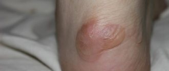
The key symptom of the disease is acute pain in the heel area that occurs when walking and putting weight on the leg. In the initial stages of development, the disease manifests itself only during intense physical activity, and at rest, discomfort and pain disappear completely. As the osteophytes grow, the spur increases in size, causing significant discomfort even at the slightest pressure on the foot.
Children with this disease complain of a feeling of a foreign object in the sole of the foot and ask their parents to pull out the non-existent splinter. A characteristic symptom of heel spurs in children is the cyclical occurrence of pain, which can be explained quite simply: at rest, the shortened fibers of the fascia begin to gradually recover, and by standing up sharply, putting a load on the foot, the child causes new microdamage to the tissue, which is accompanied by acute pain. During active physical activity, the pain dulls, the sensations weaken, but some discomfort remains. After the child calms down and rests, the injured fibers will again make themselves felt with a new round of pain.
Another telltale sign of a heel spur is a change in gait. Children, feeling pain, unconsciously begin to avoid putting pressure on the damaged area, which is why the gait becomes uncertain, the axial tilt changes, and the main load from the heels moves to the nose. As a result, the child literally walks “on his toes,” due to which coordination suffers and the risk of developing transverse flatfoot increases. In particularly advanced cases, children with heel spurs cannot move without additional support.
TYPES OF KERATOSIS
Most often, the phenomena of hyperkeratosis are localized on the legs (heels, feet, knees) and arms (palms, elbows), less often - on the scalp, face in the area of the nose, cheeks, forehead. Basically, this pathology is formed in areas of irritation by any external mechanical or environmental factors. However, hormonal imbalances, heredity and improper skin care can also provoke hyperkeratosis.
The most common types of manifestations of hyperkeratosis are:
- Actinic (solar) keratosis on the face and body (keratomas). It develops due to excessive exposure of the skin to ultraviolet rays and photodamage. Appears as flat, rough yellow or brown spots;
- Palmar and plantar keratoderma - calluses, corns on the soles of the feet and palms, heel cracks. Formed in areas exposed to prolonged friction or pressure, during infectious and fungal diseases;
- Follicular or pilaris keratosis (“goose bumps”). Dermatosis occurs as a result of both accelerated keratinization and disruption of the physiological desquamation of horny scales. Against this background, the normal secretion of sebum is disrupted and local inflammation of the hair follicles on the body occurs. It looks like a rash of small multiple nodules of bright pink and gray color on the face, shoulders, legs, buttocks;
- Seborrheic keratosis is the most common form of hyperkeratosis. It is a hyperpigmented “plaque” with clear boundaries from light to dark brown, covered with keratinized skin. It can be either single or multiple.
- Actinic keratosis - develops in old age in the form of beige-brown spots and is localized on the face, shoulders, back, and back of the hands.
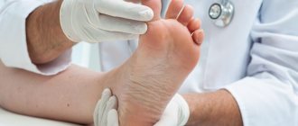
Diagnostics
The diagnosis of heel spur is based on:
- complaints from the patient or his representatives;
- visual inspection data;
- characteristics of clinical manifestations;
- objective data from a radiographic examination of the foot;
- information obtained during ultrasound examination of the soft tissues of the sole.
In some cases, laboratory tests may also be needed to identify signs of inflammation, assess uric acid levels, as well as magnetic resonance or computed tomography. Sometimes, in case of disturbances in the blood supply to the lower legs, duplex scanning of the vessels is performed.
Treatment of heel spurs in children
Plantar fasciitis is treated by orthopedists, traumatologists and surgeons. For children, treatment for heel spurs is selected individually, taking into account the personal parameters of the little patient, general health characteristics, and the presence of acute or chronic pathologies. The main criteria for choosing treatment tactics remain the cause of the spur, the degree of damage to the fascia and the nature of the clinical manifestations.
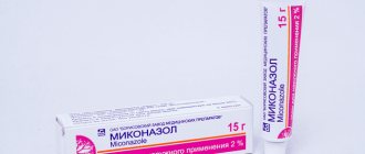
The treatment package may include:
- medications, the action of which is aimed at relieving pain and suppressing inflammatory processes;
- medicines that improve blood flow;
- drugs necessary to control concomitant pathologies;
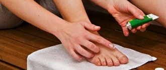
- physiotherapeutic techniques that help improve blood supply to damaged tissues and accelerate regeneration, enhancing the effect of medications;
- massage techniques that help stimulate metabolism and blood flow in the foot.
Additionally, specialists can prescribe a course of shock wave therapy, the purpose of which is to destroy salt deposits and reduce the rate of formation of new osteophytes. In some cases, laser techniques are used to improve the supply of nutrients to tissues.
Additionally, to strengthen the muscular-ligamentous apparatus and connective fibers, a course of gymnastic exercises and therapeutic massage is recommended. It is also advisable to maintain a healthy diet, avoid foods rich in artificial flavors, and monitor the child’s weight.
In situations where medications in combination with other therapeutic methods do not bring the desired result, surgical treatment is indicated. It involves manually removing the bony protrusion by excision of the damaged fascia. In childhood, this method of solving the problem is extremely rarely used, since in most cases, after surgery, the anatomy of the patient’s foot changes significantly.
Treatment
The nature of treatment depends on the stage of the disease being diagnosed. If the disease is in the first or second stage, then the question arises of surgery, which can help in the fight against cancer. Radiation therapy and chemotherapy will be auxiliary at this stage.
If the disease has progressed to the third or fourth stage, then the operation will no longer be effective. The main thing here will be to prevent the spread of metastases to other organs and slow down the growth of the malignant tumor. Therefore, the main treatment options will be radiation therapy and chemotherapy. The patient is also given strong painkillers to reduce pain.
Prevention
With adequate and timely treatment, the disease can be managed without any consequences for the body.
If therapy is not carried out or is chosen incorrectly, if it is not carried out in full, due to the shift in load, the development of arthritis, arthrosis of the lower extremities, flat feet, scoliosis and other orthopedic diseases is likely. As preventive measures that can significantly reduce the risk of plantar fasciitis in childhood, experts advise:
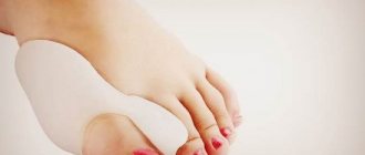
- choose comfortable shoes that correspond to the size of the foot with good leg support, high, dense instep support and orthopedic soles;
- teach children to alternate physical activity with rest, so that not playing sports and other active disciplines does not lead to foot injuries;
- control the healthy nutrition of schoolchildren and adolescents, preventing obesity;
- provide children with adequate physical education;
- prevent injuries;
- promptly treat spinal diseases, endocrine disorders and other diseases;
- immediately contact orthopedists for help if you suspect limb deformities and other orthopedic pathologies.
Experts also strongly do not recommend using any traditional medicine methods to treat heel spurs in childhood. The use of untested products can cause burns and mechanical injuries to the skin, and unpredictable allergic reactions.
Make an appointment at the SM-Doctor clinic to get more reliable information about plantar fasciitis, undergo an examination and choose a rational treatment regimen. Calls and online applications on the website are accepted 24 hours a day.