April 5, 2020
Neoplasms (neoplasia) is the medical name for tumors, i.e., excessive growth of any tissue in the body. Tumors are the result of uncontrolled proliferation of cells that have not yet reached maturity and therefore have lost their ability to fully perform their functions.
Tumors can occur in internal organs and on the surface of the skin. Many people, not knowing what types of skin tumors there are, when any skin tumor appears, mistakenly believe that it is cancer. In fact, this is not always the case.
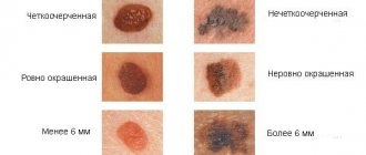
According to the main classification, skin tumors are divided into benign and malignant. There are also precancerous formations - borderline between the two main types. Each type has its own subtypes and characteristics, and correct diagnosis is needed to make an accurate diagnosis.
Why do benign formations appear on the skin?
Cosmetologists and dermatologists do not know the exact mechanism of their formation. Most often the cause is:
- injuries;
- viruses;
- systemic diseases of the body, for example xanthomas, occur due to an excess of fat in the blood;
- long-term skin diseases;
- exposure to aggressive substances;
- excessive exposure to ultraviolet radiation;
- x-rays;
- heredity (for example, seborrheic dermatosis).
Most skin lesions are benign
Benign and malignant neoplasms on the skin: what are the differences?
Benign pathologies do not pose a threat to human life. If they reach large sizes, they can interfere with the adequate functioning of various body systems. In contrast, malignant ones grow quickly and aggressively, penetrate into surrounding tissues, and form metastases over time. Some damage vital organs and cause death.
Sometimes benign skin tumors change due to external or hereditary causes. They acquire the ability to degenerate into malignant pathologies. Such conditions are called borderline or precancerous. They pose a great danger to health and life, although they do not always have pronounced symptoms.
Diagnosis of cheek cancer
If a tumor of the mucous membrane of the cheek is suspected, oncologists at the Yusupov Hospital conduct an examination using a mirror, palpation of the tumor and lymph nodes. If a long-term non-healing ulcer is detected, a biopsy is performed. If a negative result of histological examination of the material obtained during the biopsy is obtained, the suspicion of the malignant nature of the tumor remains, the biopsy is performed again.
Oncologists clarify the stage of the tumor process, assess the extent of spread of the malignant tumor from the buccal mucosa to adjacent tissues, and find out whether there are metastases to regional lymph nodes and distant organs. If the presence of metastases is suspected, computer and magnetic resonance imaging, ultrasound, and scintigraphy are used. Distant metastases are found in 20% of patients at the time of diagnosis.
Computed tomography and magnetic resonance imaging make it possible to assess the condition of the deeper anatomical structures of the oropharynx and surrounding tissues. If there is a suspicion of metastases to the lymph nodes or tumor infiltration of the floor of the mouth, a cytological examination of the aspirate obtained under ultrasound guidance is performed. To exclude distant metastases, a chest x-ray in two projections and an ultrasound examination of the abdominal organs are done.
Considering that the prognosis for cheek cancer is serious, a tomography of the neck, chest and upper abdominal cavity is performed. Using bone scintigraphy, I exclude bone metastases. Positron emission tomography allows one to identify the source of metastases in cases of undetected primary tumors.
What is the structure of benign neoplasms
The growths consist of cells that have partially retained their original functions and are capable of growing slowly. They are similar in structure to the tissues from which they originated. They can put pressure on nearby tissues, but do not penetrate them, since they have a capsule in their structure. They respond well to hardware and surgical treatment and, as a rule, do not cause relapses.
There are always congenital formations on the skin - moles or warts, as well as acquired ones. The latter are formed on the surface or in the subcutaneous layer as a result of metabolic disorders, decreased immunity, or under the influence of a virus.
Types of acquired red moles on the body
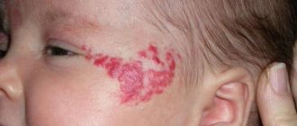
- Simple (capillary). Proliferation of newly formed capillaries, small venous and arterial vessels. Looks like a red spot.
- Cavernous. A spongy cavity with blood - a red or bluish nodule. Often forms under the skin.
- Branched (racellose). A plexus of tortuous dilated capillary trunks. They pulsate, noise and trembling are detected. It is rare and occurs on the extremities or face. If injured, life-threatening bleeding may occur.
How to identify hemangioma? Press on top of it and it should fade or disappear.
Types of benign formations
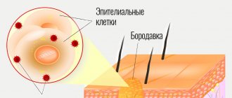
- Warts and papillomas
Papilloma is a small tumor on a stalk or broad base, which has clear boundaries. Flint is visible on an uneven, grainy surface. The growth is painted in any color - from white to dark brown. It is found both individually and in large quantities.
Both papillomas and warts appear as a result of the papilloma virus entering the body, live in different parts of the body, and reach a diameter of several centimeters. They are activated on the skin as a result of nervous tension, stress, decreased immunity or vegetative disorders.
Papillomas and warts are not dangerous if they are not injured
- Birthmarks
They are not prone to degeneration, but such cases do occur, so it is necessary to pay attention to changes in contour, color, and size, especially if the area is constantly injured.
- Lipoma
A round, soft-to-the-touch tumor of adipose tissue that does not disrupt the functions of the body, but only creates a cosmetic defect. Wen appears in various areas where there is a fat layer. Reaching large sizes, they grow into tissue and approach bone surfaces, often spreading to muscles and blood vessels.
- Atheroma
Atheroma is a residual cyst caused by blockage of the ducts of the sebaceous glands. It contains products produced by the sebaceous glands in the form of an odorless curd mass. The pathology has clear contours and a dense consistency. Superficial atheromas occur mainly on the back, head, neck, face, and limbs - due to poor hygiene, metabolic disorders and unprofessional depilation.
When subjected to mechanical action, the atheroma becomes inflamed, swells and becomes red. When an infection gets inside, pus is formed and a greasy consistency is erupted. There is a risk of degeneration into liposarcoma, so it is recommended to remove this pathology.
A seemingly harmless atheroma can degenerate into liposarcoma
- Nevus
More often than others, melanoma degenerates into a malignant formation, especially with prolonged exposure to negative external factors or hereditary predisposition.
- Lymphangioma
The pathology is mainly congenital, developing from lymph nodes in the skin, but can also spread to fiber or muscles. It is most often localized on the head, face and upper body. The blue tumor-like growth rises above the skin, has a dense consistency and clear boundaries, dimensions are 1-5 mm. Since it puts pressure on vital organs (lungs, larynx, trachea), surgical removal is resorted to.
- Hemangioma
It is formed on the basis of blood vessel cells in the subcutaneous layer, rises above the surface, and is characterized by rapid spontaneous growth. When pressed, the growth decreases. It is mainly localized in the head and neck area, mainly in young children. Cavernous hemangioma is colored blue and resembles a node, located deep in the tissues. Capillary - in the epithelium, has a red or blue color. If it becomes inflamed, it can provoke open bleeding, and if it is close to vital organs, it disrupts their functions.
Hemangiomas are diagnosed more often in children under three years of age.
- Fibroma (dermafibroma)
A tumor of dense consistency, light pink in color, is formed from connective tissue. Mild varieties occur more often on the neck and chest, in the groin folds and armpits in women. Hard - in different areas near the upper layers of the skin. Dermatologists recommend removing fibromas, since under favorable conditions they can transform into fibrosarcoma.
- Keratoma
This skin tumor is formed from keratinocytes that make up the stratum corneum. A growth in the form of a spot or node is formed from dead cells and is localized on the back, head, face, and limbs. Such a benign neoplasm requires urgent treatment.
- Neurofibroma
Formed from nerve sheath cells, it looks like a hard tubercle up to three centimeters in diameter. Causes discomfort and pain because it puts pressure on nerve endings. Most often located on the face, back, abdomen, arms and legs. If there are many formations, they talk about neurofibromatosis - a disease that is mainly inherited.
Why does my cheek swell?
Periostitis of the jaw
It is the most common dental cause of the symptom. Develops against the background of dental diseases: periodontitis, pulpitis, alveolitis, periodontitis, suppurating jaw cyst. It is provoked by open fractures of the jaws, infected facial wounds, operations, and tooth extraction. In some cases, it becomes a consequence of hematogenous or lymphogenous spread of infection from distant foci. Depending on the form, it is accompanied by the following manifestations:
- Acute serous periostitis.
Moderate swelling of the soft tissues of the cheek, redness of the mucous membrane, and enlargement of regional lymph nodes are detected. The general condition suffers slightly, sometimes there is an increase in body temperature to subfebrile levels. - Acute purulent periostitis (flux
)
.
The contours of the face are sharply changed, the predominant localization of edema is determined by the location of the inflammatory focus in the periosteum. Patients complain of sharp pain radiating to the temple, eye, ear, and neck. General hyperthermia, chills, weakness, headache, and regional lymphadenitis are observed. When examining the oral cavity, an area of swelling with a fluctuation in the center is detected - a subperiosteal abscess. - Chronic periostitis.
Slight swelling of the cheek, thickening of the jaw, and enlargement of the submandibular lymph nodes are typical. The patient complains of periodic moderate pain. The mucous membrane of the affected area is swollen, hyperemic, with a bluish tint.
Other dental diseases
Other possible dental causes of cheek swelling include the following:
- Perimaxillary abscess.
It occurs as a result of infection of the soft tissues of the perimaxillary area due to boils, tonsillitis, wounds, abrasions, and some dental diseases. Swelling is preceded by toothache, which gets worse when biting. Then dense swelling appears, the temperature rises, and appetite disappears. After spontaneous opening of the abscess, the condition improves, but subsequently the pathology may recur. - Vincent's stomatitis.
Develops when immunity decreases due to diseases, injuries, and stressful situations. The leading symptom is the formation of multiple ulcers on the mucous membrane. Swelling of the cheek is detected in severe cases of the pathology. - Noma.
An area of ulcerative-necrotic lesions of the lip or oral mucosa appears. Necrosis covers the gums, tongue, cheek tissue, and facial bones. The tissues around the area of necrosis are swollen; in severe cases, swelling from the cheek spreads to the chin and infraorbital area. - Salivary gland adenoma.
The parotid gland is most often affected. Local swelling with clear boundaries forms on the outer surface of the cheek, in the parotid zone, and spreads to the angle of the jaw, the area under the earlobe. Characterized by slow growth, painlessness at the initial stage, progression of discomfort and dry mouth as the formation increases. - Tumors of the salivary glands.
Along with adenomas, benign connective tissue neoplasia, intermediate and malignant neoplasms can form in the area of the salivary glands. The area of swelling is located in the same place as with adenomas. The swelling slowly increases in benign tumors and spreads quickly in malignant ones. - Purulent parotitis.
Swelling occurs in the parotid area, which spreads to the adjacent part of the cheek. There is severe pain, difficulty when trying to open the mouth, severe intoxication, and severe hyperthermia.
Cheek swelling
Traumatic injuries
All facial injuries are accompanied by rapidly increasing soft tissue swelling, spreading to adjacent anatomical structures. Swelling of the cheek of traumatic origin can occur with the following injuries:
- Injury.
Swelling without clear boundaries. Along with edema, pain, hyperemia, and sometimes hemorrhage are detected. The pain intensifies when opening the mouth and active facial movements. Speech and the ability to eat were preserved. - Hematoma.
Formed against the background of a bruise. A compaction appears in the area of diffuse swelling, which, as a rule, resolves on its own within 1-2 weeks. - Fracture of the upper jaw.
The most pronounced swelling is observed in Le Fort type 1 fractures, combined with neurological symptoms, hemorrhages in the conjunctiva and periorbital area. Type 2 is manifested by edema, hemorrhages in the periorbital zone, and changes in facial parameters. In type 1 fractures, swelling is more noticeable in the area of the upper lip and medial cheek. - Fracture of the lower jaw.
Injuries to the ramus, lateral and angular fractures of the bone body are characterized by swelling of the lower or outer edge of the cheek. Facial asymmetry, hematomas, bruises, articulation disorders, and stepped dentition are observed. - Fracture of the zygomatic bone.
Swelling appears in the cheekbone area, quickly spreads down the cheek, up the infraorbital region. Bruises and hemorrhages form in the conjunctiva. Along with pain, victims are sometimes bothered by nosebleeds and double vision.
Allergic reactions
Swelling of both cheeks is observed with angioedema, combined with swelling of the eyelids and lips, and breathing problems. The condition develops acutely, within a few minutes, less often – hours. It is provoked by contact with an allergen, insect bites. Along with the listed symptoms, angioedema in children may be accompanied by abdominal syndrome, and sometimes by neurological symptoms.
The cause of swelling of the cheek from the oral cavity may be an allergy to prosthetic materials. The pathological condition occurs several months or years after the installation of prostheses and is characterized by a burning sensation in the area where the prosthesis is fixed, tongue, cheeks, soft palate, changes in taste sensitivity, thirst, and dry mouth. When using metal products, a metallic taste may appear.
Ophthalmic diseases
In patients with acute dacryocystitis, swelling of the lacrimal sac area is complemented by swelling of the cheek, eyelid, and dorsum of the nose. In the chronic form of the pathology, swelling is noticeable along the upper inner edge of the cheek and covers the inner edge of the lower eyelid. Cellulitis of the lacrimal sac is characterized by sharp pain and swelling along the inner edge of the eyelid, combined with fever, weakness, weakness, headache, swelling of the cheek and paranasal area.
Neurological pathologies
Local symptoms of cavernous sinus thrombosis are exophthalmos, blurred vision, swelling and pain in the eyeball, swelling of the temple, part of the forehead, cheek, upper lip, and mastoid process. The clinical picture also includes headache, nausea, vomiting, and in case of infectious genesis of the pathology - hyperthermia, intoxication syndrome.
Swelling of the cheeks can be detected with one of the types of angioneurosis - rosacea. There is constant redness of the cheeks, nose, forehead, chin, and the formation of spider veins. The cause of swelling is persistent dilation of blood vessels, which, with a long course of pathology, leads to skin changes.
Skin lesions
Minor swelling of the cheek may result from simple contact dermatitis. The severity of the symptom increases against the background of prolonged contact with the irritant and secondary infection. With allergic dermatitis, the swelling is more noticeable and is combined with itching of the skin. Atopic dermatitis is characterized by mild swelling combined with the formation of vesicles.
Due to the abundant blood supply to the face and the structural features of the soft tissues, a boil on the cheek is accompanied by significant swelling. In the center of the swelling area there is a limited round or cone-shaped formation with a black rod in the center. After the boil has matured, yellowish pus appears around the shaft. Increasing twitching pains are noted.
In young children, swelling of the cheeks is often provoked by superficial pyoderma and occurs against the background of pustular rashes. A severe form of facial skin lesions in adults and children is erysipelas. The disease manifests itself with itching, bloating, and a burning sensation. Subsequently, the cheek swells, and a focus of clearly defined hyperemia with uneven edges, reminiscent of a geographical map, forms on it. Fever and intoxication syndrome are observed.
ENT diseases
Slight swelling of the cheeks is possible with the development of acute sinusitis or exacerbation of chronic inflammation of the maxillary sinuses. Other symptoms include pain, impaired nasal breathing, nasal discharge, weakness, fever, and signs of intoxication. Patients with large odontogenic cysts of the paranasal sinuses are bothered by a feeling of tension and heaviness. Objectively, diffuse swelling of the cheek on the affected side and protrusion of the bottom of the nasal cavity are detected.
Other reasons
Swelling of one or two cheeks is observed with the following pathologies:
- Myxedema.
The swelling is bilateral, uniform, covering the forehead and chin, making the face look puffy. In severe cases, swelling spreads to the entire body. Symptoms of hypothyroidism are observed. - Parotitis.
Due to inflammation of the salivary glands, the perimaxillary area, the outer part of the cheeks, swells. The deformation is bilateral, often uneven. The disease manifests itself acutely, accompanied by fever, chills, and signs of general intoxication. - Melkersson-Rosenthal syndrome.
The leading manifestation is periodic swelling of the lips. Possible swelling of the tongue, cheeks, eyelids. Neuritis of the facial nerve is often detected. - Phlebolith.
Stones in the veins of the cheek are often asymptomatic, but can manifest as pain and swelling, and sometimes inflammation of the affected area.
Removal of benign skin tumors
Dermatologists are convinced: it is necessary to get rid of benign formations, with the exception of small scatterings of moles or other minor defects throughout the body. This is especially true for the face, because the growth attracts attention, spoils the overall impression and gives a lot of unpleasant emotions to its owner.
There are several methods for removing benign skin tumors:
- Electrocoagulation
Under local anesthesia, the growth is cut off with a special surgical coagulator, which creates a high-frequency current. Simultaneously with removal, the tissues are soldered together, which avoids bleeding and infection. The crust at the treatment site disappears after 7-10 days, sometimes leaving a slightly noticeable scar. The method is effective if the defect is small.
- Cryodestruction
Liquid nitrogen can only be applied to formations in the upper layers of the epidermis. If it is flat, apply applications with liquid nitrogen. In case of deeper occurrence, a cryodestructor is used. After the procedure, the body begins a reaction of rejection of the treated defective tissue. The resulting crust disappears and heals within a month and a half.
- Laser removal
A powerful beam of light destroys neoplasm cells and evaporates them from the skin. The laser does not affect healthy neighboring tissues. The manipulation is carried out under local anesthesia, it is low-traumatic and bloodless. Laser treatment parameters are selected individually. Damaged tissues are evaporated layer by layer until the beam reaches healthy skin. The formed crust disappears on its own after 1-2 weeks.
- Radio wave method
High frequency radio waves cut and coagulate tissue. The crust disappears after a week. The method is contraindicated in patients with a pacemaker, herpes, or elevated body temperature.
- Surgical removal of benign skin tumors
This method is used when the growths are too large and other technologies cannot cope. The formation is excised with a scalpel and removed, capturing a small area of healthy skin. The scar after surgery takes several weeks to heal, but the wound requires long-term careful care. The description of this traumatic technology is not encouraging, so it is better to choose another method to eliminate defects on the face.
On the face, neoplasms are removed using laser or cryodestruction
Treatment for cheek cancer
Malignant tumors are successfully cured using radical radiation therapy while preserving the function of the oral cavity. Radiologists implant radiation sources because it is possible to irradiate a small volume of tissue at a high dose. Radioactive isotopes of cesium, gold, radium, iridium, tantalum, which have the same efficiency, are used as sources.
For small tumors, the size of which does not exceed 1 cm, they are limited to implantation of a radiation source, without resorting to additional external influence. In cases of slightly larger tumors that are not suitable in size for implantation, external irradiation is used along with source implantation.
Traditionally, large cheek tumors are treated with external beam radiation. Recently, oncologists have been using a combination of radiation and chemotherapy. Mobile lymph nodes are radically excised. During prophylactic removal of lymph nodes without signs of damage, in a significant number of cases, foci of micrometastasis are found in them. For this reason, radiologists prefer to perform prophylactic irradiation of the neck in patients without evidence of lymph node involvement by the external beam, sometimes in combination with surgical treatment.
After surgery for cheek cancer, a cosmetic defect is formed. The results of surgical intervention are improved using the technique of microvascular free skin grafting. If a patient develops dry mouth after radiation therapy, he is prescribed oral pilocarpine. The drug increases salivation, which is usually accompanied by minor side effects - sweating and increased urination. For primary or secondary treatment of tumors of the buccal mucosa, patients are prescribed chemotherapy drugs.
Early diagnosis of a cheek tumor allows for effective treatment. If the tumor process is at a late stage, the prognosis worsens. If you experience unpleasant sensations in the oral cavity or identify ulcers of the buccal mucosa, undergo an examination at the clinic by calling the Yusupov Hospital.
Prevention of skin tumors
Unfortunately, medicine has not yet learned to prevent the appearance of various formations on the skin. But dermatologists give their patients the following preventive recommendations:
- do not delay contacting a doctor if a tumor appears on the skin;
- remove formations only after a specialist and diagnostics confirm their benign nature;
- avoid excessive exposure to the open sun;
- use sunscreen, especially if you are prone to moles and hyperpigmentation;
- do not come into contact with chemically active and carcinogenic substances;
- do not eat foods that contribute to the development of cancer (smoked meats, sausages, animal fats, meat products with food stabilizers).
Do you have a lot of moles? Forget about tanning in the open sun
Causes of cheek cancer
Cancer of the buccal mucosa develops under the influence of the following provoking factors:
- Use of tobacco in any form (cigarettes, cigars, pipes, chewing tobacco);
- Alcohol abuse (the risk of developing cancer increases when the use of alcohol and tobacco is combined);
- Infection with carcinogenic forms of human papillomavirus.
One risk factor is exposure to sunlight. Both family history and genetic predisposition, as well as exposure to mutagenic environmental factors, play a role in the development of cheek cancer. The formation of a malignant tumor occurs in several stages. The most important is the disruption in the functioning of oncogenes and genes that inhibit tumor growth. The development of malignant neoplasms of the cheek is associated with inactivation of the p16 gene, mutations in the p53 gene, and the introduction of the human papillomavirus.
Do benign formations hide the danger?
Benign neoplasms are unpredictable structures that can manifest themselves at any time or not at all. The process of their transformation into malignant ones has not been fully studied. There is no clear answer to the question of what exactly activates this process. It is believed that mechanical trauma, excess ultraviolet radiation, metabolic disorders and other factors contribute to degeneration. One way or another, if you have a benign skin lesion, you should not experiment and rely on chance. Moreover, today removal does not cause difficulties.