The facial skull has several cavities called paranasal sinuses. They are located in the ethmoid, frontal, sphenoid bones and the upper jaw. All of them are involved in cleaning, warming and humidifying the inhaled air, and regulate pressure in the respiratory tract. Inflammation of the mucous membrane of the paranasal sinuses is called sinusitis. Factors contributing to the development of sinusitis are:
- hypothermia of the body;
- untimely treatment of runny nose and ARVI;
- congenital and acquired defects in the structure of the bones of the facial skull;
- reduced immunity.
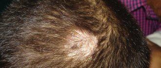
All these factors can contribute to the development of frontal sinusitis - inflammation of one or both sinuses of the frontal bone. It is characterized by intense headache and the risk of developing serious complications.
This disease affects both adults and children.
Features of frontal sinusitis in children
Newborns and babies in the first months of life do not suffer from frontal sinuses at all, because the frontal sinuses are absent at birth and develop throughout life as the bones of the skull grow. The disease begins to occur in children over 6 years of age.
This disease should be suspected if a child complains of a severe headache, mainly in the forehead. Moodiness, tearfulness and irritability appear. The child loses interest in games and cannot concentrate on studies.
Since the frontal sinuses are involved in the formation of voice timbre, their swelling often leads to nasal sound.
Classic symptoms of frontal sinusitis:
- pain and heaviness in the forehead, on one or both sides, often throbbing;
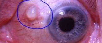
- pain in the inner corner of the eye, worsens in the morning and with pressure;
- photophobia, excessive tearing as a reaction to light;
- decreased sensitivity to smells up to complete loss of smell;
- redness and thickening of the upper eyelid, swelling of the nose and brow ridges;
- mucous discharge at the beginning of the disease, later acquires a purulent character and an unpleasant odor;
- nasal congestion to the point of impossibility of nasal breathing, especially at night;
- increase in body temperature to 37.5-38.5 ° C, sometimes higher;
- phenomena of general intoxication - weakness, malaise, sweating, loss of appetite, poor sleep.
Based on these signs, you may suspect acute frontal sinusitis yourself. However, meningitis, encephalitis, neurological pathology and other sinusitis may have similar symptoms. In any case, if a child presents such complaints, he should be shown to a pediatrician or ENT doctor as soon as possible.
“White” and “red” fever in children: what is it and why is it dangerous?
“White” fever is a very dangerous condition in which you urgently need to call a doctor. Anna Khoperskova, an infectious disease specialist at the Russian Railways Medicine children's clinic, spoke about the difference between “white” hyperthermia in children and the usual “red” one.
There are two conditions for fever: “white” and “red” fever.
“White” hyperthermia in children is the most dangerous. Therefore, if a child has pale, marbled skin, with a bluish tint to the nail beds and lips, the limbs are cold to the touch, the “white spot” symptom is positive when pressing on the skin, behavior is disrupted (the baby becomes indifferent, lethargic), delirium and convulsions are possible, immediately call a doctor, or better yet, an ambulance or emergency care,” the doctor recommends.
Under no circumstances should you give your child medications on your own, especially those containing acetylsalicylic acid and metamizole.
As for “red” hyperthermia, it is characterized by reddish skin - hot and moist to the touch, warm extremities, rapid pulse and breathing against a background of rising temperature. With “red” fever, the child’s behavior does not change.
“An increase in temperature or fever is a protective mechanism of the body, provided by nature as a reaction to exposure to pathogenic stimuli. During an illness, the essence of its occurrence is to increase natural protection by restructuring thermoregulation processes with the formation of higher body temperatures than in a healthy state,” explained Anna Khoperskova.
The causes of fever, in addition to a reaction to infection, can also be: overheating, metabolic or endocrine disorders, conditions after transfusion, allergic reactions, and the use of a number of drugs.
An infectious disease specialist at the Russian Railways Medicine children's clinic warned: playing games on the phone and watching TV should be avoided during treatment for “red” hyperthermia. Audio fairy tales and reading books can serve as a substitute. It is recommended to reduce the temperature using physical methods:
- undress the baby as much as possible;
- apply a cool, wet bandage to the forehead;
- regularly ventilate the room (without creating drafts);
- blow with a fan;
- wipe the body with water at room temperature;
- apply cold to the projection of large vessels (in the axillary and groin areas, on the side of the neck) and the liver area;
- to drink a lot of water.
It is recommended to take antipyretics at temperatures above 38.5 degrees. You don’t have to give medications to a child if he drinks a lot of fluids, is active, eats food as usual, has no toxicosis, and feels normal.
“It is necessary to give the child’s body the opportunity to develop that level of protective antibodies, which in the future, when encountering this infection, will help the child not get sick again or suffer from a mild form of the disease,” the doctor concluded.
Antipyretics must be given to those children who are at risk for developing complications due to high fever.
Types and classification of frontites
The symptoms described above are characteristic of the acute form - when the disease develops in a few days and disappears without a trace in less than 1 month.
With a weakened immune system and insufficient treatment, the inflammatory process may become chronic. With a continuous course, the disease takes on a less pronounced form, its symptoms weaken, but do not disappear:
- headache occurs at night;
- runny nose and nasal congestion do not go away, vasoconstrictor drugs lose their effectiveness;
- cough with sputum is often associated;
- In the morning, redness and swelling of the eyes and watery eyes appear.
All this is accompanied by symptoms of asthenia - the child becomes lethargic, lethargic, and his academic performance drops.
The recurrent form is characterized by alternating stages of exacerbation and remission. In this case, the signs of frontal sinusitis may completely disappear, but with any hypothermia and colds, as well as in the off-season, the picture of an acute illness returns.
A special variant of the disease is allergic frontal sinusitis. This form develops as a complication of chronic allergic rhinitis. When the mucous membrane of the nasal cavity is constantly inflamed and swollen, the ventilation of the paranasal sinuses and the outflow of secretions from them are disrupted. This promotes stagnation and increases the likelihood of infection. The main treatment in this situation is aimed at combating allergens.
Skin pigmentation in children: freckles, urticaria
Disorders in the melanocyte system are divided into hypermelanosis (an increase in melanin in the epidermis or dermis) and hypomelanosis (a decrease or absence of melanin in the dermis, leukoderma), which in turn can be generalized or localized. Some of these disorders are caused by hormonal changes (Addison's disease), others are local developmental defects (white spots in tuberous sclerosis) or the result of skin inflammation (post-inflammatory hypo- or hyperpigmentation).
Consultation with pediatric dermatologist Moscow - Markushka clinic.
Hypermelanosis in a child, children
Hypermelanosis is divided into epidermal (brown color) and dermal (blue, bluish-gray, gray color).
Brown hypermelanosis (melanoderma) in children is associated with an increase in melanin content in the epidermis as a result of increased activity of melanocytes, an increase in the number of secretory melanocytes, the number of melanosomes or their size. Bluish-gray hypermelanoses (ceruloderma, blue skin) are similar to false tattoo melanin and are explained by the presence of melanin in the dermis, in ectopic dermal melanocytes or dermal macrophages, which, as a result of the Tyndol effect, give the skin a characteristic gray, grayish-blue or blue color.
Hyperpigmentation of the skin in a child or children can be generalized uniform, generalized spotty or limited in certain areas of the skin. Diffuse congenital melanosis is characterized by hyperpigmentation already at birth (other organs are unchanged), later hyperkeratosis of the hands is added. Constitutional hyperpigmentation is observed in dark-blond children, mainly of Mediterranean peoples.
Diffuse brown hypermelanosis in a child is characteristic of adrenal insufficiency (Addison's disease), in which hyperpigmentation of the skin is expressed in places of pressure on it (vertebrae, interphalangeal, elbow and knee joints), in the folds of the body, on the palmar surfaces, and in the mucous membrane of the gums. Adrenalectomy, pancreatic and lung tumors lead to hypermelanosis. In these conditions, hyperpigmentation is caused by overproduction of melanocyte-stimulating hormone and ACTH, which have the same amino acid sequence.
Generalized hypermelanosis in a child is a typical sign of hemochromatosis, late cutaneous hematoporphyria, and variegated porphyria. In hemochromatosis, the hyperpigmentation is grayish-brown or brown and indistinguishable from that of Addison's disease, and the diagnosis can be made by skin biopsy, which reveals hemosiderin deposits in the sweat glands and melanin deposits. In case of late cutaneous porphyria, the diagnosis is established by vesicles, blisters, atrophic spots, sclerodermoid changes and millet-like rash on the skin of exposed parts of the body, and is confirmed by the presence of an increased content of uroporphyrin in the urine (the ratio of uroporphyrin and coproporphyrin is normally more than 3: 1) or red fluorescence of acidified urine . Skin changes in variegated porphyria are identical to those in porphyria cutanea tarda; they are differentiated by their different response to treatment.
Hyperpigmentation of healthy skin in children is observed in chronic renal failure and primary biliary cirrhosis.
With chronic nutritional deficiency (kwashiorkor, nephrotic syndrome, malabsorption syndrome, etc.), hyperpigmented spots appear on the skin of the body. With pellagra, the pigmentation zone is limited to areas of the skin exposed to light or trauma; Vitamin B12 deficiency is accompanied by premature graying of hair and hypermelanosis, especially pronounced around the small joints of the hands. Hypermelanosis can be a consequence of treatment with myelosan, cyclophosphamide, methylurea, aminazine, etc.
Hyperpigmented skin elements. Freckles, hives
Hyperpigmented skin elements . Freckles (ephelides) in a child or children are small pigment spots located at the level of the skin on the face on both sides of the nose, on the shoulders. Larger café-au-lait spots may be a manifestation of Recklinghausen's neurofibromatosis, in which neurofibromatosis of the skin and peripheral nervous system, hypertension, and precocious puberty are detected. Leopard syndrome is an autosomal dominant condition with a generalized distribution of dark brown spots in combination with neurosecretory deafness, growth retardation, heart defects, and genital abnormalities. In Peutz-Jeghers syndrome (inherited in an autosomal dominant manner), melanotic spots on the lips and mucous membranes are combined with polyposis of the small intestine. This syndrome must be differentiated from other syndromes associated with multiple pigment spots and common freckles, Gardner and Cronkhite-Canada syndromes (gastrointestinal polyposis, alopecia, onychodystrophy and skin pigmentation).
Blue nevus is a group of pigment cells accumulated in the dermis; the epidermis visible above them looks like bluish spots; when localized in the sacral area, they are called Mongolian spots and disappear after the age of 3 years.
In children, there are also benign and malignant variants of melanoma (tumor-like, growing pigmented nevus).
Mastocytosis, or urticaria pigmentosa in a child, children. A disease characterized by paroxysmal rashes of spots, papules, pink-red blisters of round or oval shape, localized on the torso, limbs, scalp, face and rarely on the palms and soles.
It usually begins before the age of 2 years. The child is restless due to severe itching, the mucous membranes are not affected. The Unna-Darye symptom is considered pathognomonic for mastocytosis, when, when rubbing a spot or papule with a spatula, or after touching it with a warm object, redness and swelling of the element soon appears - it takes on a blister-like appearance.
Rashes in the progressive stage may periodically disappear and reappear, which ultimately leads to a darker color of the rash elements, even brown spots, and an increase in their number - from single to hundreds. Regression of the disease begins at the age of 6-7 years or by puberty and is characterized by gradual blanching and sometimes even resolution of the elements. Spontaneous involution occurs in approximately 50% of patients at puberty; in 25%, partial resolution occurs in adulthood. Systemic signs of histamine release during mastocytosis (episodic hot flashes, tachycardia, respiratory distress, headache, intestinal colic, diarrhea, hypotension) are almost constant.
Hypomelanosis in a child, children
Hypomelanosis in a child or children is observed with albinism, Hermansky-Goodluck syndrome (tyrosinase-positive albinism with platelet defects and hemorrhagic diathesis), Cross-McCusick-Breen syndrome (tyrosinase-positive albinism with microphthalmia, developmental delay, muscle hypertonicity and athetosis).
Partial albinism in a child or children is characterized by amelanotic plaques in the frontal region, the front of the head (resulting in the appearance of a white strand of hair), on the chest, in the area of the knee and elbow joints. Plaques are caused by a local absence or decrease in the number of melanocytes and do not disappear.
Wardenburg syndrome is inherited in an autosomal dominant manner and is characterized by white strands of hair, pigment defects and hypopigmentation of the skin, heterochromic irises, wide bridge of the nose, dystopia of the corners of the eyes (consultation with a pediatric ophthalmologist, Markushka Clinic), and congenital deafness.
Tuberous sclerosis in a child is inherited in an autosomal dominant manner, characterized by small white leaf-type spots (1-3 cm), localized mainly on the torso, fibromatous nodules on the skin of the forehead, torso, arms and legs, mental retardation, epilepsy (e.g. child - polyclinic "Markushka"), tuberous nodules in the cortex and subependymal areas, retinal phakomatosis, cardiac rhabdomyomas, cysts of the kidneys, lungs and bones.
Ito's hypomelanosis (achromatic pigment incontinence) is a congenital disease characterized by oddly shaped hypopigmented spots forming clearly demarcated patterns, stripes and plaques over the entire surface of the body that remain throughout childhood and disappear in adulthood. Hypopigmentation is not preceded by either the inflammation or vesicular lesions characteristic of Bloch-Sulzberger syndrome.
Vitiligo (acquired pigment defect) in a child or children occurs at any age and is characterized by depigmented spots of various shapes and sizes with clear boundaries, localized on the skin of the face (around the eyes and in the mouth), in the genital area, hands and feet, elbows and knee joints, upper half of the chest. Skin lesions may disappear spontaneously, new spots may appear, or depigmentation may continually progress.
Causes of the disease
Frontal sinusitis does not occur in isolation from other ENT pathologies. Rhinitis, pharyngitis, sinusitis and other inflammatory diseases can trigger the mechanism of its development. Predisposing factors:
- weak immunity - the child suffers from colds often and for a long time;
- the presence of allergic rhinitis or bronchial asthma;
- polyps and other neoplasms of the mucous membrane of the upper respiratory tract;
- defects of the bones of the facial skull - can be congenital or acquired as a result of injuries, surgical interventions, or foreign bodies.
The most common traumatic pathology of the nose is a deviated nasal septum (VNS). Even a minor blow to the head or face as a result of a fall from a height or a fight is enough to deform it. Boys and young men are most susceptible to the disease.
PPI may also have physiological causes. The uneven growth of the brain and facial parts of the skull, as well as different parts of the septum, leads to the fact that the septum bends in one direction or another, some parts of it thicken, and bone growths appear - spines and ridges. Such defects form most intensively in adolescence (12-16 years), but congenital deformities that arise in the prenatal period also occur.
A typical sign of IPN is impaired nasal breathing. A deformed nasal septum can be corrected with surgery under local anesthesia.
Another unobvious cause of frontal sinusitis and other inflammations of the ENT organs is caries and poor oral hygiene. From an early age, it is necessary to teach your child to brush their teeth at least 2 times a day, and at least rinse them after meals. Every 6 months it is necessary to take your child to see a dentist and have caries treated in a timely manner.
Some parents prefer to save money on a dentist for their child, believing that the baby teeth will soon change, which means there is no need to treat them. This is a very dangerous misconception. Firstly, baby teeth affect the health of the molars, and secondly, the focus of chronic inflammation contributes to the development of other diseases throughout the body. But above all, the respiratory organs are at risk.
How to treat frontal sinusitis for children
The main diagnostic method is x-ray of the paranasal sinuses. More expensive methods are ultrasound and MRI.
Treatment of frontal sinusitis in children should be aimed at eliminating the pathogenic factor and alleviating the severity of the condition.
During an acute period of illness, you should refuse to attend kindergarten or school. It is necessary to provide the child with bed rest and plenty of fluids.
If you have a headache, you should take painkillers. The dosage is selected according to the age and body weight of the child.
In order to reduce swelling of the mucous membrane, vasoconstrictor drugs are indicated, as well as drugs containing hypertonic solutions of sea water.
To determine the pathogen, the doctor prescribes diagnostic smears. If the disease is caused by bacteria, an antibiotic must be included in the treatment regimen. To select the drug, a culture of the flora is done to determine sensitivity. The prescribed medication should be taken in its entirety.
In case of purulent frontal sinusitis and the ineffectiveness of medications, the doctor performs a therapeutic puncture. The purulent contents are removed from the inflamed sinus, washed with an antibacterial solution and drainage is installed. This method is used as a last resort, since it is traumatic and leaves behind a cosmetic defect - a scar on the eyebrow. Endoscopic operations exist as a modern and gentle alternative.
The use of Aqualor in the treatment of frontal sinusitis in children
The Aqualor line of drugs helps alleviate the condition of frontal sinusitis, promotes a speedy recovery and is used to prevent colds.
The main component is a solution of sea water. Salts have natural bactericidal properties without causing toxic effects on the body. Due to its ability to attract liquid, hypertonic sea water relieves swelling of inflamed tissues and restores nasal breathing.
Aqualor Soft and Aqualor Norm are suitable for the prevention of colds and exacerbations of chronic infections in children. They are made on the backbone of an isotonic seawater solution. They act as gently as possible and do not dry out the mucous membrane of the upper respiratory tract. By clearing inflamed tissues of mucus, allergens and bacterial waste products, Aqualor increases the body's availability of other medications for topical use.
Aqualor Forte will help during the period of severe runny nose, characteristic of acute sinusitis. It contains sea water of hypertonic concentration, which, due to the osmotic effect, reduces swelling of the mucous membrane.
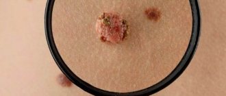
What is infantile hemangioma?
This is a tumor-like formation of a qualitative nature, which is formed by fragments of blood vessels of varying thickness. The nature of the origin is as follows: during the process of intrauterine development, the formation of blood vessels is disrupted. As a result, almost immediately after the birth of the child, or some time (about a month) later, a hemangioma appears on his body.
Cavernous hemangioma
By the way. Most often, this formation appears on the skin, at any point. But in 20% of cases, the tumor can localize itself to internal organs, muscle and even bone tissue.
The neoplasm looks like this:
- a thickened tumor-like spot of various sizes and degrees of elevation above the surface of the skin or depression;
- the color is either pale burgundy, closer to pink, or bright burgundy;
- the edges are uneven;
- the structure is dense;
- composition – atypical and normal cells.
New growth on a child's cheek
Important! The key point is that although this is a full-fledged tumor, it is always benign. Therefore, there is no need to do anything. Over time, in most cases, the hemangioma subsequently resolves on its own, without a trace.
In children, quite often hemangioma simply resolves over the years.
Of course, exceptions, deviations and anomalies occur, since each person’s body is individual, including a newborn baby. The most important thing for parents when a neoplasm is detected is not to panic and carefully study the issue of how the local pediatrician and this material can help them.