Almost all women diagnosed with breast cancer undergo surgery and/or radiation therapy. Surgery and radiation lead to significant changes in the skin. At first, many patients take this skin condition for granted: the main thing is that they got rid of a malignant tumor, and everything else is not so important. But then comes the understanding that life goes on, and there is a desire to reduce the external signs of cancer treatment, which for many women can immediately or over time cause emotional distress and more serious medical problems.
What you need to know about mastectomy scars
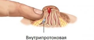
After a mastectomy - an operation to remove part or all of the breast with or without lymph nodes, even if it is performed by the most experienced and technologically advanced oncologist surgeon, scarring of the skin tissue occurs at the site of the incisions in the chest, armpit, and collarbone. The extent of scarring depends on the type of surgery—some types of mastectomy have more scarring, while others have less scarring. Most mastectomy scars run horizontally across the chest, diagonally, or sometimes in a crescent shape.
Due to the fact that after surgery for breast cancer, most patients receive chemoradiotherapy, and they develop lymphostasis to varying degrees, normal tissue healing is disrupted and slows down. In addition, the sutures after a mastectomy are in places of friction with clothing, so there is a very high (50-93%) probability of the formation of not ordinary (normotrophic), but pathological scars (“rough”) - atrophic, hypertrophic and keloid.
Such scars are not only unaesthetic, but they also cause physical suffering (itching, discomfort, pain) and make it difficult to perform delayed breast reconstruction surgery or worsen the result of one-stage breast reconstruction with a prosthesis.
Therefore, after a mastectomy, it is very important to ensure normal healing of the postoperative wound immediately after removing the sutures and to prevent the formation of “rough” scars using special means.
General complications
They arise due to the damaging effects of radiation, as well as due to intoxication with tumor decay products. The severity depends on the technology used and the dose received. In general, the symptoms are mild, so a break or cancellation is not required. Most often noted:
- from the gastrointestinal tract - nausea, vomiting, loss of appetite, taste perversion, stool disorders;
- chest organs - depression of respiratory function, cough and shortness of breath;
- central nervous system - dizziness, fatigue, sleep disturbance, memory impairment, irritability, depressed emotional state, headaches;
- with organic changes in blood vessels and the heart - a drop in blood pressure, arrhythmia, increased heart rate, and the appearance of systolic murmur.
Such signs are reversible, the disrupted processes are gradually restored and return to normal 3-6 months after the end of the manipulations.
To prevent deep leukopenia and thrombocytopenia, decreased blood clotting, and increased capillary permeability, laboratory tests of biological material are performed weekly.
What you need to know about radiation therapy burns
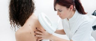
Vital in the treatment of breast cancer, radiation therapy in most women causes skin damage: ranging from dryness, redness, swelling, itching, to severe inflammation - “radiation dermatitis”, and from darkening of the skin, as with excessive tanning, to severe burns, even the appearance of blisters with the formation of weeping ulcers.
The area of such burns can be quite large - on the chest and under the chest, neck, shoulder and armpit. Forming postoperative scars are especially sensitive to rays - for many they swell and hurt. Such changes do not begin immediately, but after 10-15 irradiation procedures, so initially many women do not carry out prevention of such burns and are faced with a developed adverse skin reaction to irradiation.
These skin changes require timely treatment using special products. After radiation therapy, the skin needs to be helped to recover as quickly as possible so that it can fully perform its functions during further therapy and rehabilitation.
Skin reactions during radiotherapy
Skin changes are common and common during radiation therapy. Each patient responds to treatment differently.
The type of skin reaction that occurs depends on:
- the part of the body that is affected by the therapy;
- type and dose of radiation received.
Tell your doctor or nurse if you smoke and if you have:
- high blood pressure;
- diabetes;
- any connective tissue disease such as rheumatoid arthritis or dermatomyositis (a disease whose symptoms include skin rash and muscle weakness);
- previously detected skin cancer in the irradiated area.
These conditions may affect wound healing and the degree of response to radiotherapy.
Types of skin reactions during radiotherapy
During radiotherapy, the area of skin exposed to radiation may turn pink or tan. As therapy continues, your skin may become bright red or dark brown and may develop swelling. Additionally, your skin may become dry and you may feel tight, itchy, or flaky.
Some people may develop a rash or blisters where they receive treatment. These blisters may rupture or peel off. If skin reactions develop, they will most likely become most intense 2 weeks after the last therapy session. After completing radiotherapy, it may take several weeks for your skin to improve.
During therapy, you will be assessed weekly by your treatment team. They will examine your skin and make new skin care recommendations if necessary.
to come back to the beginning
Two problems - one solution
Modern medicine offers many different mechanisms for the normal healing of skin damage, prevention and treatment of pathological scars.
Among them there is one unique remedy, unlike others: the main active ingredient in it is a natural compound with high biological activity - epigallocatechin-3-gallate (one of the catechins of green tea). It is obtained by a special extraction method from the leaves of green tea Camellia sinensis.
This molecule has already been well studied: 3,500 studies have been conducted and its powerful anti-inflammatory effect has been scientifically proven, as well as the ability to normalize the process of formation and growth of new blood vessels, which is very important in wound healing without pigmentation and pathological scarring. Epigallocatechin-3-gallate accelerates and shortens the growth phase of new vessels, which leads to faster production of collagen in the first days of healing and suppresses its excessive production in the future, which prevents the formation of pathological scars.
Classification of radiation proctitis
Depending on the severity, the following types of radiation proctitis are distinguished:
- Catarrhal proctitis. During the examination, a hyperemic, loose, edematous intestinal wall is discovered. There may be increased mucus production. This form of proctitis is considered mild.
- Erosive-desquamative proctitis. This form is manifested by foci of destruction of the epithelium with the formation of erosion - a tissue defect within the epithelial layer.
- Ulcerative proctitis - destruction spreads to deeper layers of tissue and involves the mucous membrane and submucosa in the process.
- Fistulous proctitis. A through perforation of the intestinal wall is formed with access to the pelvic cavity or with the involvement of nearby organs, for example, the bladder or vagina.
There is a classification of radiation proctitis based on the rectoscopic picture:
- There is local redness and friability of the intestinal mucosa, telangiectasias and dilated blood vessels are noted).
- Against the background of hyperemia and edematous intestinal mucosa, ulcers covered with a gray scab are found.
- Against the background of inflammatory lesions, strictures of the intestinal wall are detected.
- Against the background of ulcerative lesions, strictures and fistulas or intestinal perforation are detected.
Egallochit - a reliable source of epigallocatechin-3-gallate for topical use
A large amount of epigallocatechin-3-gallate (70% substance) is contained in Egallohit gel and cream. Egallohit is used in cases where it is necessary to accelerate skin healing, prevent the formation of pathological scars and pigmentation, relieve itching and inflammation in a fresh scar, improve the condition of an old scar, eliminate inflammation and restore skin in case of radiation dermatitis and burns.
As a product of natural origin, Egallohit is completely harmless and does not cause side effects.
Side effects
Unfortunately, the breakdown products of a cancerous tumor cause a number of undesirable effects:
- dryness, peeling, itching, redness of the skin, alternating with pigmentation. To prevent it from scratching and infection, moisturizers - ointments and creams - are prescribed;
- soreness, swelling and tightness in the muscles;
- limitation of range of motion in joints.
The attending physician should be informed of all unpleasant manifestations associated with treatment.
How to use Egallohit during and after radiation therapy
For radiation dermatitis and radiation burns of 1.2 degrees, Egallohit is recommended to be used from the 1st day: it should be applied in a thin layer to the affected areas of the skin 2 times a day for 1-2 months (if necessary, continue use until the skin is completely restored).
For radiation burns of degree 3A, Egallohit can be used after the formation of a “crust”: if it is wet - in the form of a gel, if dry - in the form of a cream. The total recommended duration of treatment is at least 8 weeks.
Help with complications
Patients are prescribed consultations with relevant specialists, who, after examination, prescribe hematopoietic stimulants, anti-inflammatory, absorbable drugs according to indications, taking into account age and general condition. Therapy is aimed at alleviating symptoms and should be comprehensive.
In cases of tumor recurrence, the question arises about the need for an additional course of radiation. This is most often done for palliative (maintenance) purposes using low doses. Such an attempt usually ends in weak destruction of cancer cells and the formation of a trophic ulcer. Repeated palliative radiation does not provide any guarantee of success.
What you need to remember about Egallohit
- Egallohit is an effective local remedy that ensures rapid healing of the skin and prevents the formation of all types of pathological scars, and also improves the condition of old scars after mastectomy and radiation therapy, which has been proven in clinical studies;
- Egallohit is a concentrate of the biologically active substance epigallocatechin-3-gallate, which has an anti-inflammatory and wound-healing effect on tissue;
- Egallohit is of natural origin, therefore it is harmless, does not cause side effects and can be used for a long time.
List of scientific works:
- Epigallocatechin Gallate (EGCG) is the most effective cancer chemopreventive polyphenol in green tea (Du GJ, Zhang Z, Wen XD, Yu C, Calway T, Yuan CS, Wang CZ);
- Study of the effectiveness and safety of epigallocatechin-3-gallate (galaderm cream) during photodynamic therapy of basal cell skin cancer (A. S. Yusupov);
- Results of an open comparative randomized controlled study of the clinical effectiveness and safety of epigallocatechin-3-gallate gel in patients with acne (Z. R. Khismatullina, G. S. Kantyukova, V. I. Kiselev, V. M. Drukh).
PHOTOS: unsplash.com
Share
Diet
To prevent the occurrence of a general reaction or eliminate its manifestations, it is necessary to consume foods rich in microelements and vitamins, and drink at least 2 liters of fluid per day. These tips apply in almost all cases, but it is advisable to first discuss them with your doctor:
- avoid using spicy, fatty, fried foods, smoked foods, marinades;
- reduce salt intake or replace table salt with mineralized salt;
- cook food using vegetable oils;
- give preference to chicken and fish;
- drink only filtered water.
It is also worth considering the characteristics of individual injuries. For example, after irradiation of the abdominal organs, loose stools, nausea, and vomiting are noted. Therefore, if you have diarrhea, it is advisable to exclude dairy products from the menu. Vegetable soups, pureed porridge, mashed potatoes, boiled fish, steamed cutlets, and jelly are very healthy.
Possible treatments for keloid scars
Despite the abundance of advice on the Internet, folk remedies cannot get rid of keloid scars; they can only be used in combination with medications, physiotherapy or cosmetology.
The most popular ways to get rid of keloid scars are drug treatment, that is, the use of gels, ointments, creams and injections in combination with physiotherapy, for example, ultraphonophoresis or electrophoresis and the injection of corticosteroid hormones under the skin. Mesotherapy is also effective - injections into the scar tissue of vitamin complexes and medicinal substances that absorb excess collagen and excess hyaluronic acid.
If conservative methods do not give the expected result, then surgery is resorted to.
Drug treatment
Pharmacy and cosmetic products come in different directions:
- contain interferon;
- corticosteroids;
- enzymes or enzyme-containing preparations.
Interferon-containing products inhibit collagen production. In other words, the scar stops growing in size, however, it remains at the stage to which it has grown now. A similar method of treating keloid scars is used after surgery in the form of injections of alpha and beta interferon.
Injections are given every centimeter along the entire length of the scar; the course lasts 4 months.
Corticosteroids can be administered either on their own or in combination with other substances and any therapy. They are injected not into the keloid scar itself, but into the nearest place next to it. This protects from further thickening of the scar, and, despite the course of treatment - 5 weeks, relapses are observed in 20-30% of patients.
To prevent re-formation of the scar, therapy is supplemented with laser or surgical removal of the scar. These methods are very painful and do not exclude relapse (re-formation of a scar). Laser resurfacing requires a long recovery period.
Enzyme-containing preparations break down excess collagen and excess hyaluronic acid - the main components of scar tissue. Due to this, the relief and color of the skin are restored. The scar becomes elastic and its active growth is prevented.
Pressotherapy
The method is more close to prevention than to treatment, but some experts note a positive effect.
Various silicone dressings, bandages and plates are applied to the problem area. It is believed that a scar that is constantly compressed decreases in size. Pressotherapy means include:
- cotton underwear and special bandages (recommended to be worn for six months, made to individual measurements);
- silicone and gel pressure plates;
- gel-based liquids - collodion with polysilicone or silicone.
All this can be found in any orthopedic salon or pharmacy, but this method alone will not be able to completely remove the keloid scar. The pressotherapy method is effective only in complex therapy in combination with other methods of scar correction.
Microcurrent physiotherapy
During the procedure, the body is exposed to a weak current, which stimulates metabolic processes in the tissues of the epidermis, the keloid decreases in size and smoothes out.
Stages of therapy:
- treating the scar with an antiseptic;
- applying a drug that destroys the scar;
- connecting the device, applying electric current to the scar;
- remove remaining medication with a tissue.
The procedure is not difficult to perform, however, there are contraindications to it:
- acute viral diseases;
- poor blood clotting;
- pathologies with the heart;
- exacerbation of chronic diseases;
- neurological abnormalities.
This procedure is considered ineffective compared to other types of physiotherapy. Moreover, it is not cheap.
Radiation exposure
Involves controlled x-rays that destroy fibroblasts within scar tissue. The intensity of the rays is assigned based on the severity of the problem: after all, 90% of the total flow will be absorbed by the epidermis, and only 10% will reach the deeper layers of the skin.
However, therapy is carried out only in combination with other treatment, otherwise the risk of relapse increases by 50%.
Contraindications for use:
- scars on the face, neck and chest;
- oncology;
- kidney diseases;
- impaired blood circulation.
The usual radiation dose is 15–20 Gy. The procedure is repeated once every 2 months, but no more than 6 times.
However, radiation exposure is considered one of the most effective methods in the fight against scars, regardless of the cause of their appearance.
Keloid removal with laser
There are several types of laser resurfacing: argon , carbon and dermabrasion . The purpose of the procedure is to evaporate fluid from the scar's connective tissue, causing it to dry out and shrink in size. Dead cells are removed surgically, and the laser procedure itself is performed under local anesthesia.
Advantages of laser removal:
- during the first session, up to 70% of the scar disappears, which indicates a quick visible result;
- The duration of therapy is from 20 minutes to one and a half hours, depending on the complexity of the problem.
The procedure is quite painful and requires a long period of rehabilitation.
To avoid relapse, doctors advise combining laser with other types of treatment for keloid scars: the use of anti-scar gels will be an excellent assistant on the path to healthy skin.
Cryotechnique
Effect of liquid nitrogen on keloid. It burns out scar tissue cells, in their place healthy skin is formed. The contact time of the scar with nitrogen is 10–30 seconds; in case of an overdose, pigmentation is possible, and there is also a high risk of developing an atrophic scar. You need to be extremely careful with this correction method!
A visible effect is achieved in 1–3 sessions, but for better results cryotherapy is combined with hormonal injections with glucocorticosteroids.
However, for large scars, it is better to combine nitrogen cauterization with surgery. The main disadvantage of the method is pain.
Cosmetology
Cosmetic procedures will help make the scar less noticeable. They will not be able to completely get rid of the scar, but in combination therapy these methods are very effective:
- dermabrasion;
- peeling;
- mesotherapy.
Peeling. With the help of peelings, you can polish the scar, even out the skin texture and eliminate pigmentation. As a result, the skin becomes smoother and the scar more elastic.
Deep dermabrasion - exfoliation of the stratum corneum of the epidermis. The procedure is sensitive and involves the use of hardware technology. The cosmetologist decides how deep and long the session should be.
Mesotherapy - injections of heparin, an immunomodulator or a vitamin complex into the problem area. Has an anti-inflammatory and softening effect.
For small scars, dermabrasion or mesotherapy is recommended, while large and old keloids are removed in combination with drug therapy.
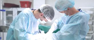
Surgery
Prescribed as a last resort when other therapies are not effective enough. Excision of the keloid is carried out a couple of years after its formation and in several stages:
- a small incision with a scalpel on the scar under local anesthesia;
- the edges of the scar are sewn together with cosmetic stitches for better healing of the incision;
- after resorption of the sutures - hormonal injections and enzyme therapy.
After surgery, prevention against relapse is indicated, because a fresh scar is more amenable to correction. During the rehabilitation period, radiation therapy, injections with immunomodulators and hormones, as well as external agents in the form of gels and ointments are often prescribed.
Prevention
It is impossible to predict the behavior of a scar after an injury , but you can reduce the risk of keloid scars. Silicone gels and patches can help with this; they help prevent the growth of scar tissue. These methods are effective provided that the scar is fresh (no more than six months). Silicone products help maintain the correct water balance in scarred skin and create additional pressure, due to which the blood vessels in the scar are reduced.
It is recommended to start preventing scar formation after 3-4 weeks, when the crusts have completely come off from the wound. The healing area must be kept clean, washed with soap and under no circumstances remove the stratum corneum from the wound - this will cause infection, and this is a sure way to the formation of a keloid scar!
Stages of development of a keloid scar
An annoying defect is formed in 4 stages:
- Epithelialization . An epithelial layer forms on the injured area; after 7–9 days it thickens and turns pale. After 2–3 weeks, the damaged area turns red and swells.
- Swelling . In 3–4 weeks, the damaged epidermis thickens. Injured skin takes on a red tint.
- Seal . The injury becomes much denser. A bulge is noticeable on the skin.
- Softening . The scar fades and softens. Pain may occur.
The sooner treatment begins, the sooner and easier it is to get rid of a keloid scar. However, any external medicinal and cosmetic products should be used only after the wound has completely healed.
Causes of keloid scars
Some people have a genetic predisposition to the formation of keloids, especially blacks. But most often, a scar is formed due to insufficient sterile care of a wound or burn, as well as under the following circumstances:
- with “lacerated” wounds;
- with wound suppuration;
- with strong tension of the skin in the area of injury;
- in case of hormonal imbalance;
- in case of traumatization of an already forming young scar;
- reduced immunity.
The appearance of a scar can be avoided by following the doctor's recommendations, as well as by establishing a healthy diet and rest regime.
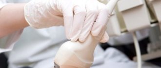
How is radiation treatment performed?
Radiation therapy is a wide arsenal of methods that can be divided into two large groups: external and internal (brachytherapy). In the first case, radiation is generated by a special device that moves near the problem area and sends rays to the tumor at different angles. The patient lies motionless on the table in the position chosen at the planning stage. The exposure time may vary. On average, one session takes 10-30 minutes. In most cases, the patient is prescribed several of these procedures. After some time, the course is repeated. If the purpose of radiotherapy is pain relief, it can be performed once.
The procedure itself is completely painless, but causes anxiety for some people. Irradiation rooms are equipped with audio equipment. With its help, the patient can tell doctors about any problem or just talk to relax. The doctors themselves are in the next room at this time.
Brachytherapy involves irradiating the tumor with radioactive substances, which are injected directly into the tumor or adjacent tissues. It has two varieties: temporary and permanent. In the temporary version, radioactive drugs are placed inside a special catheter, which is injected into the tumor for a while and then removed. Permanent brachytherapy uses a tiny implant that is placed directly into the tumor, where it gradually releases radioactive substances. Over time, they run out, and the grain of the implant remains in the body for life, without causing any inconvenience.
Consequences and prognosis
The consequences of eye burns are determined by the depth of damage, the type of damaging factor, the severity of tissue damage and the timeliness of treatment.
Burn injuries to the eyes of II – IV degrees are accompanied by certain complications and do not go away without leaving a trace. In the tissues of the eye, scarring may occur, disruption of the lacrimal ducts, decreased transparency of the cornea, development of dry eye syndrome, cataracts or glaucoma. The outcome of burns can be damage to retinal cells in the macular area and the occurrence of irreversible blindness.
In the medical department, everyone can undergo examination using the most modern diagnostic equipment, and based on the results, receive advice from a highly qualified specialist. The clinic is open seven days a week and operates daily from 9 a.m. to 9 p.m. Our specialists will help identify the cause of vision loss and provide competent treatment for identified pathologies.
You can make an appointment at the Moscow Eye Clinic by calling 8 8 (499) 322-36-36 (daily from 9:00 to 21:00) or using the online registration form.
Patient immobilization means
In order to accurately deliver ionizing radiation to the irradiated target, it is necessary to clearly reproduce the position in which the process of preparation for radiation treatment took place, i.e. computer topometry and dosimetric planning. This is provided by a variety of means for positioning and immobilizing the patient. They can be in the form of different standard decks with headrests, fastenings, bolsters and supports for arms, legs, and pelvis. There are also individual means. For example, vacuum mattresses and thermoplastic masks that fix the individual shape of the patient’s body in the irradiation position. These devices make it possible to avoid displacement of the irradiated area due to involuntary movements of the patient.
Labor activity
Many citizens, after successful radical therapy, after a certain time are considered practically healthy and, if the diagnosis allows, can return to their workplace. However, they also remain under medical supervision to prevent the development of complications. Control is carried out over the cardiovascular system, morphological picture of the blood, immunity, bioelectrical activity of the brain, etc.
Our specialists will help you get back to your favorite job faster and feel in demand in your professional field. Do you want to ask clarifying questions about rehabilitation? Dial the specified hotline number or request a call back, and our staff will answer them.
Thermal burns
Thermal are eye burns that occur when exposed to high or extremely low temperatures, including exposure to hot liquids, fire, steam, burning or hot solid particles, dry ice, as well as cryogenic liquids and liquefied gases. Such burns, as a rule, are localized in the anterior part of the eye, and damage to the deep ocular structures can be observed only in severe cases.
If a thermal burn occurs, traces of the traumatic substance should be removed if possible and the eyes should be rinsed under a weak stream of water. The skin around the eye is lubricated with an antiseptic or antibiotic ointment (for example, tetracycline ointment), the same ointment should be placed behind the eyelid. The eye is covered with an aseptic bandage, and scratching or rubbing it is strictly prohibited. The patient is hospitalized as quickly as possible.