Almost every owner of a mole that rises above the level of the skin has encountered unpleasant sensations in the area where it is located.
More often this is due to trauma.
Especially if the mole is located on an area of the body that is often subject to friction from clothing.
For example, in the collar area or on the back.
In some cases, a mole hurts due to other reasons not related to injury.
There is a change in its appearance, which can become symptoms of the degeneration of a benign process into a malignant one.
A mole (nevus) is a pigmented neoplasm that appears on the skin from birth or throughout life.
It has various shades (brown, red, blue, purple and others).
Moles are formed when pigment accumulates in the skin.
In increased quantities, it turns into melanocytes and forms moles.
These tumors in normal condition do not cause pain or discomfort.
But under certain conditions and under the influence of certain factors, pain occurs in a state of calm or when pressing a mole.
This may be a consequence of inflammatory processes or hormonal disorders.
In any case, you should consult a qualified healthcare professional.
Since in some situations it may be necessary to remove the disturbing tumor.
Why does a mole hurt?
There are several main reasons why a mole hurts.
Most often, pain occurs as a result of injury.
This is also accompanied by minor bleeding, swelling and hyperemia of the surrounding soft tissue.
If the mole hurts and itches, and new nevi appear, hormonal changes are suspected.
This occurs more often in women during pregnancy.
Painful moles are caused by the following reasons:
- Injuries - a hanging mole in particular often hurts, which can easily be touched by clothing, a washcloth or jewelry, depending on its location. But other types of moles can also be injured, sometimes this happens in beauty salons when performing intense massage, using anti-cellulite cups, or performing depilation. More often, nevi located in the armpits, pubis, collar area, and on the folds of the lower and upper extremities are injured. Often the cause of pain is an injury to a mole while using a razor. Any damage to a benign formation should not be ignored, since even minor scratching can provoke malignancy, which poses a direct threat to human life.
- Increase in size - the cause of such changes is often hormonal changes, but sometimes the mole grows and hurts when the process becomes malignant.
- Prolonged exposure to direct sunlight or staying in a solarium leads to burns. In this case, the mole hurts and may itch, but after 3-5 days the discomfort goes away on its own. If after being under the hot sun the pain does not disappear for 7 or more days, you should see a doctor and undergo an examination, the process of malignancy (malignancy) may have begun.
- Inflammatory processes - most often occur when a mole is damaged and infection enters, which causes hyperemia, pain and even swelling.
- Thermal burn of the skin - exposure to hot steam or liquid, fire, high temperatures causes burns of varying degrees of severity, the most sensitive areas are moles and age spots, where increased pain is felt.
- Hypothermia or overheating of the body - with any deviations in body temperature from the norm increases the sensitivity of the skin. Not only existing nevi can hurt, but also the site of a mole removed in the recent past.
Genetics
Genetics is rightfully considered one of the main reasons for the appearance of moles. If parents have a large number of moles or a predisposition to them, then their children will probably have them too.
Moreover, moles have one interesting feature - they can appear in the same places as on close relatives.
Signs of malignant degeneration of a mole
How to assess the danger if a mole hurts and determine the need for surgical intervention?
Unpleasant sensations can be the first symptoms of malignant degeneration.
How quickly and rapidly malignancy occurs depends on several factors.
For example, frequent exposure to sunlight significantly accelerates the process of malignancy.
Therefore, it is very important to periodically examine pendulous or other moles located in areas of increased trauma.
If any pathological changes are detected, you should consult a doctor.
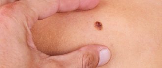
Signs of malignancy:
- violation of symmetry (all benign moles are completely symmetrical, with the exception of congenital ones)
- uneven contours (malignant nevi are characterized by blurred contours and unclear edges; in a healthy state, a mole has clear contours and smooth edges)
- change in color (if dots, stripes appear on a mole, or it becomes multi-colored, this indicates its malignancy)
- increase in size (in a normal state, this is possible in adolescence or in women during pregnancy; in other cases, a sharp, causeless growth of benign formations requires examination and, in most cases, removal)
- any pathological changes (the occurrence of bleeding, cracks or the formation of crusts for no particular reason are the causes of degeneration)
In some cases, additional symptoms of malignancy of the process may include:
- swelling and pain of a mole
- peeling
- thickening of the tumor
- a feeling of burning and itching, periodic tingling in the area where the mole is located
- causeless formation of rashes around the nevus, similar to an allergic rash
Sometimes a mole hurts after removal, which can also mean the development of a cancerous process or the growth of a new benign nevus.
More often this is due to incomplete excision of melanocytes, cells that accumulate as moles.
I monitor one mole, remove another: how not to miss dangerous neoplasms on your skin
Where do they come from
On the Internet you can find a version that our moles initially grow... due to tanning. They say that a person is born with clear skin, and over time it is because of the sun that moles appear on it.
“In fact, the formation of moles and their number is determined primarily by heredity,” says dermato-oncologist at the Clinic of Skin Diseases, assistant at the Department of Skin Diseases at Sechenov University, candidate of medical sciences Ekaterina Vertieva.
- That is, those who have a lot of new growths on the skin in their family - mom, dad, grandmother or grandfather - will have more moles.
The second factor is the sun. Under the influence of its rays, the number of moles increases.
At the same time, there are several peaks in a person’s life when most moles appear. Such surges are associated with hormonal changes: at a younger age (3 - 6 years), adolescence and from 25 to 35 years. Women may experience additional peaks during pregnancy.
— Is it true that there is a “safe limit” of moles, and if there are more than normal, then a person has an increased risk of skin cancer?
— If there are more than a hundred moles on the skin, then the risk of malignant neoplasms is really higher. But not so much that those with a rich scattering of moles should immediately sound the alarm. The logic is simple: the more tumors there are, in principle, the higher the likelihood that among them there will be nevi with malignant potential (how to protect yourself from danger - see below).
STAY IN TOUCH
What is what
Among the names of growths on the skin you can find the classic, familiar word “mole,” as well as nevus and keratoma. What is the difference?
“ Nevus is the medical name for the same mole,” explains Ekaterina Vertieva. - Essentially, this is a benign skin tumor. A mole consists of pigment cells of melanocytes, which produce the coloring pigment melanin (because of it, our skin darkens when tanning. - Ed.) A mole is a cluster of such melanocyte cells. That is why the appearance of nevi is associated, among other things, with exposure to the sun.
— Keratoma is a benign superficial formation on the skin that can never degenerate into something malignant. Keratomas occur more often in old age. They consist of keratinocyte cells (these are the surface cells of the upper layer of the skin of the epidermis). Externally, a keratome usually looks like a brown formation rising above the surface of the skin. Keratomas often become crusty and flaky. Pieces can fall off, which scares people. In fact, this is a natural desquamation process. There is no need to be scared, the doctor reassures.
On a stem and with hairs - what can you expect from them?
- There are moles that seem to move away from the skin and rise above it - is this dangerous?
- Look: we have superficial layers of skin, the epidermis, and deep layers, the dermis. Common, classic dark moles are superficial, so-called border nevi. They are found in the epidermis. Over time, some superficial nevi absolutely physiologically, that is, safely, naturally grow into the deep layers of the skin, the dermis. And then they begin to rise above the surface of the skin. Nothing wrong with that.
Moreover, over time, by about 50-60 years, dermal nevi completely lose their pigment. They cease to be brown and acquire the color of healthy skin. This is the so-called mole involution. All this is absolutely safe.
- When do hairs grow from a mole? They say this is a good sign, such nevi are definitely safe and will not degenerate into melanoma.
- This is a misconception. Melanoma can easily grow hair. Therefore, if a nevus has suspicious signs (see below), then the hairs are not a reason to calm down and not check the mole.
By the way, doctors categorically do not advise pulling hair out of a nevus; this is unnecessary trauma. You can only cut it carefully.
Meyerson phenomenon
This is the name of the condition when redness appears around the mole, like an inflamed pink corolla, our expert explains. And it reassures: as a rule, it is not dangerous. Most often, the Meyerson phenomenon occurs in patients with a tendency to skin diseases: eczema, atopic dermatitis. If you experience any discomfort or itching, you should consult a doctor. Usually, dermatologists prescribe atopic corticosteroids to relieve such symptoms; with their help, the symptoms are easily removed.
— If you damage a mole and it bleeds, do you need to see a doctor urgently?
— It is necessary, but not necessarily right on the same day or the next day, up to 7 days is quite normal. There is no need to be afraid that after injury, even with bleeding, your mole will definitely degenerate into something bad. Do not believe the stories about a grandmother who picked off a mole, it immediately metastasized into the blood, and death quickly occurred. It is a myth. If a person injures a benign mole, then nothing bad will happen to it. Adverse consequences occur if it was initially melanoma.
— What to do immediately after a mole is damaged, before going to the doctor?
- Treat with a clear antiseptic. Hydrogen peroxide, for example. Just don’t smear it with fucorcin, iodine or brilliant green. Because it will then be difficult for the doctor to examine and assess the condition of your mole due to staining with these substances.
How often to check with a doctor
There are tips on the Internet to show your moles to a doctor every 6 to 12 months.
“If a person has 100 or more nevi and is advised to visit a doctor every six months or a year, then this is still too much,” says Ekaterina Vertieva. - In most cases, moles will not turn into anything bad. But, to be sure of this, it is important to undergo a full examination once by a dermatologist, or even better, an oncodermatologist. Have a specialist carefully check all moles and confirm that none of them are cause for concern. Or he indicated those that could potentially be dangerous, and then told how often they need to be monitored.
If the doctor, after examination, says that all moles are calm, then, as a rule, it is recommended to see the next time in 3 - 4 years. At the same time, no one canceled the self-examination.
THIS WILL BE USEFUL
The golden rule for self-examination
This is the international standard for assessing moles. It can be used as a test at home. The mole is tested according to the ABCDE criteria.
A (asymmetry, asymmetry) - has the mole become asymmetrical?
B (border) - have the borders of the mole become uneven?
C (colour) - has the color of the mole changed: several shades of brown, black, blue, pink have appeared?
D (diameter, diameter) - has the mole grown more than 6 mm in diameter?
E (evolution, evolution) - does the mole change intensively over 4 - 6 months (in shape, color, size)?
Important : if the answers to all questions are “yes,” this is reason to suspect the mole is malignant and immediately consult a doctor. If you have several of the symptoms, then the risk is lower, but you also need to see a doctor.
Delete cannot be saved
“There are three reasons for removing moles,” says Dr. Vertieva. — Cosmetic removal — if a person simply doesn’t like the way a mole looks. We also recommend removing benign moles if they are located in places where they are often injured. For example, for men in the beard area, for women in the bra clasp area. The third option is if we see suspicious symptoms. In these cases, the doctor conducts an examination using a dermatoscope (a device similar to a microscope. - Ed.). If a mole is causing concern, removal is done for medical reasons.
— What are the current methods for removing moles?
- Laser, radio wave method, electrocoagulation (cauterization of the nevus with high-frequency current) are used - these methods destroy the mole. As well as surgical excision.
If the patient has a suspicious nevus, then surgical removal is necessary. Only in this case, the tumor tissue is fully preserved, and histologists will be able to give a reliable conclusion as to whether there are malignant cells in the mole.
In other cases, when the mole is definitely calm, the least traumatic method is the radio wave method or laser (coagulation is a rougher method). After removing the nevus, a neat wound remains; it must be treated with a solution of potassium permanganate or fucorcin. A crust forms, which falls off after an average of two weeks. You can wet the wound site with water from 3 to 4 days.
I strongly do not recommend self-medicating at home and using celandine to remove tumors. At best, this technique leads to a chemical burn, and sometimes to more serious consequences in the case of malignant potential of the formation.
QUESTION ON THE TOPIC
Is it possible to sunbathe if you have a lot of moles?
“Yes, this is not a contraindication for tanning,” our expert rejoices. — The main thing is to follow safety rules: stay in the sun before 11 a.m. and after 4 p.m. And use sunscreen.
BREAKTHROUGH AND PROSPECTS
“Now there is a real revolution in the treatment of melanoma, the most aggressive skin cancer,” says Ekaterina Vertieva. — Previously, chemotherapy was used, which was very toxic and at the same time extended the life of patients by only a couple of months. Since the beginning of the 2010s, targeted drugs have been used. They do not hit all cells of the body, like chemotherapy, but selectively hit specific targets. These drugs are available in Russia under compulsory medical insurance and prolong the life of our patients very significantly. The main trend now is the study of new targets for the fight against melanoma. And also immunotherapy (the method for which the Nobel Prize in Medicine was awarded this year. - Ed.).
CONGRATULATIONS!
The oldest department of Sechenov University is 150 years old
On May 27, the Department of Skin and Venereal Diseases named after. V.A. Rakhmanov Sechenov University - 150 years. Already in the first years of its work, the department was recognized as the best in Europe. Over a century and a half history, vast experience in teaching the science of dermatovenereology has been accumulated here. Today, about 2,500 skin and venereal diseases are known, say department employees. The most common are psoriasis, atopic dermatitis, eczema and fungal infections; In recent decades, there has been a steady increase in the incidence of allergic dermatoses.
The head of the department, Professor Olga Olisova, lists modern promising scientific developments of the department's employees: improving the early diagnosis of lymphoproliferative skin diseases (T-cell lymphomas) using genetic markers; development of hardware methods for diagnosing melanocytic skin neoplasia; studying the pathogenetic connections of HPV with the development of skin tumors using molecular genetic detection methods; development of diagnostic markers for severe bullous dermatoses and orphan skin diseases (using the example of mastocytosis).
BY THE WAY
At the Department of Skin and Venereal Diseases of Sechenovka, a unique plaster museum has been created, containing more than 3,000 wax plaster casts with images of various skin and venereal diseases. In terms of the quality and quantity of exhibits, the dummy museum is a national treasure that has no equal not only in Russia, but throughout the world.
Why does the process of malignancy of moles occur?
Any changes in the functioning of the body require consultation and examination by a qualified specialist.
It is very important to undergo diagnostics for timely detection of cancer and factors that provoke malignancy.
First of all, direct sunlight has a bad effect not only on moles, but also on the skin in general.
The aggressiveness of ultraviolet radiation leads to the development of cancer processes quite quickly.
It is no less dangerous to scratch the areas where moles are located.
Any damage to their integrity can trigger the process of degeneration or cause the introduction of an infectious pathogen.
Also, in areas where moles rise above the surface of the skin, you should not use hard washcloths.
Because there is a high risk of damage.
Melanoma
This is a type of cancer that occurs due to direct exposure to ultraviolet rays on the pigment cells of the skin. Melanoma is a dangerous disease. This is due to two reasons:
- the ability of a nevus to become an atypical neoplasm in a short time,
- the appearance of such a neoplasm leads to the appearance of a disease such as cancer.
If a mole becomes inflamed (red and hurts) or there is redness around the mole, this is a direct reason to seek help from a specialist.
The general clinical picture can be supplemented by:
- uneven edges,
- one half of the nevus is different from the other,
- heterogeneous coloring of the nevus,
- redness around the mole,
- nevus compaction.
In case of untimely diagnosis or unwillingness to consult a doctor in a timely manner, symptoms appear that indicate the active activity of the malignant process:
- the formation of numerous cracks on the surface of the nevus,
- blood comes from the nevus,
- significant weight loss.
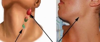
There are also multiple enlargements of the lymph nodes and the appearance of corresponding pain upon palpation.
Dangerous moles that degenerate into melanoma
What does a dermato-oncologist do?
The specialist studies nevi and prescribes tests to determine the nature of the neoplasm.
Conducts differential diagnostics, assesses the likelihood of transforming a benign process into a malignant one.
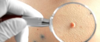
First, a visual inspection is carried out.
Then specialized equipment is used for a more detailed study - a dermatoscope.
If there are risks of malignancy or the process of malignancy has already started, surgical intervention is prescribed.
The operation is performed by a surgeon using general or local anesthesia.
Risk factors
According to recent, numerous scientific experiments, it was found that women, unlike men, have a greater tendency to develop melanoma. It is important to highlight that a malignant tumor can be localized separately on an area of the skin or on the nevus itself. Most often, the legs are affected by the pathological process, and a little less often the upper limbs and abdomen. A mole on the face and neck can also become inflamed, swollen, fester and cause many other problems.
People who are characterized by fair skin covered with freckles, as well as patients with a hereditary predisposition, constitute the main risk group. It is in these people that inflammation of the nevus or redness of the mole can result in malignant degeneration.
The above category of people should undergo regular examination by an oncologist and pay maximum attention to any change in the birthmark.
Pale skin and freckles are one of the risk factors for melanoma
Mole removal methods
If a mole has enlarged and hurts for no apparent reason, as well as if there are pathological changes in its structural structure, its removal is prescribed.
If the development of melanoma is suspected, a surgical method is used.
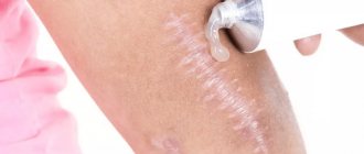
During the operation, not only the mole itself is excised, but also the healthy tissue surrounding it.
The disadvantage of this procedure is the formation of postoperative scars.
This is due to the need to remove the neoplasm together with adjacent structures, the diameter of which should be at least 3-5 cm.
After the operation, the removed material is sent for histological analysis.
The use of liquid nitrogen at low temperatures – cryodestruction.
This technique is often prescribed to remove birthmarks that are at the level of the skin and represent a cosmetic defect.
During the procedure, the nevus is frozen, after which it shrinks and a crust is formed, which prevents infection.
Over time, the crust falls off and healthy structures are regenerated in its place.
Sometimes, if melanocytes are not completely eliminated, a repeat procedure may be required.
Since there is a high probability of relapse.
Moles that rise above the surface of the skin are sometimes cauterized using a needle electrode.
As a rule, the removed tumor is sent for histological examination to determine its nature.
After the procedure, a crust forms; after a while, an almost imperceptible scar may remain.
Removal of a nevus using laser radiation is considered one of the most effective and least traumatic operations.
Used to remove benign formations located on open areas of the skin, for example, on the face.
Laser equipment is highly accurate due to which the surrounding healthy tissue remains undamaged.
When eliminating small moles after restoration of tissue structures, there are no scars or scars.
Radiosurgery is also used to remove tumors protruding above the surface of the skin.
The technique is based on the influence of radio frequency waves, is the safest and completely non-contact.
After the procedure, the adjacent tissues with the surgical field are restored quite quickly.
Good equipment allows you to make cuts of various depths and any configuration.
Pus from a mole
There are often cases in which the nevus can become inflamed and fester. With light palpation of the nevus, a significant compaction is identified, which in itself is alarming. When consulting an oncologist, the doctor assures that there is nothing wrong, and the strange lump is just an inflamed pimple under a mole, which explains the presence of purulent discharge and lump. This seemingly absurd and illogical explanation is only relevant in relation to a healthy mole. During the cancerous process, no inflammation can be observed under the spot, since the epidermis underneath dies.
Healing after mole removal: can it hurt and for how long?
What to do if a mole hurts after removal, as well as the area around it?
Sometimes after surgery you may experience discomfort, pain and even a burning sensation.
More often this is observed after surgical removal of the nevus and adjacent tissues.
Minor soreness and discomfort may be present for up to one week, and an itchy sensation may also occur.
After removing the nevus, the wound and surrounding areas are treated with antibacterial ointments or a weakly concentrated solution of potassium permanganate twice a day for 5-7 days.
After surgery, a scab (crust) is formed at the site of mole removal, which protects the damaged area from infection.
In order not to injure the healing surface in the first 7-10 days, the use of a washcloth, taking a bath, sauna, or applying cosmetics is strictly prohibited.
After 8-9 days, the scab disappears, and healthy pink skin appears underneath.
Very often, patients are interested in whether it can hurt and how long it will take for the skin to heal after removal of a mole?
Pain may be present, but not always, and as a rule, if all medical recommendations are followed, it disappears within 2-3 days.
The main condition for rapid recovery is the complete exclusion of direct sunlight.
If the operation is planned to be performed in the summer, it is necessary to avoid going to the beach for 3-4 weeks.
Also reduce the frequency of being outside in the open sun.
What to do if your mole becomes inflamed and red?
The first thing to do is to stop further independently studying the Internet on this topic. The fact is that on the Internet you can almost always find confirmation of your worst suspicions about your health. This is not what we need now.
In addition, it must be remembered that this symptom alone is extremely rarely a sign of melanoma. In the vast majority of cases, there should be other symptoms - uneven (geographic, scalloped) edge, asymmetrical shape, bleeding, etc.
Then just follow these recommendations:
- Wet a regular gauze pad (not sterile) with a solution in the ratio of 1 part chlorhexidine, 1 part dimexide, 2 parts water
- Apply the resulting compress to the area of the inflamed mole for 30 minutes, 3 times a day, for 5 days.
In most cases, redness, pain, and enlargement of the mole disappear under the influence of these compresses within 5 days.
If the inflammation does not subside within 5 days, you should urgently see an oncologist. In this situation, the likelihood that these changes were caused by transformation into melanoma is very high.
What complications may arise after mole removal?
If the scar hurts after removing a mole, a relapse is possible.
This happens in cases where, during surgery, not all melanocytes were eliminated; new birthmarks begin to form from the smallest particles remaining.
If moles grow again, they may need to be removed again.
If the wound becomes infected during the postoperative period, the following symptoms may occur:
- increased feeling of itching, weeping
- bleeding
- purulent contents are released from the wound
- the place of the mole is swollen and hurts
- swelling of surrounding tissues (lasts more than three days)
- increase in body temperature (over 38 degrees)
When do moles appear?
Pigment formations, moles, birthmarks or nevi appear on the human body even when the child is in the womb. From the first days of birth, brown moles can be found on a child’s body, but they are usually few and small in size.
During puberty, small hanging moles may appear, the number of which can gradually increase or decrease - it all depends on the characteristics of the body.
Is rubbing a mole dangerous?
Rubbing a nevus causes damage. During friction, the upper layers of tissue of the formation are damaged, resulting in redness, inflammation, swelling, pain and other unpleasant symptoms.
The more regular the friction, the higher the likelihood of bleeding. There is a danger of bacterial contamination. It will lead to suppuration, an acute inflammatory process, and sometimes tissue necrosis.
If a person rubs the formation once, then the action provokes only unpleasant symptoms. If it happens daily, the risk of developing dangerous consequences increases. An injured nevus is prone to cellular degeneration. This must be taken into account when detecting formations in places subject to mechanical action. More often, moles located on the scalp, neck, arms, legs, under the seams of clothing are removed.
Causes of pus
An abscess, purulent inflammation in a nevus appears as a result of penetration of bacteria or infection into the skin. A birthmark is not a vulnerable spot, but a special condition of the skin with a high content of melanin pigment.
Reasons preceding the decay of a mole:
- A cut, scrape or abscess that has been exposed to dust, dirt or harmful germs.
- A skin disease characterized by inflammatory processes (acne, furunculosis, etc.).
- Burns, skin trauma, causing necrosis of the layers of the epidermis and basal cell carcinoma.
- Excessive exposure to the sun in hot weather is fraught with the harmful effects of ultraviolet rays.
- Skin irritation caused by rubbing clothes, scratching with nails.
People with freckles and unusually pigmented skin are especially susceptible to the disease. It is recommended to spend hot daytime in the shade and avoid long stays in stuffy rooms.
It is possible for a mole to spontaneously transform into a malignant tumor. Preceded by hormonal imbalances or nervous breakdowns.
The main danger is that degeneration is difficult to recognize. Symptoms appear after the disease develops. Treatment will require radical surgical intervention.
A boil under a mole is recognized by an increase in the size of the pigment spot and a sharp change in the usual color.
Preventive measures
It is easy to injure a mole due to carelessness. Therefore, it is important to monitor the neatness of your manicure and the comfort of your clothing. Rough, uncomfortable clothes and shoes with straight collars have a negative impact on skin tumors, and long nails can cause scratching of the birthmark. Constant trauma can provoke mutation of nevus cells and further degeneration into a malignant formation.
The proposed treatment methods are effective when it is necessary to treat a festering mole. But, I would like to draw your attention once again to the fact that the inflammatory process, and even more so the decay process, cannot be ignored and left to chance, so at the first signs you need to go to the doctor as soon as possible. Modern treatment allows you to get rid of the problem quickly and efficiently.
How is the diagnosis done?
The most accurate method for diagnosing moles is dermatoscopy. The digital device dermatoscope allows you to “enlighten” the formation and enlarge its image. It becomes possible to study the texture and nature of skin pathology. Diagnosis is painless, safe and does not take much time.
Interesting: After dermatoscopy, all examined formations are recorded. At a subsequent visit to the doctor, you can monitor changes in nevi, their size, and shape.
During the examination, the dermatologist decides on the need for histological analysis. Most often, there are no contraindications for mole removal. It remains for the doctor to choose the optimal method.