Treatment of allergic dermatitis in adults, symptoms and prevention
Allergic dermatitis is a specific skin reaction to contact with a substance perceived by the immune system as an allergen. Its manifestations - itching of the skin, redness and rashes, often causing weeping - are only the external manifestation of the complex process of allergic sensitization.
Treatment of skin manifestations of an allergic reaction includes a course of desensitizing therapy, which is supplemented by the use of folk remedies and a hypoallergenic diet.
Causes
Contact dermatitis, like allergy itself, is an increased reaction on the part of the body to allergens affecting it in one form or another.
Most often, these allergens include the following types of substances:
- nickel (earrings, chains, rings, jewelry, etc.);
- latex (baby pacifiers, gloves, condoms, etc.);
- clothing (in particular, certain materials based on it: synthetics, rubber, latex, etc.);
- cosmetic products used in skin care (shampoos, soaps, creams, gels, etc.);
- certain medications (antibiotics, corticosteroid creams, etc.);
- other types of substances (ink, paints, etc.).
In general, this disease can develop due to exposure to absolutely any substance on the body, and the determining factor in this matter is based not on the chemical composition of these substances, but on the sensitivity of the body to them in each specific case.
Development mechanism
The essence is a delayed-type tuberculin-like allergic reaction. Sensitivity develops over a certain period of time after the first contact with the allergen.
When a critical level of the immune response is reached, the disease manifests itself. Allergen molecules are very small to be recognized by immune cells, but this is helped by protein components that attach to them when they enter the body and give them the properties of an antigen.
The allergic properties of a substance depend on the ability of its molecules to penetrate the body and create stable bonds with its proteins. In this case, a major role in the immune response does not belong to antibodies, as usual, but to lymphocytes and Langerhans cells. From the moment of first contact with the allergen, T-lymphocytes that recognize the antigen accumulate within two weeks. After this they become so-called. memory cells and migrate to all peripheral areas of the immune system.
Upon repeated contact with the allergic factor, rapid activation of immunological memory and cellular response occurs.
Allergic contact dermatitis: basic approaches to diagnosis, treatment and prevention
Allergic contact dermatitis is a classic form of delayed-type hypersensitivity reaction mediated by sensitized lymphocytes. According to a number of authors, this pathology affects 1% to 2% of the population in various regions. The prevalence of the disease is higher in industrialized countries. It is increasing as more and more new chemical substances are introduced into use in medicines, cosmetic products, medical implants, household chemicals, and industrial reagents.
Unlike simple contact dermatitis, in which an irritant causes inflammation in all people when exposed to the skin, allergic dermatitis occurs only in sensitized individuals, that is, in people who have immune cells specific to this substance - T-lymphocytes. Contact dermatitis is often caused by harmless chemicals that, under normal conditions, do not cause any clinical symptoms in healthy people. But allergic dermatitis is also known when in contact with aggressive agents - components of hair dyes, hair growth products, dyes for fabrics, fur and leather, detergents, medications, juice of poisonous plants.
A classic example of allergic contact dermatitis is dermatitis caused by plants of the genus sumac (in particular, poisonous sumac - Rhus toxicodendron), in which the rashes often have a linear shape and are located on exposed areas of the body.
The pathogenesis of allergic contact dermatitis is based on a tuberculin-like delayed (cellular) hypersensitivity reaction, the inductive phase of which begins with local exposure to the skin of low molecular weight chemicals of organic or inorganic nature. Their sensitizing (allergenic) properties depend on their ability to penetrate the skin and form stable covalent bonds with the proteins of the host organism. Thus, dinitrochlorobenzene forms complexes in the epidermis with proteins containing a lot of lysine and cysteine. Skin lipids can also serve as an adjuvant.
In the formation of hypersensitivity, the leading role is played by professional macrophages of the epidermis - multi-processed Langerhans cells. The resulting delayed hypersensitivity is directed not only to the chemical itself, but also to the carrier protein.
Typically, at least 10–14 days pass from the moment of skin contact with the allergen until the development of the first clinical manifestations. The duration of the sensitization period is usually shorter for aggressive chemicals. Thus, according to our observations, drug allergens when applied to the skin can cause manifestations of contact dermatitis as early as 7–8 days. The most common allergenic medications are local forms of antibacterial drugs; contact allergic reactions to local anesthetics, antiseptics and latex are less common.
The location and configuration of the lesion is determined by the causative factor. The most common form of the disease is eczematous dermatitis. The disease is easy to diagnose and, as a rule, is characterized by a favorable course. The rash disappears when exposure to the pathogenic factor ceases. To accelerate the regression of clinical manifestations, local anti-inflammatory drugs, mainly topical glucocorticosteroids, can be used.
Etiology
According to our observations, the most common cause of allergic contact dermatitis is stainless metal alloys from which household products are made - kitchen utensils, jewelry, watches, denim rivets, zippers, keys, as well as medical supplies - dental crowns, braces , devices for focal and extrafocal osteosynthesis. Thus, having analyzed 208 cases of allergic contact dermatitis that we encountered in practice in the period from 1999 to 2009, we came to the conclusion that the metals nickel, cobalt and chromium, which are part of stainless alloys, were the cause of inflammation in 184 (88.5% ) patients.
A list of the most common, according to our data, causes of allergic contact dermatitis is given in
.
Pathogenesis
Allergic contact dermatitis is a delayed-type allergic reaction. An allergen that gets on the skin binds to tissue proteins, forming a compound that can cause an allergy - an antigen. Langerhans cells absorb antigen in the membrane molecules of the major histocompatibility complex class 2 by T lymphocytes. Activated T lymphocytes and Langerhans cells produce gamma interferon, interleukins 1 and 2, which enhance the immune response and inflammatory response. Activated T lymphocytes migrate through the lymphatic vessels to the paracortical zone of the regional lymph nodes. In the lymph nodes they undergo antigen-dependent proliferation and differentiation. Some of the “specialized” T-lymphocytes take part in the immune response, while the rest turn into memory cells. They cause the appearance of a rapid, pronounced response after repeated contact with the allergen. After the first contact with the allergen, T-lymphocytes recognizing it accumulate, which usually lasts 10–14 days. After this, T-lymphocytes leave the regional lymph nodes into the blood and populate all peripheral organs of the immune system. Upon repeated contact with the allergen, memory cells are activated and rapid accumulation of delayed-type allergic reaction effector cells—macrophages and lymphocytes—occurs.
Histological picture
The histological picture of allergic contact dermatitis is characterized by infiltration of the dermis with mononuclear cells, primarily near blood vessels and sweat glands. The epidermis is hyperplastic and also infiltrated with mononuclear cells. Typically, the formation of vesicles in the epidermis combines with the formation of bullae. The serous fluid filling them contains granulocytes and mononuclear cells.
Clinical manifestations
The disease, according to our data, is more common in young and middle-aged people. However, exceptions are possible. Thus, of the people we examined, the youngest was a one-and-a-half-year-old girl with an allergy to cobalt, and the oldest patient was an eighty-year-old man, sensitized to chromium and nickel.
In the clinic, allergic contact dermatitis is distinguished into acute, subacute and chronic forms, as well as mild, moderate and severe.
The interval from the initial exposure to the allergen to the formation of skin hypersensitivity can vary: from relatively short (2–3 days when exposed to a strong sensitizer, for example, urushiol from the juice of plants of the genus sumac) to very long (several months or years in the case of a weak sensitizer, for example, chromic acid salts or chloromethylisothiazolinone). As a rule, in an already sensitized organism, the disease develops acutely 12–72 hours after exposure to the allergen and is manifested by itching, bright hyperemia and swelling of the skin at the site of contact, against which papules, small blisters or blisters are visible, opening and leaving weeping erosions (wetting) . Sometimes skin necrosis occurs.
The subsiding inflammation leaves crusts and scales. In a chronic course, peeling and lichenification appear.
Acute allergic contact dermatitis is characterized by the following stages of development of rashes: erythema => papules => vesicles => erosions => crusts => peeling. For a chronic course: papules => desquamation => lichenification => excoriation.
In severe allergic contact dermatitis (for example, caused by sumac poison), the patient may experience symptoms of intoxication - headache, chills, weakness and fever.
The localization of dermatitis can be any and depends on the place of contact with the allergen. Thus, occupational allergens more often form foci of inflammation on the palmar and lateral surfaces of the hands and fingers, forearms, and metal allergens sensitize the skin and mucous membranes at points of contact with rings, bracelets, zippers, and denim rivets (“jean rivet disease”). , metal dental crowns.
Different areas of the skin are characterized by varying susceptibility to allergic dermatitis. Inflamed and infected tissues become sensitized more often. Friction, squeezing, maceration and increased sweating contribute to the formation of allergies. In this regard, the skin of the eyelids, neck, perineum, and anterior abdominal wall in the area of contact with fasteners and buckles is often sensitized. Often patients do not realize that they suffer from allergies, believing that they simply “rubbed” the skin in the area of inflammation.
Allergic contact dermatitis always begins at the site of exposure to the allergen. Therefore, at the beginning of the disease, the lesion is clearly demarcated, although it often extends beyond the area of skin in contact with the allergen. In sensitized patients, the lesion may spread to other areas of the body or become generalized.
With a single contact, the disease lasts several days or weeks. With frequent and regular contacts - months and years.
Diagnostics
Based on the location of skin lesions, as a rule, possible causative allergens can be assumed. In the future, their role in the pathological process is determined when performing skin patch tests. To conduct a patch test, the test material is applied to the skin for 48–72 hours, and then the size of the reaction caused by the allergen is assessed.
Since allergies are always a systemic process, the skin and mucous membranes of the entire body are sensitized. Consequently, inflammation develops when an allergen is applied to any area of the skin. However, it is technically more convenient to carry out patch skin tests in the interscapular region, the outer surface of the shoulder and the inner surface of the forearm, when fixing the material on which the patient feels most comfortable during the study.
The test materials are applied to dry skin treated with alcohol, covered with pieces of gauze and then attached with adhesive tape (that’s why the test is called “plaster”). It is convenient to use a standard test system with standardized allergens already applied to the adhesive base. Thus, the Allertest system for diagnosing allergic contact dermatitis to 24 reagents has been registered in Russia. It is sold in a pharmacy and allows you to diagnose contact allergies to nickel sulfate, lanolin, neomycin sulfate, potassium dichromate, a mixture of local anesthetics - caine derivatives, a mixture of flavoring substances, rosin, epoxy resin, a mixture of quinolines, Peruvian balsam, ethylenediamine dihydrochloride, cobalt chloride, p-tert-butylphenol formaldehyde, parabens, carbamate mixtures, black rubber mixtures, chloromethylisothiazolinone, quaternium 15, mercaptobenzothiazole, paraphenylenediamine, formaldehyde, mixtures of mercaptans, thiomersal and mixtures of thiuram derivatives. This is a simple and ready-to-use patch skin testing system. Allergens are included in a hydrophilic gel, from which the allergen is then released when soaked. "Allertest" contains two plates that are adhesive to the skin, each of which contains 12 allergens. All 24 antigens can be tested simultaneously, or the desired allergen can be cut from the plate with scissors and applied independently.
After 48–72 hours from the start of placement, the flaps are removed, wait 20–30 minutes for the nonspecific mechanical irritation to subside and take into account the severity of the reaction. Changes at the site of skin contact with the allergen are taken into account quantitatively. The gradation of a positive result is carried out as follows: (+) - erythema; (++) - erythema and papules; (+++) - erythema, papules, blisters; (++++) - erythema, papules, blisters and severe swelling.
A true allergic reaction lasts 3-7 days, while a reaction caused by skin irritation disappears within a few hours. Therefore, in doubtful cases, the severity of the reaction should be re-evaluated the next day.
H1 blockers do not affect the results of application tests. Topical use of corticosteroids on the skin area selected for testing should be discontinued at least one week before the test. Taking systemic corticosteroids in a daily dose exceeding 15 mg of prednisolone can suppress even sharply positive reactions, therefore skin patch tests are carried out no earlier than 7 days after the cessation of immunosuppressive therapy. In rare cases, skin tests are performed in patients chronically taking corticosteroids if the dose of prednisolone does not exceed 15 mg/day. However, it should be borne in mind that in this case there is a risk of obtaining false negative test results.
When performing a patch test, it should be remembered that the procedure itself may cause sensitization in the patient. Among the substances that have the ability to cause sensitization even at the first contact, it is worth noting plant resins, paraphenylenediamine, and methyl salicylate. Therefore, the application test must be justified. In addition, when conducting the test, it is necessary to exclude the possibility of nonspecific inflammation - primary irritation of the skin by the tested substances. To do this, the test materials, if they are not included in the standard test system, must be used in concentrations that do not cause irritation in the majority of healthy people (in the control group). The test should not be performed in cases of acute or extensive contact dermatitis, as increased skin reactivity may lead to a false positive result. In addition, testing with the causative allergen can cause a sharp exacerbation of the skin process. Therefore, before conducting the study, the patient must be instructed in detail, drawing his attention to the fact that if severe irritation occurs, he must remove the bandage with the allergen and contact the doctor.
When receiving a positive result from a skin patch test, it must be remembered that it only indicates sensitization to the test substance, but is not absolute proof that this particular allergen caused the dermatitis, because the possibility of long-term and polyvalent sensitization always remains. In other words, another antigen that you have not tested may also be the cause of your allergy. Therefore, when making a diagnosis, it is also necessary to take into account the history and physical examination.
Differential diagnosis
Allergic contact dermatitis must be differentiated from simple contact dermatitis, seborrheic and atopic dermatitis.
Simple contact dermatitis can develop due to damage to the epidermis by irritating chemicals (croton oil, kerosene, phenol, organic solvents, detergents, caustic soda, lime, acids, etc.) or physical impact (overheating, squeezing, compression). There is no primary sensitizing effect. Symptoms of inflammation occur immediately after exposure to the irritant, rather than 12 to 48 hours later, as with allergic contact dermatitis. The presence of papules in acute contact dermatitis indicates its allergic nature. Occupational simple contact dermatitis is similar in appearance to allergic dermatitis. The patch test allows you to differentiate these conditions.
The distinctive signs of seborrheic dermatitis include oily skin, as well as other signs of seborrhea and typical localization - the scalp and nasolabial folds. The affected areas are covered with greasy crusts and peel off profusely; Itching is usually not typical.
Atopic dermatitis usually begins in early childhood. The skin is dry. Itching is characteristic that appears before the rash, and not after it, as with allergic contact dermatitis. The flexor surfaces are most often symmetrically affected. The edges of the affected areas are indistinct; There is no consistent development of the elements of the rash: erythema => papule => vesicle.
In our practice, we encountered combined skin lesions when allergic contact dermatitis developed in response to ointments and other topical dosage forms for the treatment of dermatoses. Thus, in a 45-year-old woman suffering from microbial eczema, aggravated by the use of Zinerit (erythromycin, zinc acetate), we identified sensitization to erythromycin, an antibiotic from the macrolide group. 3 days after stopping this medication, the symptoms of exacerbation disappeared.
Three of the patients we examined, who received topical Celestoderm-B with garamycin for a long time, complained of the lack of therapeutic effect from the use of this medication. That is, despite the use of an anti-inflammatory drug, the itching and intensity of the rash not only did not decrease, but sometimes intensified some time after applying the medicine. During an allergological examination using the patch testing method, sensitization was established - a drug allergy to the antibiotic gentamicin (Garamycin), which is part of the drug. Replacing the drug with the topical glucocorticosteroid Elocom after a few days led to complete regression of dermatitis symptoms in all three patients.
When carrying out differential diagnosis, it is also necessary to remember about photocontact, phototoxic and true photoallergic dermatitis.
Photocontact dermatitis is caused by the interaction of a chemical and ultraviolet light in the skin. With it, rashes appear only on open, insolated areas of the body. The sensitizing agent is most often drugs (tetracyclines, sulfo compounds, griseofulfin, hormonal contraceptives) or locally applied resinous extracts. In phototoxic dermatitis, skin damage is caused by the action of substances (for example, hogweed juice) that acquire toxic local irritating properties under the influence of ultraviolet rays. In true photoallergic dermatitis, the sensitizing allergen undergoes chemical changes under the influence of ultraviolet rays. In the absence of insolation, it is harmless to the patient's body.
One of the rare variants of contact allergies is contact urticaria. Depending on the pathogenesis, allergic, non-immune and combined forms of this disease are distinguished. The non-immune form develops due to direct exposure of the skin or mucous membranes to an agent, most often nettle, leading to the release of mediators from mast cells. Allergic contact urticaria is caused by the production of specific IgE antibodies and, according to the mechanism of development, is classified as type 1 hypersensitivity. Most often it is caused by food products (fish, milk, peanuts, etc.), pet allergens (saliva, fur, epithelium) and penicillin antibiotics. Little is known about the combined form of contact urticaria, caused by the influence of both immune and nonspecific factors. It is believed that ammonium persulfate, an oxidizing substance found in hair bleach, often causes this type of reaction.
Treatment
The treatment of allergic contact dermatitis is based on eliminating contact of the body with the allergen that caused the disease. In the acute stage, with swelling and oozing, wet-dry dressings are indicated, followed by topical application of glucocorticoids. If the rashes are represented by large blisters, then they are punctured, allowing the liquid to drain; the bladder cover is not removed; every 2–3 hours, change bandages moistened with Burov's liquid. In severe cases, systemic corticosteroids are prescribed.
Prevention and treatment of staphylococcal and streptococcal skin infections play an important role.
Allergic contact dermatitis generally has a favorable prognosis. With timely identification of the causative allergen and elimination of contact with it, the symptoms of the disease completely regress within 1–3 weeks, and sufficient patient awareness of the nature and causative factors of the disease significantly reduces the possibility of chronicity and recurrence of dermatitis.
Prevention
To prevent the formation of allergic contact dermatitis, the local use of medications with a high sensitizing ability, primarily beta-lactam antibiotics, furatsilin, antihistamines, sulfonamides and local anesthetics, should be avoided.
In case of frequent and professional contact with low-molecular compounds, it is necessary to use personal protective equipment for the skin, mucous membranes and respiratory tract - special protective clothing, gloves, and protective creams.
Once the cause of allergic contact dermatitis has been identified, the patient must be carefully instructed and all possible sources of the allergen discussed with him, drawing his attention to the need to stop contact with this reagent and cross-reacting substances (the most common allergens, their sources and cross-reactive substances are listed in
). For example, patients with nickel allergies are not recommended to wear stainless steel jewelry or use nickel-plated cookware. For such patients, implants containing nickel are contraindicated, including dental crowns and white metal braces, and steel structures for osteosynthesis. It is also recommended that steel rivets and fasteners on jeans or other underwear be sealed on the inside with adhesive tape or cloth to prevent their contact with the skin.
If dermatitis is caused by rubber gloves, they can be replaced with vinyl ones. It is also necessary to remember that rubber drains and other medical supplies should not be used in such patients. The use of latex condoms is contraindicated for them.
If you are allergic to formaldehyde, the patient should not use certain medications and cosmetics containing this preservative. The patient should be explained that before using medications and cosmetics, it is necessary to familiarize themselves with their composition indicated on the packaging.
In the case of occupational dermatitis, it is necessary to recommend acceptable types of work to the person.
Literature
- Harrison T.R. Internal diseases. Ed. E. Fauci, J. Braunwald and others. In two volumes. Per. from English M., Practice - McGraw-Hill (joint ed.), 2002.
- Patterson R., Grammer L.K., Greenberger P.A. Allergic diseases: diagnosis and treatment. Per. from English edited by A. G. Chuchalina. M., GEOTAR MEDICINE, 2000.
- Popov N. N., Lavrov V. F., Soloshenko E. N. Clinical immunology and allergology. M., REINFOR, 2004.
- Luss L.V., Erokhina S.M., Uspenskaya K.S. New possibilities for diagnosing allergic contact dermatitis // Russian Journal of Allergology. 2008. No. 2.
- Fitzpatrick T., Johnson R., Wolfe K. et al. Dermatology. Atlas-directory. Per. from English edited by E. R. Timofeeva. M., Praktika, 1999.
E. V. Stepanova , Candidate of Medical Sciences, Research Institute of Vaccines and Serums named after. I. I. Mechnikova RAMS, Moscow
Key words: allergic contact dermatitis, patch skin tests, preventive dermatitis, allergic dermatitis, drug allergens, occupational allergens, contact allergens, metal allergy, contact dermatitis, metal dermatitis, contact urticaria.
Classification
There are several types of skin pathologies that can occur as a consequence of an allergic reaction and occur with significant inflammation of the skin.
Types of allergic dermatitis:
- Atopic. Characterized by neuro-allergic etiology. Symptoms of this form of allergic dermatitis resemble a combination of respiratory manifestations and eczema. Among all varieties of the disease, this one has the mildest course.
- Toxidermy. It can occur when the allergen penetrates the digestive tract, is inhaled, or is injected. This often occurs due to medication, and clinical symptoms depend on the type of active substance. Lyell's syndrome is a dangerous form of toxicoderma with characteristic acute necrolysis of the integument, deterioration of general health and the appearance of specific blisters in the armpit area. The opening of these neoplasms provokes the occurrence of erosions. About 20-40% of the skin area may be subject to peeling.
- Contact. May develop after secondary contact with a specific irritant. The body's response to its influence is an expansive external reaction. The main feature of this type of dermatitis is that the skin rashes disappear spontaneously after eliminating contact with the allergen.
- Phytodermatitis. The pollen and sap of plants of different families (chenopodiaceae, ranunculaceae, primroses, lilies, euphorbias), as well as citrus fruits, contain substances that, if exposed to the integument, can cause an acute reaction.
General information
Allergic diseases have been occupying a leading position in the structure of general morbidity among the population for a long period. Among allergic pathologies, a special niche belongs to allergic dermatoses . According to the literature, the prevalence of allergic dermatitis in the human population varies between 15-25%, with young people and children more often affected, while in older people, due to age-related involution of the immune system, allergic dermatoses develop relatively rarely. Allergic dermatoses are represented by several types. The most common ones include:
- Allergic contact dermatitis develops when the mucous membrane/skin is exposed directly to an allergen. It develops predominantly on the skin in the area of contact with the allergen (on the face or on the arms or legs), but can extend beyond the site of action of the external allergen. Disseminated/generalized rashes may develop much less frequently.
- Toxic-allergic dermatitis (allergens enter the body through the digestive tract, respiratory tract or by injection through the blood).
- Atopic dermatitis (a chronic relapsing disease caused by the genetic predisposition of the human body to a certain type of allergen).
The ICD-10 code for allergic dermatitis is determined by the type of dermatitis: L23 Allergic contact dermatitis; L20 Atopic dermatitis; L27 Toxic-allergic dermatitis. Due to the specific etiology, pathogenesis, clinical picture and treatment of each type of allergic dermatitis, it is not possible to consider them in the scope of one article, so we will consider only allergic contact dermatitis (ACD), which in most cases is a manifestation of a cell-mediated delayed (late) allergic reaction. type (type IV hypersensitivity reaction), which occurs in response to contact with a specific skin allergen. In fact, ACD is the result of sensitization (hypersensitivity) of the body's immune system to one/several specific allergens, which leads to the occurrence (relapse) of an inflammatory reaction on the skin.
The number of visits to dermatologists by patients with signs of contact allergic dermatitis is at least 10% of all visits to a dermatologist. Moreover, in 4-5% they are due to the influence of professional factors. Contact allergic dermatitis is more often registered in women, which is due to their more frequent contact with skin allergens (jewelry, detergents/cosmetics, etc.). The development of allergic dermatitis can occur as a reaction to exposure to any substance. In this case, the leading importance is not the nature of the stimulus, but the individual’s individual sensitivity to it. The concentration of the irritant, the area of its impact and the route of penetration into the body are not decisive.
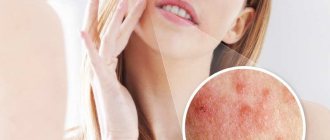
Allergic dermatitis on the face
Allergic dermatitis on the face most often worries women. The fact is that its main cause is contact with the skin of allergens that are part of cosmetics and care products. Although men may well encounter this problem, because irritants are also present in shaving lotions.
The following substances can be identified as striking examples of sensitizers that lead to allergies on the face and neck:
- metals (especially nickel, which is one of the most common materials for making jewelry);
- rubber (part of the sponges used to apply makeup);
- acrylates (used in the manufacture of eyeglass frames);
- pine resin (a strong allergen that is an ingredient in many cosmetics).
Allergic dermatitis on the face can also be caused by plant pollen (both indoor and outdoor), frostbite, chapping, prolonged exposure to direct sunlight, and taking certain medications.
Stages of allergic dermatitis on the face and neck:
- rashes in the form of bubbles and blisters;
- redness, itching and swelling of the affected areas of the skin;
- bullous stage, characterized by the formation of scars and death of the skin.
A mild form of allergic dermatitis on the face is manifested by blistering rashes that form crusts when dry.
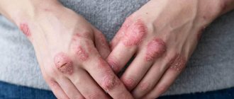
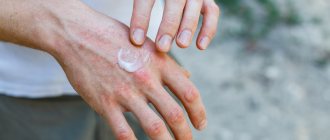
Treatment of dermatitis on the hands - selection of therapy
The selection of therapy for inflammation of the skin of the hands depends on the form and severity of the disease. The first step to recovery is identifying the allergen and stopping contact with it. If the reaction is caused by food products, a hypoallergenic diet is prescribed; if household chemicals or inks or paints are used, then rubber gloves must be used when working with them. If dermatitis on the hands appears upon contact with a certain metal, it is worth replacing it with another or completely abandoning jewelry. Often, for mild contact dermatitis, this treatment is enough to relieve symptoms. More serious cases of the disease require drug treatment and observation by a dermatologist.
The second stage of therapy is to relieve the acute stage of inflammation, during which severe itching, swelling and redness of the skin, and even pain are observed. Drugs are prescribed for internal and external use.
Allergic dermatitis in children
Often in infancy, food intolerance and allergic reactions are observed in a child when eating a particular food. This is largely due to the immaturity of some organs and systems (for example, gastrointestinal), insufficient enzyme production, and a hereditary predisposition to allergies.
- There is an opinion that the regular intake of certain foods or medications by the mother during pregnancy can influence the occurrence of allergic dermatitis in the newborn, even when irritants penetrate through breast milk.
- When allergens enter a child’s body, the immune system produces and accumulates specific antibodies that provoke inflammatory processes on the skin. Rashes on the face, called “diathesis” by parents, are not uncommon. The localization area of dermatitis may affect the shoulders, legs, and buttocks.
By 1.5-2 years of age, dermatitis can move to the next stage of development. Childhood dermatitis, unlike infantile dermatitis, is characterized by rashes all over the body with especially strong manifestations on the knee-elbow folds, upper chest and back. Often, by the age of 7-10 years, allergic dermatitis enters the chronic stage, and occasionally a relapse occurs, especially in spring and autumn.
Symptoms of allergic dermatitis in adults, photos
Symptoms of allergic dermatitis in adults appear depending on the form of the disease (see photo).
1) In the contact form of the disease, symptoms are expressed:
- bright red spots of rashes in various areas of the skin that have contact with the allergen;
- subsequent replacement of spots with bubbles filled with liquid;
- constantly itchy process of focal lesions;
- spread of the rash to healthy tissue, with prolonged exposure to the irritant, accompanied by joint aches, headaches and increased temperature.
Symptoms of diathesis (atopic form) are manifested:
- severely itchy rash on various parts of the body;
- insomnia and nervous disorders due to continuous itching;
- lethargy or severe agitation;
- the addition of a staphylococcal or streptococcal infection in areas of scratching;
- the formation of purulent foci, swelling, cracks and dry yellowish crusts at the site of burst combed blisters.
Signs of toxicoderma are accompanied by:
- general weakness with possible loss of consciousness;
- cold sweat and swelling;
- joint pain and pain in the lumbar area;
- bloody blisters on certain areas of the skin;
- damage to the brain and spinal cord;
- dysfunction of the lungs and liver.
Signs of epidermal toxic necrolysis appear within a very short period of time after contact with an antigenic pathogen. Within one or three days the patient may die.
Symptoms appear:
- a sudden increase in temperature to very high levels, for no apparent reason;
- skin rashes on the torso, arms and legs;
- multiple swollen red spots, gradually merging into large lesions;
- the formation of blisters of various sizes on the lesions (the size of the palm of the sick person), the skin covering them becomes thin, flabby, easily torn under mechanical stress;
- involvement in the process of damage to the mucous membranes of the internal and genital organs.
Stages of development of allergic dermatitis:
- Spicy. After 1-2 days or immediately after close contact with the irritant, swelling, inflammation, and a rash appear on the skin, and the patient complains of severe itching.
- Subacute. At the site of the rash, signs of weeping form, later - scabs and signs of peeling.
- Chronic. Skin that is subject to frequent inflammation becomes rough and thick.
The skin of babies is especially susceptible to negative factors. External signs of dermatitis in children of the first year of life manifest themselves in different ways.
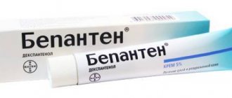
Losterin cream for the treatment of dermatitis on the hands
Natural remedies must be used every day for several weeks or even months, depending on the severity of the disease. It is important that they do not have synthetic fragrances, and that their active components quickly penetrate deep into the epidermis. These requirements are fully met by the “Losterin ” cream, intended for daily care, moisturizing and protecting the skin of the hands. Almond oil regulates water balance. Sophora japonica extract reduces the intensity of inflammation and peeling, and deresined naphthalan is a natural product that has been used in the complex treatment of dermatitis for more than 100 years. Naftalan has bactericidal, vasodilating and regenerative properties, and in combination with urea and salicylic acid it has a powerful antipruritic effect. For hygiene procedures, people with dermatitis are offered the “Losterin” shower gel, which does not contain alkali, does not irritate the skin and has a desensitizing effect.
Treatment of allergic dermatitis
Waiting for the skin reaction to disappear on its own is absolutely not the right approach. No one is immune from its repeated, more violent manifestation and possible complications.
Treatment regimen for allergic dermatitis:
- Eliminating the allergen.
- Antihistamines. Tavegil, diphenhydramine, suprastin have serious side effects: drowsiness, impair coordination, and reduce attention. Therefore, antihistamine treatment should be carried out with new generation medications that do not cause such effects (Zyrtec, Erius, etc.). Allergies that cause severe skin itching are perfectly relieved by fenkarol.
- Antipruritic treatment (sedatives - novopassit, motherwort tincture, valerian tablets).
- Detoxification preparations and enzymes (activated carbon, Mezim-Forte, Linex for dysbacteriosis).
- Local therapy. Ointments and gels with anti-inflammatory, antifungal and antipruritic effects are used (combined action drug - Akriderm). Hormonal drugs are not recommended for children and are used only when other drugs are ineffective; the duration of their use should not exceed 5 days.
When treating with folk remedies, it is worth knowing: the use of medicinal herbs only neutralizes the skin reaction and reduces the body's allergic mood, but does not get rid of the allergy itself.
Nutrition rules
Compliance with nutritional rules for dermatitis is included in the treatment of the disease and allows you to achieve stable remission. A specially developed diet for allergic dermatitis in adults with a daily rational menu eliminates allergens and speeds up the process of getting rid of the disease.
Highly likely irritant foods include:
- nuts;
- coffee;
- sauerkraut;
- seafood;
- citrus;
- legumes;
- chocolates;
- strawberry.
Also, the presence of foods containing preservatives, emulsifiers and dyes should not be allowed in the diet. Dangerous foods for allergy sufferers are rich broths, all fried, salty and spicy foods, which increase the permeability of the gastrointestinal tract to the absorption of irritating substances.
When preparing dishes, it is important to follow the technology and not use expired products. Vegetables and fruits should be purchased from those that were grown without fertilizers. If dishes are prepared from cereals, they must be soaked in water for at least 10 hours. It is recommended to reduce the consumption of sugar and salt by 2 times. As for meat, it is recommended to boil it twice.
List of sources
- Ivanov, O. L. Allergic contact dermatitis and associated allergic dermatoses: modern ideas about etiology, pathogenesis and diagnosis / O. L. Ivanov, E. S. Fedenko // Ros. magazine leather and venereal diseases. 2010. -No. 4. - P. 47-51.
- Korsunskaya, I. M. Therapy of contact dermatitis in adults and children / I. M. Korsunskaya, O. B. Tamrazova, T. A. Shashkova // Vestn. dermatology and venereology. 2006. - No. 4. - P. 46-46.
- Yu. Ilyina, N. I. Allergic skin diseases in clinical practice / N. I. Ilyina, E. S. Fedenko // Ros. allergol. magazine 2005. -№3.-S. 55-67.
- Luss L.V., Erokhina S.M., Uspenskaya K.S. New possibilities for diagnosing allergic contact dermatitis // Russian Journal of Allergology. 2008. No. 2. P. 28-35.
- Immunological mechanisms of development of allergic dermatoses / R.T. Kazanbaev, V.I. Prokhorenkov, T.A. Yakovleva, E.Yu. Vasilyeva // Siberian Medical Review. - 2013. - No. 4. — P.9-13.
Folk remedies
Traditional medicine methods help relieve exacerbation of allergic dermatitis, itching and weeping, and also contribute to the speedy restoration of the skin:
- Baths and rubbing from infusions of string, chamomile, viburnum bark, elecampane root.
- A cream made from sea buckthorn oil mixed with goose fat in equal proportions heals and softens the skin well.
- Grind 5 plantain leaves in a meat grinder, pour 70 ml of the pulp. dry white wine, leave for a day. Lubricate manifestations of dermatitis 2 times a day.
- Fresh basil leaves are crushed into a paste and then applied under a gauze bandage to the affected areas of the skin.
- Gruel from crushed celery is an effective remedy for weeping processes on the skin.
However, in some cases, folk remedies can only aggravate the situation, so such treatment should be undertaken with caution.
Hormonal drugs
Allergic dermatitis in both adults and children is accompanied by unpleasant symptoms, so treatment is largely aimed at eliminating them. Hormone-containing products cope well with inflammation, redness, swelling and itching of the skin. These ointments and creams are based on topical glucocorticosteroids (TGCS). [8] These external agents differ in the strength of the active substance. If the disease has become complicated due to the addition of a microbial infection, it is advisable to use local combination medications, which include, in addition to a glucocorticosteroid, an antibiotic and an antifungal component. [9]
Among the drugs for the treatment of allergic skin diseases is Akriderm GK. The composition includes three active components:
- betamethasone is a topical glucocorticosteroid that has antiallergic, anti-inflammatory, and decongestant effects on the skin;
- gentamicin is a broad-spectrum antibiotic that is active against aerobic gram-negative bacteria, inhibits bacterial protein synthesis and has a bactericidal effect;
- Clotrimazole is a broad-spectrum antifungal component.[10]
The drug is applied to the affected areas of the skin twice a day and begins to act after the first application.[9] Akriderm GK can be used with caution to treat children over two years of age.[10]
ICD 10 code
Contact dermatitis of an allergic nature is classified in the ICD 10 by the following types of diagnoses:
- disease caused by exposure to metals – L23.0;
- allergy caused by adhesive substances – L23.1;
- dermatitis caused by cosmetics – L23.2;
- drug dermatitis – L23.3;
- allergic contact dermatitis caused by exposure to dyes – L23.4;
- dermatitis caused by the influence of various chemicals – L23.5;
- food dermatitis caused by contact of products with skin – L23.6;
- dermatitis caused by non-food plants - L23.7;
- dermatitis caused by exposure to other substances – L23.8;
- dermatitis of unknown etiology – L23.9.
Tests and diagnostics
The diagnosis of allergic contact dermatitis is made on the basis of the clinical picture, medical history, physical examination and the results of skin patch tests. Of particular importance is the medical history (in dermatovenereology), according to which it is necessary to carefully study the history of the development of the disease and especially the factors contributing to the disease. To identify a specific allergen, skin patch tests with allergens (standard test kits) are used. Differential diagnosis is made with atopic and seborrheic dermatitis , simple contact dermatitis , herpetic skin lesions, psoriasis , eczema .
Prevention
To prevent allergic dermatitis, possible sensitization in any form should be avoided (occupational necessity, medications, etc.).
Frequent local use of drugs containing components with highly sensitizing ability is not recommended:
- beta-lactam antibiotics;
- sulfonamides;
- local anesthetics, etc..
If there is a production need for contact with low-molecular substances, it is necessary to use personal protective equipment (gloves, protective suit, mask).
If you are allergic to rubber gloves, they should be replaced with vinyl ones. When planning surgical interventions in a patient with a latex allergy, this must be taken into account. Latex condoms are contraindicated for such people. If you are hypersensitive to formaldehyde, you must be very selective in choosing cosmetics. It is necessary to exclude cosmetics containing this preservative.
If a person is diagnosed with allergic dermatitis, then in order to prevent relapses and complications of the disease, he should be familiarized in detail with the sources of the allergen, as well as with cross-reacting substances.