The white front surface of the eye is the sclera. In a healthy person, its color can range from whitish-blue to whitish-pink. A change in the shade of the sclera indicates the appearance of pathology. This may be due to both systemic diseases of the body and a local ophthalmological problem. Most often, the sclera changes its color to yellow: yellow spots appear on the even color of the white.
Causes
The eye has a rather complex structure; its structure includes the macula, ciliary body, cornea, lens, iris (iris), sclera, and conjunctiva. Pathologies of the visual organs can affect different parts and develop for various reasons.
For your information! A cataract is often confused with a cataract. But with cataracts, the lens becomes cloudy, and the cataract is a clouding of the cornea of the eye.
Eye sores, which may appear as white or yellow spots, affect the cornea. But why does such a disease appear? The main reason for the development of a cataract is cicatricial changes in the structure of the cornea (sometimes the lens). Normally, the cornea consists of special endothelial cells that are not able to regenerate, that is, repair themselves. The cornea is transparent, allows light to pass through to the lens and provides a person with the ability to see clearly.
With various injuries and other pathological influences that can appear under different circumstances, the process of inflammation or scarring starts. During inflammatory reactions, keratocytes included in the structure of the cornea synthesize special substances: chemoattractants and enzymes. The stroma swells, the vessels dilate, and leukocytes move from the bloodstream into the affected area, triggering cellular infiltration. Normal corneal cells are destroyed and replaced by connective tissue cells that are denser and more opaque.
It is because of such processes that a cloudy area appears, which at first has a white or whitish tint. And as the tissues degenerate and fat cells are included in their structure, the thorn becomes yellow or yellowish in color.
So why do eyesores appear? The reasons are as follows:
- injuries to the cornea, for example, from blows, bruises to the eyes;
- injuries to the cornea when hit by sharp foreign objects;
- thermal effects, burns;
- chemical tissue burns that develop when aggressive substances that damage tissue come into contact with the cornea of the eye;
- inflammatory diseases of the cornea (keratitis) of a bacterial or viral nature, which may appear due to the activity of pathogenic microorganisms;
- ulcers localized on the cornea;
- previous surgical interventions on the eyes (especially unsuccessful ones);
- persistently high intraocular pressure, glaucoma;
- pterygiums (recurrent or false).
Risk group
The risk group includes categories of patients suffering from the following conditions and diseases:
- cataract;
- glaucoma;
- diabetes;
- hypertension;
- atherosclerosis;
- autoimmune diseases (cells of the immune system are directed against the body’s own tissues);
- ischemic disease;
- ulceration and erosion on the surface of the eyes;
- chronic infectious diseases.
Also at risk are persons whose work involves the risk of foreign substances or objects getting into the eye area. These include builders and welders.
Types of cataracts
An eyesore can be congenital or acquired. The congenital form is usually caused by intrauterine infection of the fetus during gestation and is often accompanied by other pathologies, for example, clouding of the lens or complete blindness of the baby. Acquired cataracts are the result of various eye injuries or diseases.
Also, cataracts can develop separately and have characteristic symptoms without complications or be combined with other eye pathologies: clouding of the lens, synechiae (adherence of the cornea to the iris or soldering of the iris to the lens and cornea).
In addition, the eyesore may be adhesive and ectatic. In the first case, the cornea fuses (sticks together) with the iris due to significant damage or severe inflammation. Ecstatic leukoma is accompanied by curvature of the cornea, its protrusion.
Symptoms
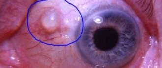
If a cataract appears, it will look like a spot on the cornea: white or yellow. In the first stages of development of leukoma, it has a white tint, and as the tissue degenerates into fat, it becomes yellow or yellowish in color.
The thorn can be located anywhere. If it appears on the iris or on the pupil in front of the lens, it will be more noticeable. This leukoma looks like a whitish spot or cloud, clearly visible against a dark background. When localized in the peripheral zones of the cornea above the sclera, cataracts often remain invisible and have virtually no visible external symptoms.
The main symptom is direct clouding of the cornea with the formation of a whitish, white, yellowish or yellow spot (depending on the degree of tissue damage and the stage of development of leukoma). But concomitant symptoms may also occur, depending on the underlying disease or pathology that provoked the development of the eyesore.
Important!
The thorn may affect only one eye (left or right). Sometimes cataracts appear on both the right and left eyes at the same time.
The clinical picture may include the following manifestations:
- decreased visual acuity (up to its complete loss) when the cataract is located on the iris or pupil (this symptom develops if the clouded area blocks the natural lens of the lens and makes it impossible to see);
- blurred vision, veils, spots before the eyes (any such symptom can occur when the cataract is localized in the optical zone, when the lens is partially or completely covered by a clouded area);
- blurred vision and changes in visual perception (objects look blurry, without clear contours, distorted);
- swelling of the cornea and other parts of the eyes, redness, increased lacrimation (such symptoms indicate inflammatory diseases);
- sensation of a foreign body in the eye (this symptom occurs when the cataract grows and thickens).
Diagnostics
Diagnosis of the patient’s condition consists of several stages:
- Anamnesis collection. This is data obtained from the words of the patient or his close relatives. Based on these, the doctor may prescribe further examination.
- General examination of the patient's condition. The doctor evaluates the extent to which the superficial tissues of the eyes are affected. The skin of the eyelids and surrounding tissues may be involved in the process.
- Submission of laboratory tests. These include general blood and urine analysis, blood biochemistry, glucose determination, lipid profile. The presence of an inflammatory focus is detected using indicators of leukocytes and ESR, the amount of glucose and fat deposits.
- Fundus examination. A solution is first instilled into the patient’s eyes, which temporarily eliminates eye accommodation. The doctor looks at the condition of the lens, vitreous body, retina, and eye cameras. Evaluates the transparency or opacity of these areas, the presence of damage, hemorrhages.
- MRI, . Not only the structures of the eyeball, but also the brain are assessed. The doctor can reveal the condition of these structures layer by layer, since many tissues are visible through fundus examination.
Based on the data obtained, the doctor can make a reliable diagnosis. Only after diagnosis is treatment prescribed individually for each patient.
Treatment
How to get rid of a thorn? If the leukoma is not located in the optical zone, does not cover the lens, does not in any way make vision worse and is not accompanied by obvious manifestations, then treatment is not required. But regular visits to an ophthalmologist are required to monitor the dynamics. If the condition worsens, for example, the growth of the cataract and the involvement of other parts of the visual organs in pathological processes, therapy is prescribed.
Conservative treatment
Conservative treatment is effective only in the earliest stages and usually includes the use of drugs that promote the resorption of scars and trigger regeneration. Korneregel, Lidaza, Actovegin are prescribed.
Physiotherapeutic procedures can also be prescribed for leukomas that eliminate progression, stop pathological changes and help restore the normal structure of the visual organs. Ophthalmologists use the following techniques:
- iontophoresis – exposure to low-frequency currents;
- diathermocoagulation – cauterization of damaged and altered tissues with high-frequency alternating currents;
- Diathermy – heating of tissues with high-frequency currents.
To eliminate an obvious visible external defect, the doctor may recommend that a patient with a cataract wear a colored contact lens, which will cover the cloudy area and create a normal iris color.
Important! If the thorn grows and becomes more cloudy, then you will not get rid of it with conservative treatment. Only radical methods will be effective.
Surgery
The surgical treatment method is the most effective and is indicated when the cataract interferes with vision and the person sees worse due to leukoma. Only surgery can completely get rid of the cataract. Keratoplasty or keratoprosthesis is performed. In the first case, the surgeon makes incisions in the cornea with special instruments or a laser beam, and then replaces the clouded area of the cornea with donor material. During keratoprosthesis, instead of the excised area with a cataract, an artificial prosthesis is installed, which has all the properties of the natural cornea.
Usually, combined operations are performed on the right or left organ of vision, during which, in addition to removing the cataract, other manipulations are performed, for example, installing an intraocular lens, replacing the lens, and others.
Our services in ophthalmology
The administration of CELT JSC regularly updates the price list posted on the clinic’s website. However, in order to avoid possible misunderstandings, we ask you to clarify the cost of services by phone: +7
| Service name | Price in rubles |
| Ultrasound scanning of the anterior segment of the eye | 1 000 |
| Comprehensive OCT examination of the retina (one eye) | 3 500 |
| Antiglaucomatous surgery for primary glaucoma stage 1 of the disease | 55 000 — 85 000 |
| Vitrectomy with cataract phacoemulsification without IOL cost | 90 000 — 130 000 |
All services
Make an appointment through the application or by calling +7 +7 We work every day:
- Monday—Friday: 8.00—20.00
- Saturday: 8.00–18.00
- Sunday is a day off
The nearest metro and MCC stations to the clinic:
- Highway of Enthusiasts or Perovo
- Partisan
- Enthusiast Highway
Driving directions
Prevention
You will get rid of the high risks of cataract formation if you follow the rules of prevention:
- Protection of eyes from injury, thermal and chemical influences.
- Timely treatment of eye inflammatory diseases.
- Compliance with personal hygiene rules. Avoid touching your eyes with dirty hands.
- Wearing sunglasses.
- Providing timely medical care after eye injuries.
- Use safety glasses when working with welding and chemicals.
If you do not get rid of the cataract in a timely manner, then this pathology can provoke undesirable consequences: the development of myopia or astigmatism, complete loss of vision, spasm of accommodation. The external defect will become more and more noticeable, causing complexes and worries. Therefore, at the first sign of leukoma, visit an ophthalmologist and follow his recommendations.
Types and severity of cataracts
White eyesore can be congenital or acquired. The second case is more common, since people are exposed to various lesions of the corneal layer, conjunctiva, and injure and burn their eyes. According to the severity, the anomaly is mild, moderate and severe. At first, barely noticeable spots appear that do not cause any discomfort to the eyes. With an average degree of damage, the spots thicken and become visually noticeable. The severe degree is characterized by the overgrowth of the spot with a vascular network.