Risk factors (reasons for development)
Psoriasis occurs in approximately 3% of the world's population, regardless of gender. It begins at any age, but most often between 15 and 30 years. There are cases of the disease being detected in infants. The highest prevalence is among representatives of the Caucasian race, less often among the Negroid and Mongoloid races.
The disease is characterized by a wave course with periods of exacerbation and remission. Has an acute onset in most cases. A monomorphic rash of papules merges into larger plaques, which are then covered with white scales.
The etiology and pathogenesis of skin pathology have not been fully studied; both hereditary predisposition and neurogenic nature are assumed. Many patients note that the disease manifests itself and subsequently intensifies during periods of greatest stress. Some researchers believe that the cause of the disease may be an infection, but the virus has not been identified. This theory is contradicted by the fact that the patient’s immediate environment does not get sick, and the presence of psoriasis in the family history increases the likelihood of its diagnosis in descendants.
What is GWAS?
Identifying relationships between gene variants and phenotypic manifestations requires studies across multiple genomes. Until the 1990s, such work was not carried out, since there was no suitable technology yet [3]. But now genome-wide association studies, or GWAS (genome-wide association studies), have become much simpler (Fig. 1). Such studies compare genome arrays of healthy people and patients with a diagnosed disease. If one of the alleles occurs much more often in those suffering from the disease, then it is considered associated with it. Today, a huge “library” of genetic markers has already been collected in the form of single nucleotide polymorphisms, or SNPs (single nucleotide polymorphism), differences in one nucleotide. In addition to predisposition to disease, the presence of genetic polymorphisms can affect the absorption, metabolism and excretion of drugs, and therefore their overall effectiveness. Because of this, such data are also useful for pharmacokinetic studies [4].
Figure 1. How GWAS works—the process of finding single nucleotide polymorphisms (SNPs) that correlate with disease risk.
[9], figure adapted
However, GWAS is not a salvation. He rarely gives clear answers, but he encourages further work. The fact is that variants associated with a disease do not always cause the disease directly, and the cause-and-effect relationships of the development of the disease have to be checked and rechecked [1].
Manifestations of psoriasis
At the first stage, the disease manifests itself in the form of a single pink papule. It changes, increases in size, grows above the skin, and becomes covered with white scales. More often, psoriasis plaques appear in places of friction and increased pressure on the skin: buttocks, elbows and knees. As the disease progresses, they can be found absolutely anywhere on the skin, including the scalp.
Main clinically diagnosed external signs:
- Stearic stains, i.e. easy separation of flakes by scraping.
- The terminal film remaining after the scale is removed. It looks like a smooth, shiny, even pink surface.
- Pinpoint bleeding, which can be caused by removing the scale.
Psoriasis develops quite slowly; an increase in the number of plaques and their growth can be observed over several months or years. In a small percentage of patients, the disease may manifest itself more intensely. As a rule, this is preceded by severe mental stress or a serious illness requiring massive drug treatment. In this case, the papules are not pale pink, but bright red, with obvious signs of inflammation, swollen, causing itching.
The second stage of psoriasis is characterized by more extensive lesions. At the site of scratching, new papules appear, forming new plaques. As a result of peripheral growth, new growths merge with existing ones. Plaques affect symmetrical limbs and form similar patterns and lines.
At the third stage, growth slows down, changes concern mainly the structure of the rash. The boundaries between healthy and affected skin become clearer. The plaques acquire a bluish tint and begin to actively peel off. In the absence of therapy, they thicken and sometimes form papillomatous nevi (brown) and warty growths (flesh-colored).
There is another stage - regression of the disease, at which time the symptoms fade away. Peeling goes away, the clarity of the border disappears, the skin normalizes and returns to its original state.
How to understand whether we have lichen or psoriasis
To summarize all of the above, we can highlight several similar features of the two diseases:
- autoimmune nature;
- rash in the form of papules;
- itching;
- red papules;
- The flexures of the arms and legs are often affected.
The question arises of how to distinguish psoriasis from lichen. The diseases have several differences:
- psoriatic plaques peel off, scales can be seen on the surface, and with lichen planus the surface of the papules is smooth;
- psoriasis does not occur on mucous membranes, but LLP often affects the oral cavity and mucous surfaces of the genitals;
- plaques of psoriasis are larger than those that form with lichen;
- If lichen planus affects the scalp, cicatricial alopecia may occur.
Classification of psoriasis
The disease has many types, but they are all divided into two main types:
- Non-pustular psoriasis (simple, plaque, erythrodermic).
- Pustular psoriasis (generalized, palmoplantar, annular psoriasis, persistent acrodermatitis, impetigo).
A number of disease researchers identify the following types of psoriasis:
- drug-induced
- reverse type,
- seborrheic;
- Napkin's psoriasis (occurs in infants in the diaper area).
According to ICD-10, plaque psoriasis, inverse psoriasis, guttate psoriasis, pustular psoriasis, nail psoriasis (onychodystrophy), psoriatic arthritis and erythrodermic psoriasis are distinguished. Let's look at common forms in more detail.
Pustular form of psoriasis
It is characterized by the presence of plaques with cortical scales, impregnated with exudate. If damaged, for example, as a result of scratching or self-injury in the folds of the body, the rashes become wet. They cause itching and burning and cause physical discomfort. This type of disease is more often diagnosed in people with excess body weight, hypothyroidism and diabetes.
Seborrheic type
Affects the scalp. Occurs in people with dandruff and oily seborrhea. This makes diagnosis difficult at the initial stage of development. Over time, psoriasis lesions grow. They can spread to the skin of the forehead, to the ears, to the back of the neck.
Psoriasis of the hands, feet and nails
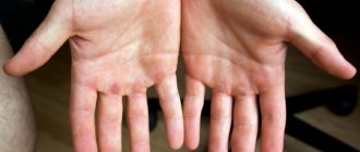
The main rash occurs on the extremities, with only isolated papules on the body. The affected nail plates become deformed, turn yellow, and thicken. They can go away painlessly. A psoriatic border may form around the affected area. There are subungual hemorrhages that look like dark red spots, which then turn black.
Pustular (generalized) form
It has a classic pattern of development, starting with a single vesicle that develops into plaques. The lesions are symmetrical and can affect any part of the body. The severe course of this form of psoriasis is characterized by the appearance of intraepidermal pustules. They can unite, forming “purulent lakes.” The pustules do not open on their own, since they are externally protected by a dense brown crust.
Arthropathic form
The most severe form of psoriasis, in which changes first affect small joints, and then large ones, including the spine. This is expressed by pain symptoms and their deformation. Joint fusion and loss of mobility are likely. Against the background of this form of psoriasis, other pathologies arise: ankylosis, osteoporosis, which leads to disability.
Modern approaches to the diagnosis and treatment of psoriasis
Currently, psoriasis (scaly lichen) is considered as a disease of a multifactorial nature.
Hereditary factors undoubtedly play a certain role in its development [1, 2, 3]. It has been proven that psoriasis is inherited in an autosomal dominant manner, with incomplete penetrance. The study of the molecular genetic basis of the development of lichen planus has made it possible to establish an important role in the determination of predisposition to psoriasis for many genes. To date, several chromosomal susceptibility loci to psoriasis have been mapped. The most significant in this group is considered to be the PSORS 1 locus in the 6p21.3 region, extending from the MICA gene to the CDSN gene, where the genes of the HLA system (Human Leukocytes Antigens) are located, among which the HLA-Cw6 antigen shows a significant association with the disease. It is possible that the PSORS 1 locus does not contain a single main gene responsible for the occurrence of psoriasis, but rather a cluster of alleles that are associated with the development of the disease.
Currently, an association of allele 2 and a set of alleles 1/2 of the M6S190 marker with severe psoriasis has been shown. An association of histocompatibility antigens HLA-B13, HLA-B17 with vulgar psoriasis, and HLA-B27 with arthropathic psoriasis (psoriatic arthropathy) has been established. Bacterial and viral theories in the etiology of psoriasis and possible changes in the genetic apparatus under their influence are considered [2].
Immune disorders play an important role in the pathogenesis of psoriasis. Skin damage is accompanied by an influx of activated T-lymphocytes. Increased synthesis of interleukin-1 (IL-1) by macrophages and activated keratinocytes induces T cells to produce IL-2, which, in turn, is a powerful stimulator of T-lymphocyte proliferation. Activation of T-helper cells is pathogenetically associated with the proliferation of epidermal cells.
Excessive migration of lymphocytes from the peripheral blood into the affected skin may cause these cells to infiltrate the epidermis by altering cytoplasmic membranes, increasing adhesion to endothelial cells, IL-1 T cell chemotaxis, and promoting migration into the epidermis.
Active mediators of inflammation are histamine, which is released during degranulation of mast cells and affects the permeability of the vascular wall; hydrolases released from neutrophils; prostaglandins, leukotrienes and other arachidonic acid derivatives. Increased local production of arachidonic acid metabolites may be caused by cytokines produced by macrophages or keratinocytes [3].
In patients with common forms of the disease, the content of antibodies to deoxyribonucleic acid (DNA) in the blood increases, with a predominance of autoantibodies to native DNA, which contributes to the formation of an autoimmune process.
Psoriasis is characterized by changes in the ratio of histone protein fractions, which have a special place in the chain of regulation of proliferative activity and DNA synthesis, which make up most of the chromatin.
In patients with psoriasis, dysfunctions of the central, peripheral and autonomic nervous systems, endocrine glands, and metabolic changes are detected [1, 3].
There are three clinical stages of psoriasis: progressive, stationary and regressive. Under the influence of provoking factors (trauma, psycho-emotional stress, infectious diseases, inadequate treatment methods), an exacerbation of the disease may develop with the appearance of abundant small nodules (papules), prone to peripheral growth, and the formation of plaques of various sizes and shapes, which can be isolated or occupy extensive areas. areas of the skin up to total skin damage. The progressive stage is characterized by a symptom of an isomorphic reaction (Koebner phenomenon), which is characterized by the fact that typical psoriatic rashes appear at the site of injury, even minor ones.
At the stationary stage, the appearance of new elements stops and the tendency for peripheral growth of existing plaques disappears.
The regressive stage is characterized by a decrease in the color intensity of the plaques, their flattening, decreased peeling, infiltration, and resorption of elements with the subsequent formation of foci of hypo- or hyperpigmentation at the site of former rashes. Around the elements you can see a narrow pseudoatrophic Voronov's rim.
There are three types of psoriasis depending on the seasonality of relapses: winter, summer, indeterminate.
Psoriasis is characterized by monomorphic rashes in the form of papules of various sizes. At the onset of the disease, in typical cases, the rash is limited in nature and is represented by single plaques in places of “favorite” localization: the scalp, the extensor surfaces of the elbow, knee joints, and the sacral area. The plaques are clearly demarcated from healthy skin, bright pink or deep red in color, and covered with loose silvery-white scales. When scraping the eruptive elements, characteristic diagnostic symptoms are revealed (psoriatic triad): “stearin stain” (scraping intensifies peeling, causing an association with the stearin of the suppository); “terminal film” (the appearance of a shiny, wet surface after removing the scales); “pinpoint bleeding” (drip bleeding with further scraping). Diffuse peeling or sharply limited layers of scales may occur on the scalp, which in some cases also extend to adjacent areas of smooth skin. Hair is not affected by psoriasis [1, 3].
Depending on the size and nature of the eruptive elements, the following clinical forms of scaly lichen are distinguished: spot psoriasis, characterized by papules the size of millet grains; guttate psoriasis, when the papules reach lenticular sizes (the size of lentils); coin-shaped psoriasis with papules with a diameter of 3–5 cm; ring-shaped psoriasis, when papules form rings, leaving areas of healthy skin in the center; figured, or geographic, psoriasis, with the rash resembling a geographical map; seborrheic psoriasis, when pinkish scaly lesions are located in places characteristic of seborrhea [1, 2, 3].
Nails are affected in about a quarter of psoriasis patients. Primary nail psoriasis develops as a result of damage to the nail matrix and manifests itself in the form of pinpoint depressions and spotting of the nail plates. Secondary psoriasis of the nails is a consequence of damage to the nail bed and ridges and is characterized by subungual horny yellowish-brown thickenings that are visible through the nail plates. The nails thicken, crumble, subungual hemorrhages and onycholysis are observed. Psoriatic onycholysis is often diagnosed without previous inflammation. When the palms and soles are affected, extensive, round, scaly, furrowed plaques with clear boundaries appear.
Exudative psoriasis is characterized by a pronounced inflammatory reaction of the skin, which is manifested by the presence of lamellar scale-crusts on the surface of plaques, sometimes multi-layered, resembling a layer cake in appearance. When the scale-crusts are removed, a wet surface is exposed.
Pustular psoriasis can be generalized (Zumbusch form) or limited, affecting the palms and soles (Barber form). The occurrence of this severe form of psoriasis is facilitated by stressful situations, infections, and irrational treatment. Generalized pustular psoriasis occurs with fever, leukocytosis, increased erythrocyte sedimentation rate (ESR), and a general severe condition. Suddenly, against the background of bright erythema, small superficial pustules appear, which is accompanied by burning and soreness; pustules can be located in the area of regular plaques and on previously unchanged skin. New foci of pustulation appear in paroxysms, occupying large areas of the skin. Merged pustules cause detachment of the epidermis in the form of “purulent lakes”, and erythroderma can develop.
Limited pustular psoriasis is more common; rashes are predominantly localized on the palms and soles in the form of pustules against a background of erythema and skin infiltration. The course, in comparison with the generalized form, is milder, but persistent, with frequent relapses with a satisfactory general condition. The provoking factor is irritating local therapy [3].
Psoriasis of the folds is more often observed in children and the elderly, especially in patients with diabetes. The lesions are located in the armpits, under the mammary glands, in the perineum, in the inguinal-femoral folds, in the navel area and are characterized by sharp boundaries, a rich red color and slight peeling.
Psoriasis of the palms and soles can exist in isolation or simultaneously with damage to other areas of the skin; it is characterized by the formation of hyperkeratotic lesions with clear boundaries, covered with scales that are difficult to scrape off, and the presence of painful cracks. The characteristic psoriatic triad is difficult to identify [3].
Verrucous psoriasis is characterized by severe infiltration, hyperkeratosis, and warty growths, often located on the skin of the trunk and limbs.
In a severe immunosuppressive state, rupoid psoriasis develops, manifesting itself in the form of plaques covered with a layer of large scales, resembling oysters, clearly defined and densely infiltrated [1, 3].
Arthropathic psoriasis, along with skin lesions, is characterized by the involvement of joints in the pathological process. Arthritis is manifested by pain, swelling, and limited mobility in the affected joints, most often small ones. Arthropathy can develop from minor arthralgia to generalized lesions and lead to disability of patients. Changes in the joints precede skin lesions or develop at different times after the debut of the skin process. The severity of skin changes may vary.
Psoriatic erythroderma is a severe form of psoriasis that develops with the gradual progression of the psoriatic process and the fusion of plaque elements into lesions of the entire skin. Psoriatic erythroderma is characterized by severe hyperemia, swelling, infiltration of the skin with abundant large- and small-lamellar, less often pityriasis-like peeling. The rash may be accompanied by itching of varying intensity, often severe. This form of psoriasis is characterized by a deterioration in the general condition (fever, weakness, reaction of the lymph nodes, heart failure, dysfunction of the liver, kidneys, changes in blood tests, hair loss) [1, 2, 3].
According to histological examination, skin lesions in patients with psoriasis are characterized by the following changes: pronounced acanthosis, parakeratosis and varying degrees of intensity infiltration of the dermis with lymph and leukocytes, as well as the presence of a small number of cells with morphological signs of apoptosis, compared to the control. In patients before treatment, a significant decrease in the intensity of keratinocyte apoptosis was recorded in comparison with the control. During therapy, the destruction of keratinocytes by apoptosis increases, which is most pronounced in patients with a short duration of the disease. The mitotic activity of keratinocytes, increased in patients before treatment, decreases somewhat during therapy, but does not reach the norm. The number of intraepidermal lymphocytes, increased before treatment, significantly decreased after therapy. Thus, it can be assumed that under the influence of treatment, some intraepidermal lymphocytes are eliminated and keratinocyte apoptosis is stimulated against the background of unchanged proliferative activity of these cells, which leads to a reduction in the severity of the pathological process in the skin. In this case, it is possible to activate the function of intraepidermal macrophages, which, as is known, can influence the process of apoptosis of keratinocytes and thereby reduce the level of lymphocytic infiltration [4].
Treatment of psoriasis is aimed at suppressing the proliferation of epithelial cells and eliminating the inflammatory process and is prescribed taking into account the anamnestic data, form, stage, extent of the process, concomitant diseases, age and gender of the patient, contraindications to a particular treatment method or drug [3]. The patient’s motivation, family circumstances and social factors are of great importance [5].
Intensive research conducted in many countries around the world has made it possible to develop a variety of methods for treating psoriasis, which, however, can only stop the exacerbation of the disease, and not cure it [5].
In case of mild psoriasis, which is limited in nature, or in the presence of single “duty” plaques, external treatment, balneo- and spa therapy, adherence to diet, work and rest are often sufficient [6].
Moderate and severe forms of the disease require complex treatment on an outpatient basis, and during exacerbations, therapy in a hospital setting. In order to prolong remission, courses of anti-relapse therapy (vitamins, biostimulants, physiotherapy) should be carried out [1, 6].
Often, in the treatment of psoriasis, hydrating agents are used to soften the flaky surface of psoriatic elements: lanolin-based creams with vitamin supplements, Unna cream, psoriatic ointment [1, 3, 6].
Among keratolytic agents, the most frequently used drugs are those containing salicylic acid. Depending on the location, degree of infiltration and hyperkeratosis of the lesions, the content of salicylic acid can range from 1 to 5%. Ointments with salicylic acid (Akriderm SK, Belosalik, Diprosalik) soften the flaky layers of psoriatic plaques and accelerate their resolution [5, 6].
In Western European countries, products containing anthralin are often used. The mechanism of action of anthralin is to inhibit the synthesis of nuclear and mitochondrial DNA, as well as to slow down the activity of cellular enzymes, polyamines and phosphorylation processes, resulting in a decrease in cell proliferation. Anthralin is applied for 1 hour and then washed off, as a result it penetrates only into the affected skin without loss of effectiveness. It is advisable to use agents containing anthralin in stationary and regressing stages [6].
Due to the likelihood of irritation and unpleasant odor, doctors are now less likely to prescribe tar preparations to patients, which have keratoplastic and anti-inflammatory effects. Most often, external products contain 2–3% tar. Coal tar and tar are often used in medicated shampoos to prevent or reduce flaking of the scalp. Tar preparations are contraindicated for exudative psoriasis and kidney diseases [3, 6].
Of the external glucocorticosteroid agents for psoriasis, the prescription of elocom is pathogenetically justified, since the drug suppresses the production of three cytokines: IL-1 and IL-6 and tumor necrosis factor alpha (TNF-α). The existence of various dosage forms (cream, ointment, lotion) allows you to choose the optimal therapy depending on the location of the lesions [7]. Combination drugs have been developed.
Irrational therapy with external glucocorticosteroids can lead to the transition of psoriatic disease to a more severe form (pustular variety, psoriatic erythroderma), cause thinning of the skin, the appearance of areas of atrophy, sluggish local infections, hypopigmentation and tachyphylaxis [6].
Similar undesirable effects have not been reported with calcipotriol, a vitamin D analogue.
In our country, Daivonex is used in the form of ointment and lotion. The drug inhibits cell proliferation and accelerates their morphological differentiation, thus normalizing cell development. The greatest effectiveness is achieved with long-term, multi-week use of the drug in patients with a PASI index from 10 to 20. For more severe forms, Daivobet is indicated, containing betamethasone dipropionate along with calcipotriol. Staged treatment of psoriasis is justified, when therapy begins with the use of daivonex, then combination therapy is prescribed and subsequently switched to treatment with daivonex. To avoid the development of withdrawal syndrome, the first and second stages should have the same duration [3, 6].
For the winter type of psoriasis, exposure to ultraviolet irradiation (UVR) is traditionally used. When taking photosensitizing drugs and irradiating with long-wave UV rays, the effectiveness of UV irradiation increases significantly. This method became possible thanks to the creation of special installations that provide irradiation in the UVA region (ultraviolet A radiation). Preparations from the group of psoralens (5-, 8-methoxypsoralen) are used as photosensitizers. The method is called photochemotherapy (PCT), or PUVA therapy.
Since 1974, vast experience in the successful use of PUVA therapy has been accumulated in world practice [5].
Installations intended for photochemotherapy are a booth or screen, on the inner wall of which special fluorescent lamps are mounted, providing long-wave UVA radiation (320–400 nm) with maximum intensity at 350–365 nm. The UV radiation density in the installations is 8–13 mW/cm2. PUVA devices have various modifications that allow procedures to be performed in a lying or standing position, and to irradiate the head, legs, palms, and soles separately. PUVA therapy is indicated for severe, widespread forms of psoriasis: exudative, widespread plaque, erythrodermic, pustular, palmoplantar. The features of photochemotherapy are the absence of addiction, the possibility of repeated courses, including outpatient ones, a significant reduction in the severity of psoriasis and an extension of the period between relapses [3, 5].
At a progressive stage of the disease, PUVA therapy can be started after detoxification therapy (hemodesis 400 ml intravenously 3–4 times), the prescription of sedatives and hypnotics [5].
Contraindications to PUVA therapy are all blastomatous processes, acute and chronic liver and kidney diseases, pregnancy, diabetes mellitus, cataracts, hypersensitivity to sunlight, indications of past use of arsenic preparations and radiotherapy [6].
Selective phototherapy has broader indications for use: it can be used at an advanced stage, in children, with limited lesions. Selective phototherapy is carried out using devices that provide medium-length radiation (280–320 nm). Photosensitizers are not prescribed. If PUVA therapy is quite effective when used as monotherapy, then selective phototherapy is usually combined with other treatment methods (detoxification agents, desensitizing agents, etc.) [5].
Psychotropic drugs are an important adjuvant for the treatment of patients with psoriasis whose psychosomatic status contains elements of asthenia and neuroticism. Correction of these conditions should be carried out with the participation of a neuropsychiatrist [6].
In the progressive stage, in addition to hemodesis, enterosorbents (activated carbon, polyphepan, enterosgel, etc.) are used [3, 6].
Patients with psoriasis and concomitant pathology of the gastrointestinal tract are recommended to be prescribed enzyme preparations and hepatoprotectors [6].
At the progressive stage of psoriasis, calcium preparations, sodium thiosulfate, and magnesium sulfate are also used. They have a hyposensitizing, detoxifying and anti-inflammatory effect, reduce the permeability of the vascular wall [6].
Synthetic derivatives of vitamin A (synthetic retinoids) are significantly effective for psoriasis: tigazon, neotigazon, etc. They affect the pathologically keratinizing epidermis, the immune system, and have antineoplastic properties. The antikeratinizing effect is manifested by suppressing the proliferation of epidermal keratinocytes, normalizing the differentiation of non-keratinizing epithelium, and reducing the adhesion of horny cells. The latter leads to faster exfoliation and prevents the formation of horny masses. Synthetic retinoids maintain the normal rate of mitosis in epidermal cells and regulate its thickness. Monotherapy with retinoids is most effective for pustular psoriasis, when pustular elements quickly disappear, epithelialization occurs, body temperature normalizes and the general condition of the patient improves [5].
The combination of retinoids with PUVA therapy (re-PUVA therapy) can significantly increase the effectiveness of treatment. This treatment method is used for severe forms of psoriasis, including palmoplantar lesions. With re-PUVA therapy, it is possible to increase the effectiveness of treatment and reduce the number of radiation sessions and thereby the total dose of ultraviolet radiation [5, 6].
The use of retinoids is limited by a number of contraindications. Retinoids have an absolute teratogenic effect and are contraindicated in cases of impaired liver function, kidney function, hyperlipidemia, neoplasms, hypervitaminosis A. When using these drugs, side effects and undesirable reactions from various organs and systems are possible. Dermatological changes include dry mucous membranes, skin rashes, itching, cheilitis, erythema, sweating, peeling of the palms and soles, paronychia, nail dystrophy, increased proliferation of granulation tissue in the affected area, and in rare cases, thinning hair, vasculitis, and photosensitivity. From the sensory organs, conjunctivitis, photophobia, decreased night vision, clouding of the cornea, decreased hearing, and nosebleeds are possible. Neurological disorders may include headaches, and in rare cases, depression and seizures. From the digestive system, nausea is possible, rarely - colitis, bleeding, and a transient increase in the activity of liver transaminases. Hematological disorders include anemia, neutropenia, changes in platelet count, and increased ESR. There is a possibility of developing metabolic disorders such as increased concentrations of thyroglobulin and glucose. From the musculoskeletal system, pain in muscles and joints is possible, and rarely - hyperostosis.
In the treatment of severe psoriasis, immunosuppressive therapy is often used. Methotrexate, a folic acid antagonist, has been used in dermatology for 30 years. A large number of complications (nausea, vomiting, ulcerative lesions of the oral mucosa, thrombocytopenia, toxic hepatitis, kidney damage) prevent the spread of this treatment method. Its disadvantages are the rapid occurrence of relapses after improvement and torpidity to other treatment methods [5]. Currently, cyclosporine A is more often used, which suppresses the development of cell-type reactions, as well as T-lymphocyte-dependent antibody formation [6]. The immunosuppressive effect of cyclosporine A is based on blocking the interleukin mechanism of immune reactions. After a single oral dose, the peak concentration of the drug in the blood occurs after 2–6 hours. The maximum dose of the drug is 5 mg/kg per day. The average maintenance dose is 1.5 mg/kg/day. The average duration of therapy is 3 months.
In some cases, a sequential combination of the drug with plasmapheresis or PUVA therapy is effective [8].
For severe refractory and disabling forms of psoriasis, infliximab (Remicade), which blocks the expression of TNF-a, is indicated. The drug contains chimeric monoclonal antibodies consisting of 75% human protein and 25% mouse protein. Infliximab is produced by genetic engineering from a line of recombinant cells accumulated through passage and perfusion. The drug is available in 20 ml bottles containing 100 mg of lyophilized powder [9].
Infliximab blocks the activity of TNF-α, which leads to a decrease in inflammation, keratinocyte proliferation and cell differentiation disorders in patients with psoriasis. In patients with an average PASI index of 48, the drug was used at a dose of 3.5–5 mg per 1 kg of body weight. As a result of prescribing the drug in the amount of three infusions, at the 1st, 2nd, 6th week, a clinical effect is achieved, the average PASI index decreases to 7.9 [9].
Literature
- Skripkin Yu. K., Selissky G. D., Fedorov S. M., Khubieva F. V. Skin diseases and sexually transmitted infections: a reference book. M.: Medical Information Agency, 2003. 544 p.
- Azarova V.N., Khamaganova I.V., Polyakov A.V. Genetics of psoriasis // Russian Journal of Skin and Venereal Diseases. 2003. No. 6. pp. 29–33.
- Samsonov V. A., Znamenskaya L. F. Psoriasis // Medicine for everyone. 2001. No. 2.
- Ruksha T. G., Salmina A. B., Prokhorenkov V. I. et al. Apoptosis and proliferation of keratinocytes in patients with psoriasis with different duration of the disease // Clinical Dermatology and Venereology. 2003. No. 2. P. 60–62.
- Chistyakova I. A. Modern problems of therapy and prevention of psoriasis // Russian Medical Journal. 1997. T. 5. No. 11. P. 709–720.
- Fedorov S. M. Psoriasis: clinical and therapeutic aspects // Russian Medical Journal. 2001. T. 9. No. 11. P. 447–451.
- Danilov S.I., Piryatinskaya V.A. New generation topical glucocorticosteroids in external therapy of dermatoses // Russian Medical Journal. 2000. T. 6. No. 6. P. 257–260.
- Smirnova L. M., Kochergin N. G. Cyclosporin A for refractory psoriasis // Clinical dermatology and venereology. 2003. No. 2. P. 45–51.
- Korotkiy N. G., Polyakova A. A. Modern therapy of severe forms of psoriasis // Consilium medicum. 2005. T. 7. No. 1. Appendix. pp. 25–26.
I. V. Khamaganova, Doctor of Medical Sciences, Professor of Russian State Medical University, Moscow
Diagnostic methods
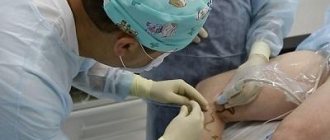
The diagnosis and treatment of psoriasis is carried out by dermatologists. Initially, an external examination of the affected areas is carried out, and an anamnesis is collected. Sometimes the disease is similar to other diseases, especially in the first stage. If the hands and nails are affected, it is important to exclude the presence of fungal infections. The seborrheic type also requires differential diagnosis. Seborrheic eczema, pityriasis rosea and papular syphilis should also be excluded.
In case of active disease and large lesions, visual analysis of scrapings is used. In the process of scraping, peeling intensifies. In place of the removed scale, a smooth, thin film is visible. When mechanically applied, it moves away, revealing a moistened surface with droplets of blood.
In most cases, diagnosing psoriasis is not difficult; it does not even require testing; it is enough to examine the skin. However, the doctor must rule out an error in the diagnosis, determine the presence of concomitant diseases and other pathologies occurring against the background of psoriasis. For this purpose, the following diagnostic measures may be prescribed:
- Blood test to detect elevated levels of rheumatoid factor titers, ESR, proteins, leukocytosis, disorders characteristic of endocrine and biochemical pathologies.
- A skin biopsy is performed to confirm psoriasis, clarify its classification and exclude other skin diseases. The nature of the thickening of skin epidermal cells, the presence of Rete accumulation, immunocompetent and other cells are checked.
Laboratory tests are also important when prescribing therapy, to monitor the effectiveness of treatment, and to identify the causes and exacerbation of psoriasis.
Where is the truth, or why the results of different GWAS often contradict each other?
It is too early to draw definitive conclusions on the genetic basis of psoriasis, despite the fact that many genes associated with this disease have been found using GWAS. In fact, the results of many GWAS are subject to inconsistencies and limitations imposed by both the study subject and the study methods.
First of all, psoriasis is a very complex, complex disease characterized by significant phenotypic heterogeneity. Many mutually influencing genetic and non-genetic factors take part in its development. It would be unreasonable to expect DNA reading to immediately cover all the blind spots in the study of psoriasis.
Many questions arise regarding the designs of the works themselves. For example, most works are retrospective, that is, they analyze the results of previous studies and source material collected by other authors. In addition, the number of patient participants in many publications is quite small. Different criteria for the success of treatment are also used. Finally, sometimes in the same study, representatives of different ethnic groups or patients taking different medications are mixed - of course, this also affects the result of the genetic analysis [71]. Let's hope that large-scale studies conducted in homogeneous populations will appear in the near future.
However, there is another explanation for why analysis of primary genetic material does not provide all the answers: epigenetics. This is precisely the reason that of two monozygotic twins (with the same genotypes), psoriasis sometimes occurs in only one. Due to external influences, gene expression can change, and there are many tools for such regulation: DNA methylation, chemical modifications of histones, microRNAs... Among the epigenetic factors responsible for the pathogenesis and/or progression of psoriasis are environmental conditions, exposure to various infectious agents (especially streptococci group A), psychological stress, smoking, obesity, excessive alcohol consumption, use of a number of medications, including non-steroidal anti-inflammatory drugs and immunomodulators, and much more [8], [11], [22]. Exciting details of the influence of epigenetics on the development of psoriasis await you in the next article of the series!
Types of analyzes
The main tests performed for psoriasis include:
- Skin biopsy, a piece of skin is examined to differentiate bacterial, fungal infections, and cancer.
- Complete blood count to detect leukocytosis and anemia.
- Erythrocyte sedimentation rate to determine the type of psoriasis. With pustulosis and erythroderma, the indicator remains normal.
- Uric acid test to rule out gout.
- An HIV antibody test is performed because sudden onset of psoriasis can be caused by HIV infection.
- Skin pH testing helps evaluate the effectiveness of the therapy.
If psoriasis affects the joints (arthropathic form), a rheumatoid factor test is performed. It allows you to differentiate rheumatoid arthritis, in which the protein level is increased. Contrast arthrography and pneumoarthrography also help to assess the degree of joint damage.
Literature
- Mahil SK, Capon F, Barker JN (2015). Genetics of psoriasis. Dermatol. Clin. 33, 1–11;
- Villarreal-Martínez A., Gallardo-Blanco H., Cerda-Flores R., Torres-Muñoz I., Gómez-Flores M., Salas-Alanís J. et al. (2016). Candidate gene polymorphisms and risk of psoriasis: A pilot study. Exp. Ther. Med. 11, 1217–1222;
- Elder JT, Bruce AT, Gudjonsson JE, Johnston A, Stuart PE, Tejasvi T, et al. (2010). Molecular dissection of psoriasis: integrating genetics and biology. J. Invest. Dermatol. 130, 1213–1226;
- Foulkes A. C. and Warren R. B. (2015). Pharmacogenomics and the resulting impact on psoriasis therapies. Dermatol. Clin. 33, 149–160;
- Psoriasis: at war with your own skin;
- Harden JL, Krueger JG, Bowcock AM (2015). The immunogenetics of psoriasis: a comprehensive review. J. Autoimmun. 64, 66–73;
- O'Rielly D. D. and Rahman P. (2015). Genetic, epigenetic and pharmacogenetic aspects of psoriasis and psoriatic arthritis. Rheum. Dis. Clin. North. Am. 41, 623–642;
- Pivarcsi A., Ståhle M., Sonkoly E. (2014). Genetic polymorphisms altering microRNA activity in psoriasis—a key to solve the puzzle of missing heritability? Exp. Dermatol. 23, 620–624;
- Ray-Jones H., Eyre S., Barton A., Warren R. B. (2016). One SNP at a time: moving beyond GWAS in psoriasis. J. Invest. Dermatol. 136, 567–573;
- Eder L., Chandran V., Gladman D. D. (2015). What have we learned about genetic susceptibility in psoriasis and psoriatic arthritis? Curr. Opin. Rheumatol. 27, 91–98;
- Chandra A., Ray A., Senapati S., Chatterjee R. (2015). Genetic and epigenetic basis of psoriasis pathogenesis. Mol. Immunol. 64, 313–323;
- van Smeden J., Janssens M., Gooris G.S., Bouwstra J.A. (2014). The important role of stratum corneum lipids for the cutaneous barrier function. Biochim. Biophys. Acta. 1841, 295–313;
- Sahle F. F., Gebre-Mariam T., Dobner B., Wohlrab J., Neubert R. H. (2015). Skin diseases associated with the depletion of stratum corneum lipids and stratum corneum lipid substitution therapy. Skin Pharmacol. Physiol. 28, 42–55;
- Kypriotou M., Huber M., Hohl D. (2012). The human epidermal differentiation complex: cornified envelope precursors, S100 proteins and the 'fused genes' family. Exp. Dermatol. 21, 643–649;
- Bergboer J. G., Zeeuwen P. L., Schalkwijk J. (2012). Genetics of psoriasis: evidence for epistatic interaction between skin barrier abnormalities and immune deviation. J. Invest. Dermatol. 132, 2320–2331;
- Sugiura K. (2014). The genetic background of generalized pustular psoriasis: IL36RN mutations and CARD14 gain-of-function variants. J. Dermatol. Sci. 74, 187–192;
- Sutherland A., Power R. J., Rahman P., O'Rielly D. D. (2016). Pharmacogenetics and pharmacogenomics in psoriasis treatment: current challenges and future prospects. Expert. Opin. Drug Metab. Toxicol. 12, 923–935;
- Menter M. A. and Griffiths C. E. (2015). Psoriasis: the future. Dermatol. Clin. 33, 161–166;
- Psoriasis: from gene to clinic congress report. (2015). J. Clin. Aesthet. Dermatol. Supplement 1. 7, 17–25;
- Teng MW, Bowman EP, McElwee JJ, Smyth MJ, Casanova JL, Cooper AM, Cua DJ (2015). IL-12 and IL-23 cytokines: from discovery to targeted therapies for immune-mediated inflammatory diseases. Nat. Med. 21, 719–729;
- Psoriasis: T-helper cells, cytokines and molecular scars;
- Hawkes JE, Nguyen GH, Fujita M, Florell SR, Callis Duffin K, Krueger GG, O'Connell RM (2016). microRNAs in psoriasis. J. Invest. Dermatol. 136, 365–371;
- Bowcock AM and Krueger JG (2005). Getting under the skin: the immunogenetics of psoriasis. Nat. Rev. Immunol. 5, 699–711.
Treatment of psoriasis
The basis of treatment for psoriasis is anti-T-cell and anti-cytokine therapy, aimed at reducing inflammation and blocking the proliferation (uncontrolled division) of skin keratinocytes. For exacerbations in winter, a course of cholecalciferol (liquid-soluble vitamin D3) is prescribed. The following medications may be prescribed. To relieve itching, burning, and eliminate skin lesions, the use of anti-inflammatory ointments, gels, and shampoos is recommended. For mild psoriasis, ointments with corticosteroids are prescribed as a course of treatment under the supervision of a physician, salicylic ointment, with naphthalan, mercury. Baths with a decoction of chamomile and/or sage are recommended. Patients undergo ultraviolet therapy and sea water treatment.
Stages of therapy
When treating mild forms of psoriasis, dermatologists try to avoid medications, allowing the body to cope with pathological processes on its own. Usually, local remedies are used first: creams and ointments. If the desired effect is not observed, and the situation continues to worsen, instrumental medicine is used. They conduct courses of UV therapy, wave therapy, and photochemotherapy. Only after the lack of results of this treatment, drugs are prescribed.
Patients with psoriasis are prohibited from drinking alcoholic beverages. A diet with limited consumption of table salt, fats, and carbohydrates is recommended. Taking herbal medicines should be under the strict supervision of the attending physician.
All patients are recommended to visit a psychologist. Often the disease worsens during periods of stress, which means the cause must be removed. Secondly, unaesthetic plaques on different parts of the body, by the fact of their existence, create stress and depression. According to studies, more than half of patients reduce social communication and experience a feeling of awkwardness and shame. Alternative methods of treating psoriasis include the prescription of antidepressants and other psychotropic drugs, diet therapy, hydrotherapy, including the use of fish that eat plaques.
Today, the prognosis for treatment of psoriasis is considered conditionally unfavorable.
The course of the disease is chronic, sluggish, and progressive. The techniques developed by modern medicine can only alleviate the condition, but not cure the disease. At the same time, refusal of medical care can lead to disability over time. Prevention of psoriasis
Based on the fact that psoriasis is considered a multifactorial disease with a share of immunopathological, genetic, endocrine, metabolic and, possibly, infectious components, there are no uniform rules for prevention.
People at risk should pay special attention to their health:
- those who have relatives suffering from psoriasis;
- those who frequently and constantly injure the skin;
- has chronic infections;
- diseases of the nervous system;
- endocrine disorders.
Increased nervousness, stress, alcohol abuse, frequent hypothermia and sunburn increases the likelihood of pathology. Unfortunately, it is impossible to completely protect yourself from destructive manifestations.
Etiology of the disease
This disorder is characterized by hyperproliferation of epidermal keratinocytes with a shortened life cycle of epidermal cells. The exact causes of psoriasis are unknown. Presumably, environmental, genetic and immunological factors play a role in the pathological process.
Environmental factors
Triggers for exacerbation of the skin disease psoriasis can be many factors, including exposure to cold, trauma, the use of certain medications (for example, iodides, acetylsalicylic acid, lithium, beta blockers), withdrawal of corticosteroids, infectious processes, including streptococcal and staphylococcal .
In addition, the role of sunlight, elevated air temperature, and pregnancy has been identified in the manifestation of psoriasis. Stress can also aggravate psoriasis. Thus, some authors suggest that psoriasis is closely related to stress disorders, which, in their opinion, is evidenced by increased concentrations of neurotransmitters in psoriatic plaques [2]. In some cases, psoriasis flares up for an unknown reason that cannot be explained by traditional psoriatic triggers.
Genetic factors
Undoubtedly, a hereditary factor plays an important role in the development of psoriasis, in particular, the presence of certain chromosomal loci in the HLA (Human Leukocytes Antigens) system [3]. Genomic analysis made it possible to differentiate 9 different gene loci, which were called “psoriasis susceptibility genes” (PSORS, psoriasis susceptibility genes). They were numbered from 1 to 9: PSORS-1, PSORS-2, etc.
The main gene that determines the hereditary tendency to develop psoriasis is considered to be PSORS-1, located on chromosome 6. Three genes in the PSORS-1 locus are associated with the development of psoriasis, and one of the most common is the allelic variant of the human leukocyte antigen HLA-Cw6. With this allele, there is often a family history of psoriatic disease, early onset of the disease, and psoriatic arthritis.
Another factor associated with psoriasis is obesity, in the development of which hereditary predisposition also plays a role. It is well established that with an increase in body weight, the course of psoriasis worsens, and with a decrease, remission occurs.
Immunological factors
Psoriasis is an autoimmune disease. The pathology is directly related to excessive T-cell activity. Studies also show increased levels of the pro-inflammatory cytokine tumor necrosis factor TNF-alpha in psoriasis patients.
Patchy psoriasis (the most common form) often develops after certain immunologically significant events, such as streptococcal pharyngitis, withdrawal of steroid therapy, and use of antimalarial drugs.
Advantages of taking tests in the laboratory of JSC "SZDCM"
- Easy registration and no queues.
- Anonymity and confidentiality of data.
- Accurate diagnostics thanks to the latest equipment.
- Quick availability of results.
- Tactful, qualified staff.
Laboratory terminals operate in St. Petersburg and the Leningrad region, Pskov, Veliky Novgorod and Kaliningrad. You can get tested at any of them, regardless of your place of registration and residence. The laboratories have a single database, which means you can get the result in any department, in a way convenient for you.
The goals of our specialists in the fight against psoriasis:
- Not just to rid the patient of skin manifestations, but to choose the most harmless treatment
- Lead to improvement of skin condition and maintain the result (achieve long-term remission)
All this is possible thanks to the use of unique methods of extracorporeal hemocorrection: photopheresis, cascade filtration of blood plasma, cryoapheresis and cytopheresis. Each of these methods has its own important place in the complex therapy of psoriasis. At the same time, the overall treatment time is significantly reduced.