Dermatomycosis is a fungal infection of the skin. This includes many types of infections that are caused by pathogenic fungal viruses.
Cost of services in our clinic
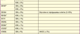
| Appointment with a dermatologist, candidate of medical sciences | 1500 rub. |
| Consultation with a dermatologist (KMS) when removing 2 tumors | 0 rub. |
| Removal of a neoplasm (wart, mole) using the radio wave method | 500 rub. |
| Make an appointment by phone: 8-800-707-15-60 (toll-free) |
| *The clinic is licensed to remove tumors |
Causes of dermatomycosis
Speaking about the etiology of the disease, it is important to take into account that the causative agents are a variety of fungi: microsporum, trichophyton, epidermophyton (the latter group includes varieties of this type of fungus, all of them are united under the common name “dermatophytes”). It is always extremely difficult to identify pathogens in body tissues, since a specialist must first isolate a pure culture for research.
Another difficulty is that the causative agents of dermatomycosis are very common, so infection occurs easily, and in some cases such manifestations may have something in common with epidemic symptoms. Most often, dermatomycosis is characteristic of countries with a tropical climate, that is, humid and hot, since fungi usually multiply actively at a temperature of 25-30 degrees Celsius. The summer season and alkaline environment are ideal for them. At the same time, experts say that children are much more likely than adults to become infected.
As a rule, the pathogen appears on the skin after a person has been in contact with the lesion for some time. Doctors distinguish three main types of pathogens:
- bestiality;
- geophilic;
- anthropophilic.
The source of infection in the first case is animals, or rather, parasites living in their bodies. This includes parasites that use domestic dogs, cats or cattle as hosts.
In the second case, we are talking about pathogens living in the ground and soil. A person can work or simply come into contact with the ground without using protective equipment, and as a result become infected.
The third case is parasites that live in the body of a person who is a carrier of the disease. The very fact of infection occurs through contact.
The development of dermatomycosis is expressed in the fact that conidia and hyphal fragments of pathogens enter the hair, upper layers of the skin, and nails (that is, those tissues that contain keratin). The degree of infection in dermatophytes is quite low, so tissue damage does not appear in healthy people. However, pathogens have a characteristic feature: they can subject keratin to complete destruction and destruction.
Pathogens located in the hair shaft can be divided into two types based on their growth. First: endothrix begin to grow from the skin into the hair and follicles, but they do not go beyond the boundaries of the hair shaft itself. Second: ectothrix grow directly into the hair directly from the hair follicle.
Much in the occurrence and development of the disease depends on the characteristics of the person’s body. Some types of dermatomycosis may be characteristic exclusively of an adult, while others - only of a child. In many ways, the disease is caused not only by age, but also by the secretion of the sebaceous glands, the composition of sweat and many other characteristic features.
Primary infection with dermatomycosis is a consequence of sensitization, after which it is time for a relapse to develop. Immunodeficiency largely determines the level of risk of dermatomycosis. In addition, various metabolic disorders, poor nutrition, problems with the hormonal system, and vitamin deficiency can also become a favorable background for infection. It is also important to remember that the fungus penetrates damaged skin faster, so people with ulcers, wounds and scratches on the skin are at particular risk.
Conidia, hyphae, and spores penetrate hair, nails and skin. Since the nutrient medium for the fungus is, first of all, keratin, the epidermis after infection begins to deteriorate at a rapid pace. In this case, healthy areas of the skin are also affected.
Athlete's foot
Athlete's foot is a fungal disease of the skin and nails. A distinction is made between athlete's foot and athlete's foot.
Athlete's inguinal
The causative agent - Epidermophyton floccosum affects the stratum corneum. The source of infection is a sick person. Routes of transmission: care items: bedpans, washcloths, sponges, oilcloths, etc.
Predisposing factors are high temperature and high humidity; hyperhidrosis. Nosocomial endemics are possible. It is observed mainly in men.
Localization - large folds, especially inguinal-femoral and intergluteal; Possible damage to other areas of the skin and nails of the feet.
Inflammatory spots in epidermophytosis are round in shape, red-brown in color, usually located symmetrically, clearly demarcated from the surrounding skin by an edematous ridge covered with small blisters, pustules, crusts and scales. As a result of peripheral growth, the spots can merge with each other, forming extensive foci with scalloped outlines. The course is chronic. Subjectively - itching, burning, pain, especially when walking.
Athlete's inguinal
Athlete's foot (foot fungus)
The causative agent is Tr. mentagrophytes var. interdigital; is located in the horny and granular layers of the epidermis, sometimes penetrating to the subulate, and has pronounced allergenic properties.
Localization – foot fungus affects the skin and nails of only the feet, usually in adults; often accompanied by allergic rashes - epidermophytis.
Infection with foot fungus occurs in baths, showers, swimming pools, and gyms, where dermatophytes get on the skin of a healthy person along with the scales of patients suffering from mycosis of the feet. Possible intra-family infection due to violation of basic rules of personal hygiene (wearing the same shoes, stockings, etc.).
Characteristics of athlete's foot (foot fungus)
Foot fungus: localization
Erased form of foot fungus Localization: folds between 5-6,4-3 toes Symptoms: slight peeling, sometimes mild itching
Squamous form of foot fungus Localization: arch of the foot Symptoms: slight erythema with peeling, sometimes thickening of the skin like callus, mild itching
Dyshidrotic form of foot fungus Localization: arch of the foot Symptoms: tense blisters of various sizes, erosions, crusts, often severe itching
Iteriginous form of foot fungus Localization: folds between the toes Symptoms: maceration, weeping, erosion, cracks, often severe itching
Athlete's foot (foot fungus)
Athlete's foot (foot fungus) can be complicated by erysipelas of the lower leg and the development, primarily on the hands, of secondary allergic rashes in which elements of the fungus are never detected.
Symptoms of ringworm
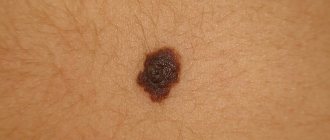
Dermatomycosis does not pose an immediate threat to the life of its carrier. Reproduction of pathogens is observed on the skin and other tissues, often all this is limited to external signs and manifestations. So what worries the patient most is not how dangerous the disease is, but how he himself now looks. If we are talking about skin lesions, then in this case for dermatomycosis the symptoms appear in the form of scaly plaques. They are usually red in color, can be located in groups or individually, each plaque is less than 5 cm in diameter. Such diseases are characterized by the brightness of the damaged areas and their itching.
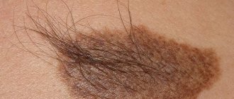
Dermatomycosis on the head is caused by the pathogen microsporum. Their symptoms manifest themselves in increased hair fragility, as the endothrix fungus penetrates the hair shaft. If the ectothrix fungus is to blame, then the hair begins to fall out as the follicles are destroyed under the influence of the pathogen.
There are other symptoms of dermatomycosis, which arise depending on what kind of disease the patient has. For example, with dermatomycosis of the mustache and beard, the clinical picture is somewhat different. The pathogen causes the appearance of pustules, and subsequently the follicles begin to be damaged. Often these affected areas become infected for the second time, so diagnosing the disease is not difficult, taking as a basis the presence of areas where swelling forms, and the lesions themselves become covered with a crust with bloody discharge. The causative agent in these cases is trichophyton.
Inguinal dermatomycosis is characterized by rashes of pustules, extensive peeling, and pyoderma appears in the groin and genital area. The causative agent is the same trichophyton in several of its forms.
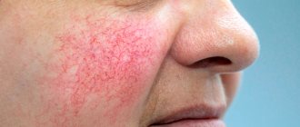
Symptoms of dermatomycosis of the body are characterized by the same manifestations: peeling in localized foci, pyoderma, erythema and pustular rashes.
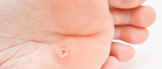
If we consider cases of tinea pedis, it should be noted that skin lesions here are usually located between the toes, as well as on the feet themselves. The pathogen trichophyton causes scales, cracks, skin erosion, and sometimes small blisters.
A disease such as favus is expressed in damage to nails, hair and skin. In this case, the course of the disease is chronic, and infection occurs through direct contact with the carrier. In the scuticular form, a week after the infection itself, erythema is observed near the follicles, which later transforms into pseudopustules and ulcers. After this, the skin begins to atrophy and cicatricial alopecia occurs. In this case, the hair does not fall out, but becomes lifeless and dull. As for the impetiginous form, in this case the clinical picture is different: brown crusts appear on the patient’s skin, under which the same alopecia is hidden. If favus of nails is observed, it is expressed in their fragility, the nails acquire a yellowish color.
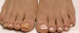
Onychomycosis affects the fingernails and toenails; they begin to peel, become rough and thicken. The causative agent is trichophyton; in some cases, the disease can be caused by various types of candida fungus.
Microsporia
Microsporia is dermatomycosis that occurs with damage to the skin, hair, and nail plates are relatively rarely affected.
The causative agent is Microsporum Canis. The incidence predominates in urban areas. Mostly children get sick. Unsatisfactory hygienic conditions, an abundance of stray animals, as well as high temperature and humidity contribute to the spread of microsporia. There is an increase in incidence in the autumn-winter period. The duration of the incubation period is 5-7 days for zoonotic microsporia, 4-6 weeks for anthroponotic microsporia.
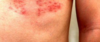
Microsporia of smooth skin
At the site where the fungus has invaded, a swollen, raised red spot with clear boundaries appears. Gradually the spot increases in diameter. A continuous raised ridge is formed along the edge, represented by small nodules, bubbles and crusts. In the central part of the spot, inflammation resolves, as a result of which it acquires a pale pink color, with pityriasis-like peeling on the surface. Thus, the focus has the appearance of a ring. The number of foci with microsporia of smooth skin is usually small (1–3). Their diameter ranges from 0.5 to 3 cm. Most often, lesions are located on the skin of the face, neck, forearms and shoulders. There are no subjective sensations or moderate itching.
In newborns and young children, as well as in young women, severe inflammation and minimal peeling are often observed.
In people prone to allergic reactions (in particular, in patients with atopic dermatitis), the fungus is often masked by manifestations of the underlying process and is not always diagnosed in a timely manner. The use of local hormonal drugs only increases the spread of fungal infection.
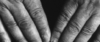
A rare type of microsporia includes damage to the skin of the palms, soles and nail plates. Nail lesions are characterized by isolated lesions of the nail, usually its outer edge. Initially, a dull spot is formed, which becomes white over time. The nail in the area of whitening becomes softer and more fragile, and may subsequently collapse.
Microsporia
Microsporia of the scalp (Ringworm)
Microsporia of the scalp (ringworm) occurs mainly in children 5–12 years old. It is generally accepted that the rarity of this form in adults is explained by the presence of organic acids in their hair, which slow down the growth of the fungus. This fact indirectly confirms the independent recovery of children during puberty, when the composition of sebum changes. Interestingly, microsporia of the scalp (ringworm) practically does not occur in children with red hair.
Microsporia of the scalp (Ringworm)
Foci of microsporia of the scalp are located mainly on the crown, in the parietal and temporal regions.
Usually there are 1-2 large lesions ranging from 2 to 5 cm in size, with round or oval outlines and clear boundaries. Along the edges of large lesions there may be screenings - small lesions with a diameter of 0.5–1.5 cm. At the beginning of the disease, a peeling area forms at the site of infection. In the first days, the fungus is located only at the mouth of the hair follicle. On the 6th–7th day, microsporia spreads to the hair itself, which becomes brittle, breaks off above the level of the surrounding skin by 4–6 mm and looks as if it has been trimmed (hence the name “ringworm”). The remaining stumps look dull and are covered with a grayish-white sheath, which is the spores of a fungus. If you stroke the stumps, they deviate in one direction and, unlike healthy hair, do not restore their original position. The skin in the affected area is usually slightly reddened, swollen, and its surface is covered with grayish-white small scales.
Diagnosis of dermatomycosis
Superficial mycoses can be detected already during an external examination. The fact is that with dermatomycosis, symptoms and treatment are closely related, so first the doctor must correctly determine which pathogen caused the disease, diagnose it, and then prescribe treatment depending on the type of pathogen. Typically, ringworm is characterized by a rash with red edges. However, the final diagnosis is manifested in clinical examinations under a microscope, or more precisely, in the examination of culture taken by scraping.
The scales of nails and skin must be examined. The patient may need to have a blood test and allergy tests. A dermatologist is not limited to an external examination, because only a thorough examination will help identify the true causes of the disease.
Treatment of dermatomycosis
Antifungal ointments and formulations are successfully used to treat such diseases; these products can be purchased at the pharmacy without a prescription. This remedy must be used 1-2 times a day, and within a week the results of treatment will become noticeable. However, in no case should the course of treatment be interrupted if the patient believes that things are getting better. Only long-term use of antifungal agents can completely rid a person of the disease.
Sometimes ointments may not help, this applies to the disease in an advanced stage. In this case, stronger oral medications are prescribed. It should be remembered that the affected areas of the skin should never be wetted; they should be dry and clean. If bubbles appear, it is forbidden to open them, as this will provoke further development of the infection. If the patient is being treated for inguinal ringworm, he must pay close attention to the quality of his underwear and change it daily. For tinea pedis, clotrimazole powder is used to treat shoes.
Dermatomycosis of smooth skin
This type of fungal disease is more common in warm countries, where the human body initially produces more sweat, which means the natural moisture of the smooth skin increases. Very often professional athletes who subject their bodies to grueling training suffer from this disease.
This type of dermatomycosis manifests itself in the formation of a lesion in the form of a ring with small bubbles inside, accompanied by severe itching.
Often a secondary infection of bacterial origin is also associated with the fungus. As a result, even scarring can be observed. After healing, areas of redness remain.