For the development of the disease, it is necessary not only to enter the pathogen, but also to have factors that contribute to the further proliferation of the fungus:
- deep skin folds of the feet;
- changes in the chemical composition of sweat;
- endocrine disruptions;
- hyperhidrosis;
- leg skin injuries;
- vegetative-vascular dystonia.
After fungi enter the skin, they can remain in saprophytic form for a long time without causing clinical symptoms. Athlete's disease is characterized by several variants of its course, some of which involve the spread of the process to the nail plate.
General information
Athlete's foot is a disease that currently occurs in residents of all countries of the world.
This is the common name for a whole group of fungal diseases, which are characterized by a common localization, as well as similar symptoms and manifestations. The disease occurs in people of any age, but it is very rarely diagnosed in children. As a rule, the disease becomes chronic and has a recurrent nature. Another important feature of the disease is its spread among people of certain professions and occupations: athlete's foot most often affects those who work in baths and showers, people working in hot shops, miners, etc. The disease most often occurs in urban residents.
Types of athlete's foot
With intertriginous epidermophytosis, the skin between the 4th and 5th fingers is affected; the lesion can also be between other fingers, but this is rare. The affected areas become red and itchy. The skin peels off and a crack runs through the center of the affected area. Small blisters may also form at the site of the lesion. This disease is most often observed in children.
Dyshidrotic athlete's foot is more common in adults than in children. With this form of the disease, small or large blisters form under the skin and feel dense to the touch. The appearance of blisters is accompanied by itching and peeling. These manifestations most often form on the inner surface of the palms and fingers. In the affected areas, swelling, skin erosion, the formation of lymphadenitis and lymphangitis, which cause a lot of unpleasant painful sensations, are possible, and there is a high probability of secondary infection.
Squamous-hyperkeratotic epidermophytosis is characterized by severe thickening and diffusion of the skin of the palms, the surface of the palms cracks and peels. This disease also occurs only in adults; the presence of this disease in children is observed in very rare cases.
Causes of athlete's foot
The disease has a high level of contagiousness. A healthy person can become infected with the causative agent of athlete's foot when visiting a shower or bathhouse, on a beach holiday, or by wearing someone else's shoes, socks or tights. The fact is that a sick person has a large number of mycelium threads and fungal spores . Consequently, they are constantly released into the environment, which provokes an unfavorable situation from an epidemiological point of view.
The fungus that causes the disease is mainly in a saprophytic state . But if certain factors influence it, the fungus becomes pathogenic. This can happen with diaper rash , constant wearing of uncomfortable shoes, sweating of the feet, abrasions, and if there is a shift in the pH of sweat to the alkaline side. In addition, the general condition of the human body is very important, because the fungus can become pathogenic in diseases of the nervous and endocrine systems, in cases of dysfunction of the body’s immune system, in the case of vascular disease, a lack of certain vitamins and microelements , etc. The disease can also provoke the manifestation of certain weather conditions, namely, too high air temperature and humidity
CLINICAL MANIFESTATIONS AND PRINCIPLES OF TREATMENT OF MYCOSIS OF THE FOOT
The collective term mycosis of the feet (MF) refers to fungal diseases that primarily affect the skin and nails of the feet (sometimes of the hands), which are similar in clinical, epidemiological, diagnostic, treatment and prophylactic terms. In terms of frequency, speed of spread and globality, MS is close to colds. The article discusses clinical issues, diagnostics, differentiation of MS from non-fungal lesions, as well as problems of therapy. Considerable attention is paid to intraconazole and lamisil, drugs that have fungistatic and fungicidal activity against dermatophytes, yeasts and molds.
The general term foot mycosis (FM) refers to mycotic diseases affecting mainly the skin and nails of foot (occasionally hands as well) which are similar in the clinical, epidemiological, diagnostic, and therapeutical-and-prophylactic context. FM is close to cold chills in terms of its incidence, rapid spread, and global prevalence.
The paper covers the problems of the clinical picture, diagnosis, differentiation from nonmycotic diseases, and those of therapy. Great emphasis is laid on itraconasole and lamisil, the drugs which have fungistatic and fungicidal activities against dermatophytes, yeasts and moulds.
V. M. Rukavishnikova, Ph.D. honey. Sciences of the Central Institute of Venereology and Venereology of the Ministry of Health of the Russian Federation. VM Rukavishnikova, Candidate of Medical Sciences, Dermatovenereological Institute, Ministry of Health of the Russian Federation
M
Icosis of the feet (IF) is a collective term. It refers to fungal diseases that primarily affect the skin and nails of the feet (sometimes also the hands), similar in clinical, epidemiological, diagnostic, treatment and prophylactic terms. In terms of frequency, speed of spread and globality, MS is close to common colds; it is no coincidence that they are called “national infection”, “retribution of civilization”.
Clinic and diagnostics
The bulk are made up of dermatophytic MS, their frequency is four times higher than the frequency of all other dermatophytoses combined. The predominant mycoses are caused by red trichophyton (rubrophytosis, detected in 80 - 93.8% of patients with MS), interdigital trichophyton (athlete's foot, which accounts for 10 - 20%). These mycoses have clinical differences (Table 1).
| Rice. 1. Squamous-keratotic rubrophytosis. a — damage to the interdigital and subdigital folds of both feet, ring-shaped peeling on the arch of the left foot; b - deep follicular-nodular element of mycosis in the lower third of the leg, ring-shaped and lamellar peeling; c - damage to the hands, floury, ring-shaped and lamellar peeling. |
Rubrophytia is characterized by pronounced clinical polymorphism and diversity. It affects both the distal parts of the extremities - the skin and nails of the feet and hands, and other areas of the skin in a variety of combinations - large skin folds, the skin of the trunk, limbs, face, neck, scalp. In other words, not a single area of the skin is immune to this fungus. Most often, squamous or squamous-keratotic rubrophytosis of the feet (hands) is observed. Initially, the fungal process is localized in the interdigital folds of the feet and under the toes, where slight peeling and surface cracks are found. Then the phenomena of keratosis increase, and the cracks become deep and painful (Fig. 1, a). The phenomena of keratosis are especially pronounced in places of pressure, for example in the heel and Achilles tendon (Fig. 1, b). The presence of three types of peeling in foci of mycosis is characteristic: floury, ring-shaped and lamellar (Fig. 1, c). Table 1. Distinctive signs of rubrophytosis and epidermophytosis
| Rubrophytia | Athlete's foot |
| It occurs against the background of reduced reactivity to the introduction of the fungus, mainly on dry, hyperkeratotic skin. | Predominant in people with a hyperergic reaction to mushrooms, with increased sweating |
| The course is chronic | The course is acute |
| A multiplicity of lesions of the skin and nails is characteristic: large skin folds, skin of the torso and limbs, nails of the feet and sometimes the hands | As a rule, only the skin and toenails are affected |
| All interdigital folds of the feet, all skin of the soles, dorsal and lateral areas of the skin of the feet are affected. | Foci of mycosis are usually localized only in the 4th interdigital fold and on the arch of the foot |
| Possible involvement of the skin of the hands (palms, back and sides) in the pathological process | The skin of the hands is not affected |
| The predominant type of squamous-keratotic skin lesions of the feet (hands) | The predominant type of skin lesions on the feet is dyshidrotic |
| Typical are multiple onychomycosis of the feet (hands) | Only the nails of the 1st and 5th toes are affected |
| There is a mixed type of nail damage with a predominance of the hypertrophic type | Nails change predominantly according to the normotrophic or dystrophic type |
| On smooth skin, the foci of mycosis are superficial, with bizarre, scalloped outlines, surrounded by a peripheral ridge that clearly outlines their boundaries | There are no lesions on smooth skin |
| Deep follicular-nodular lesions, grouped in the form of arches and rings, may appear on the lower legs (mainly in women) | Can not be |
| Infiltrative-suppurative lesions may appear (mainly in weakened children and men) on the face and scalp. They identify hair affected by the ectotrix or endoectothrix type. | Can not be |
| Allergic rashes are rare | Allergic rashes are common |
Exudative forms of rubrophytosis - intertriginous-dyshidrotic and dyshidrotic - are observed much less frequently. They usually arise as a result of inadequate treatment of erased forms of mycosis (for example, with corticosteroid creams) or develop after long walks or wearing closed, poorly ventilated shoes.
Rice. 2 Intertriginous-dyshidrotic rubrophytosis. Intertriginous-dyshidrotic rubrophytosis of interdigital folds is characterized by swelling and maceration of the stratum corneum in the folds and on the lateral surface of the fingers. When the stratum corneum is detached, erosions and rather deep cracks are formed, bordered by a fringe of exfoliated whitish epidermis. Patients are concerned about burning and pain, which intensify with movement (Fig. 2). Then a picture of dyshidrotic rubrophytosis is formed, usually on one of the feet or palms.
Rice. 3. Dyshidrotic epidermophytosis.
Athlete's foot most often occurs in an intertriginous-dyshidrotic or dyshidrotic form. The first differs from rubrophytic in that it is usually limited to damage to the closest third and fourth interdigital folds. With dyshidrotic epidermophytosis, vesicles of various sizes appear on the arch of the foot along the outer and inner edges, located either superficially or quite deep in the skin, visible in the form of sago grains. Bubbles, either isolated or merging into multi-chamber vesicles, open with the formation of erosions bordered by a fringe of exfoliated epidermis (Fig. 3). Blisters and blisters are filled with clear contents that become cloudy as they become infected. Erosion, weeping, and the addition of secondary flora lead to the formation of rough purulent-bloody crusts.
Rice. 4. Dyshidrotic rubrophytia. Damage to the nail of the second finger of the atrophic (onycholytic) type. Beak nail.
In large dyshidrotic foci of mycosis, as they resolve, three inflamed zones can be seen. In the central zone, the vesicles and blisters disappeared, the erosions became epithelialized, the scaly crusts were rejected, and the skin acquired a liquid tint. In the middle zone, erosions with a shiny, weeping, bright red surface are still visible. Blisters and blisters still continue to appear in the peripheral zone, and upon their opening, the epidermis peels off in the form of a fringe (Fig. 4.)
| Rice. 5. Types of nail damage. a - hypertrophic (nails resemble wood eaten by bugs); b - normotrophic; c - atrophic (onycholytic). |
A characteristic clinical sign of rubrophytosis is damage to the toenails (onychomycosis), observed in 80 - 100% of patients. In 20% of patients, the nails of the hands are also involved in the process. Typically, nails change after a more or less long-term lesion of the skin of the interdigital folds and soles by rubrophytosis. There is no isolated nail lesion without concomitant or previous skin lesions. Abroad, clinically, according to the location of the nail lesion, different forms of onychomycosis are distinguished, including distal, lateral and proximal.
| Rice. 6. Iris-like foci of rubrophytosis emerging from the folds on the thigh (a) and lower leg (b). | |
| Rice. 7. The focus of mycosis is clearly outlined by an intermittent peripheral ridge. | |
| Rice. 8. An extensive focus of mycosis, occupying the skin of the inguinal-femoral folds, pubis and abdomen. | Rice. 9. Psoriasiform focus of mycosis on the skin of the buttocks. |
| Rice. 10. Bizarrely shaped foci of mycosis on the skin of the buttocks and thighs. Massive peripheral ridge on the skin of the right thigh. | Rice. 11. Garland-shaped focus of mycosis in the axillary region. |
| Rice. 12. Multiple foci of mycosis from rings, arcs, garlands on the skin of the inguinal-femoral areas. | Rice. 13. Cicatricial changes in the focus of mycosis are the result of “biopsy” itching. |
| Rice. 14. A lichenified focus of mycosis similar to a focus of neurodermatitis. | |
Distal onychomycosis can be of two types: with significant total thickening of the nail and pronounced subungual hyperkeratosis and with separation of the distal part of the nail from the bed according to the type of distal onycholysis. Lethal onychomycosis is characterized by lateral onycholysis and subungual hyperkeratosis or the formation of transverse grooves of a yellowish-brown color running from one lateral edge to the middle of the nail or its other lateral edge. Proximal onychomycosis is registered in cases where the process begins from the proximal part of the nail, i.e., the area of the lunula or adjacent areas of the nail plate. There is another form of onychomycosis - white superficial onychomycosis, or mycotic leukonychia. Among the extremely rare lesions of the nails, rubrophytic melanonychia is described, i.e. the formation of brown-black stripes in slightly thickened nails.
Table 2. Differentiation of MS from non-fungal lesions
| MS | Non-fungal foot infections |
| The onset of the disease is usually associated with visiting baths, showers, swimming pools, using someone else's shoes and skin and nail care products | This connection is not visible |
| In children of nursery, preschool and primary school age, MS is rarely observed | Non-fungal lesions are common in children |
| Changes in nails are always preceded by lesions of the skin of the interdigital folds of the feet and soles | Possible isolated damage to the nails, preceding, sometimes for a long time, skin rashes |
| Unilateral, and if both feet are involved, asymmetrical location of mycosis foci | Symmetrical location of lesions on both feet (hands) (Fig. 26) |
| Intertriginous, dyshidrotic, squamous-keratotic lesions of the skin of the feet (hands) with the presence of mealy, ring-shaped and lamellar peeling in the foci of mycosis | Skin rashes typical of a certain dermatosis are usually monomorphic |
| Gradual involvement in the pathological process first of the toenails, and then (and only in 20% of patients) of the hand nails | Simultaneous and rapid involvement of the nails of the hands and feet, for example after stressful situations |
| More pronounced changes in toenails are also more common | More pronounced changes in fingernails predominate |
| Hyper-, normo- and atrophic nature of nail lesions in the same patient | Same type of nail changes (Fig. 27) |
| Foci of mycosis are often localized in large skin folds and spread centrifugally | Each dermatosis is characterized by a specific localization of the rash. Dynamics of the process characteristic of this nosological form |
| Polymorphism of rashes, increase in inflammatory phenomena from the center to the peripheral ridge | Typical morphological signs of dermatosis on the skin (mucous membranes), often monomorphic |
| Tendency to exacerbations in the summer, remissions in winter | Exacerbations do not depend on the time of year, remissions are more common in summer |
| Reduction of non-exudative manifestations of MS during antifungal therapy, worsening with the use of corticosteroid creams, adhesive bandages (“sealing method”) | Exacerbation of dermatosis during antifungal therapy, improvement with the use of corticosteroid creams and the “sealing method” |
| Microscopic detection of fungi in scrapings from skin and nails, isolation of fungal cultures on nutrient media | Negative results of laboratory tests for fungi |
In our country, it is customary to distinguish three main types of nail damage based on the nature of the damage. The most common is the hypertrophic type of damage to the toenails, apparently due to the long existence of onychomycosis. The nails thicken, hypertrophy, become deformed, easily break or crumble, lose their shine, become dull, acquire an uneven grooved surface, resembling wood eaten by bugs (Fig. 5a). The color of the nails varies from ocher yellow to dirty brown. Starting from the distal or lateral edges, the changes spread to the entire nail plate. Severe subungual hyperkeratosis is characteristic. With significant deformation and thickening, the nail becomes similar to a bird's claw (onychogryphosis).
| Rice. 15. Foci of mycosis, similar to manifestations of seborrheic dermatitis. a, b - on the skin of the forehead | |
| Rice. 16. Foci of mycosis on the face and neck, similar to foci of perioral dermatitis. | Rice. 17. Foci of mycosis in the form of rings on the forearm. |
| Rice. 18. Urticarial and papulovesicular lesions on the patient’s face (a) and forearms (b) simulate exudative erythema multiforme. | |
| Fig. 19. Widespread foci of mycosis of an erythematosquamous nature in one patient. A clear peripheral ridge bordering the lesion in front (a) is absent in places in the lesion on the back (b). | |
| Rice. 20. Foci of rubrophytosis on the forearms (a) and on the back of the neck in the zone bordering the scalp (b) resemble microsporia. | |
| Rice. 21. Almost universal mycotic lesions such as mycotic erythroderma (a, b). | |
| Rice. 22. Infiltrative-suppurative rubrophytosis. a - damage to the cheek, upper lip and chin (folliculitis, perifolliculitis, infiltrative plaques are visible); b - plaque in the upper lip; Along with the peripheral ridge, in the center of the lesion there is a ridge of follicular-pustular elements. | |
| Rice. 23. Abscessing focus of rubrophytosis, occupying the entire upper lip. | |
| Rice. 24. Vasculitis-like follicular nodular rubrophytosis of the legs in women (a, b). | |
| Rice. 25. Follicular nodular rubrophytosis of the leg (a) and buttocks (b), similar to pyoderma. | |
| Rice. 26. Strict symmetry of lesions on the feet in psoriasis. | |
With normotrophic
type of lesion, the nails retain their normal thickness and configuration for a long time, but whitish-yellow spots and stripes appear in them (usually from the lateral or distal edge, sometimes proximal), which, moving forward, increasing in size and merging, gradually cover the entire nail (Fig. 5 , b).
Rice. 27. The same type of changes in nails with lichen planus.
With atrophic
(onycholytic) form of the lesion, the nails are separated from the nail bed from the distal or lateral edges (Fig. 5, c), until they disappear from the lateral parts and form a cone-shaped, beak-shaped shape (see Fig. 4). Due to the non-simultaneous involvement of nails in the pathological process, it is rarely possible to see any one type of lesion in a patient. Usually we are talking about the predominant type of nail changes. Nail lesions in MS, as a rule, do not cause any subjective sensations. Significant pain, making it difficult to wear regular shoes and move, occurs only with nail deformities such as onychogryphosis, complications of paronychia, ingrown nails, subungual granuloma or subungual exostosis. Along with damage to the skin and nails of the feet (hands), 15 - 25% of patients with MS experience generalized forms of mycosis. They are divided into relatively superficial and deep. Among the superficial forms, localized, limited and universal (such as mycotic erythroderma) can be distinguished. Most often, foci of mycosis on smooth skin are located in large skin folds: inguinal, intergluteal, axillary. They appear for the most part symmetrically, synchronously, have irregular round or oval outlines, tend to merge and grow peripherally, and spread to adjacent areas of the skin of the thighs, buttocks, pubis, abdomen, back and chest (Fig. 6 - 13). The boundaries of the foci of mycosis are clear, the outlines are scalloped, and bizarre. Inflammatory phenomena increase from the center of the foci of mycosis towards the peripheral ridge, where they are most pronounced. Rarely, foci of mycosis have an iris-like appearance due to two or more ridges, as if inscribed one into the other (see Fig. 6). Patients are bothered by moderate to “biopsy” itching, which results in cicatricial changes that seem to repeat the round-oval outlines of previous lesions (see Fig. 13). In other cases, foci of mycosis are located on the skin of the face, forearms, the back of the hands and feet, and the back of the neck (Fig. 15 - 21). On the face they often resemble seborrheic dermatitis (see Fig. 15). Foci of mycosis located around natural openings (periorbital, perinasal, perioral) are similar to foci of perioral dermatitis (see Fig. 16). Sometimes papulovesicular and papulourticarial elements located on the skin of the face, dorsum of the hands and forearms resemble exudative erythema multiforme (see Fig. 18). Sometimes, due to significant lichenification and infiltration, foci of mycosis can be mistaken for foci of limited neurodermatitis (see Fig. 14). Rubrophytic lesions localized in areas bordering the scalp may be similar to microsporium lesions (see Fig. 20). Predominantly erythematosquamous foci of mycosis can become universal, justifying the term “mycotic erythroderma”, in patients with Itsenko-Cushing disease and syndrome, blood diseases, cancer and severe infectious diseases (see Fig. 19, 21). Deep variants of rubrophytosis are divided into infiltrative-suppurative and follicular-nodular. With infiltrative-suppurative rubrophytosis, observed more often in men and weakened children, foci of mycosis, as a rule, are single, localized on the skin of the upper lip, chin, and scalp. Hair is affected by the ectothrix or endoectothrix type. Often such lesions are mistakenly mistaken for foci of vulgar sycosis or, at best, infiltrative-suppurative trichophytosis (Fig. 22, 23). Follicular nodular varieties of rubrophytosis are more often observed in women with vascular disorders, dysfunction of the reproductive and thyroid glands. The rashes are localized on the skin of the legs or buttocks, clinically they are vasculitis-like or pyodermic in nature (Fig. 24, 25). In recent years, reports have appeared in foreign literature about unusual manifestations of deep rubrophytosis. In immunosuppressed patients, as their condition worsens, multiple painless nodules suddenly and rapidly appear in the deep layers of the dermis and hypodermis. When they are sodded, up to 10 - 50 ml of thick purulent material is obtained, consisting of a pure culture of red trichophyton. Histologically, a neutrophilic abscess is determined. If the nodes are not drained, they resolve very slowly, resulting in atrophy [1, 2]. In table Table 2 shows the characteristic features that make it possible to distinguish MS from non-fungal lesions of the feet.
Treatment
MS patients with multiple lesions of the nails, as well as widespread superficial and deep skin rashes can be cured through the use of systemic and external antimycotic drugs and pathogenetic therapy. Currently, practical medicine has 4 systemic antimycotic drugs at its disposal: griseofulvin, nizoral, intraconazole and lamisil. The chlorine-containing antibiotic griseofulvin was the only systemic antimycotic drug for a long time (about 40 years). Possessing only a fungistatic effect, and only against dermatophyte fungi, it provided cure for 40% of MS patients in monotherapy. Removing nails and repeated cleaning of the nail bed made it possible to double the percentage of those cured. A mandatory requirement for griseofulvin therapy was a thorough examination of patients and an almost month-long hospital stay. Severe hepato-, nephro- and neurotoxicity, allergic, photosensitizing properties, immunosuppressive, cocarcinogenic, teratogenic and embryotoxic effects caused numerous contraindications and a large number of adverse reactions and complications. The drug is ineffective in mixed MS; cases of resistance and intolerance have been reported. In the 70s, the first azole antimycotic drug, nizoral, was proposed, and in 1980, a drug of the new triazole generation of this series, itraconazole, was proposed. Therapy with nizoral at a daily dose of 200 mg with a treatment duration of 3 to 6 months in combination with nail removal provided a cure for 86% of MS patients. Nizoral had fungistatic and fungicidal activity not only against dermatophytes, but also yeast and mold fungi, and therefore was useful in mixed MS, in cases of resistance to and intolerance to griseofulvin, as well as in the presence of contraindications to its use. However, this drug caused a large number (up to 20 - 30%) of adverse reactions and complications, including very serious ones. In a third of patients treated with nizoral, hepatitis was observed - from subclinical to acute toxic, which developed in 1 in 10,000, and hepatic coma (in 1 in 12,000) with the possibility of death. Nizoral interacts not only with the dimethylase systems of fungi, but also with the hormones of the testes, ovaries, adrenal glands, as well as enzymes involved in their synthesis. Therefore, 11% of patients experienced endocrine status disorders. A decrease in testosterone production with a compensatory increase in the formation of luteinizing and follicle-stimulating hormones led to a decrease in potency, libido, and the development of gynecomastia. A decrease in cortisol levels and a compensatory increase in the content of mineralocorticoid hormones led to arthralgia, myalgia, and hypertension. Increased osteoporosis, including pathological fractures, was associated with a decrease in vitamin D concentrations while taking nizoral. Currently, griseofulvin and nizoral are increasingly being replaced by two effective antimycotic drugs: the azole one - intraconazole and the allylamine compound lamisil, synthesized in 1983. Both drugs have fungistatic and fungicidal activity against dermatophytes, yeasts and molds. The use of both drugs makes it possible to achieve success in 83 - 94% of MS patients with multiple onychomycosis in an outpatient setting, without subjecting patients to a painful nail removal procedure. Both drugs are keratinotropic, lipophilic, quickly enter the stratum corneum of the skin and nail plates and remain in them for a long time, which allows for relatively short courses of therapy. Preference is given to lamisil due to its greater selectivity of action against fungi and fewer adverse reactions and complications, especially affecting the liver and endocrine organs. The differences arise from the mechanism of action of the drugs. Although both of them act on the cytoplasmic membranes of fungal cells and on the processes of ergosterol synthesis, this occurs at different levels of sterol metabolism. Lamisil acts early, at the level of the squalene epoxidase cycle, inhibiting the enzyme squalene epoxidase of fungi, which is 10,000 times more sensitive than the similar enzyme in humans. Intraconazole acts at later stages of sterol metabolism, inhibiting the enzyme 14-a-dimethylase, which is involved in the synthesis of not only ergosterol, but also cholesterol, steroidogenic hormones, enzymes and vitamins. In this regard, the unfavorable effect of itraconazole on endocrine organs, the possibility of developing impotence and gynecomastia in 0.2% of patients, decreased libido (in 0.2%), hypertension, adrenal suppression, edema and hypokalemia in isolated patients were noted. Both drugs are metabolized in the liver using cytochrome P-450, but itraconazole also interacts with this enzyme, disrupting the metabolism of other medications: cyclosporine, astemizole, digoxin, rifampicin, phenytoin, phenobarbital, isoniacid. Therefore, if during treatment with lamisil, liver complications are recorded in isolated patients, and they are more likely associated with idiosyncrasy to it, then when using itraconazole their frequency ranges from 0.3 to 5%, and we are talking not only about a persistent increase in the activity of liver enzymes, but also about the possibility of developing hepatitis. The lipophilicity of Lamisil, its predominant secretion by the sebaceous glands and pronounced antibacterial properties, along with antifungal ones, as well as the connection with chylomicrons and lymphatic transport determine its special effectiveness in patients with infiltrative-suppurative, follicular-nodular forms of rubrophytosis with the formation of rubrophytic granulomas, neutrophilic abscesses, i.e. e. in patients with complicated forms of MS. Moreover, even with long-term (up to 2 - 7 years) use of Lamisil in immunosuppressed patients, no adverse effects on endocrine organs and the liver, as well as on the metabolism of other medications, were noted. Adverse reactions during treatment with Lamisil occur in the first 3 weeks of drug use, their frequency is 8% at a daily dose of 250 mg and 11% when the dose is increased to 500 mg. The most common complications are gastrointestinal and include dyspepsia, discomfort, nausea, vomiting, and rarely, a transient increase in the activity of liver enzymes. In second place in frequency are itching and skin rashes: erythematosquamous, uriticarial, eczema-like, acneiform, such as exudative erythema multiforme. Some patients experience mild headache, malaise, drowsiness, and decreased attention. Side effects usually disappear after discontinuation of the drug. Many of them are allergic in nature and their severity decreases when a hypoallergenic diet is prescribed, even if the daily dose of Lamisil is increased by 50% [3, 4]. The incidence of adverse reactions and complications during therapy with itraconazole (orungal) is 7% with weekly use and 12.5% with longer therapy [4]. These may be dyspeptic symptoms, abdominal pain, nausea, vomiting, anorexia, rarely headaches, neuropathies, menstrual disorders, itching and skin rashes (lichenoid, urticarial, angioedema), Stevens-Johnson syndrome has been extremely rarely observed. Lamisil is prescribed daily at a daily dose of 250 mg. At the same time, foci of mycosis on the skin in 100% of patients are resolved in 2 weeks, onychomycosis of the hands is cured in 94% of patients in 6 weeks, distal onychomycosis of the feet in 12 weeks [5]. A daily dose of itraconazole of 100 mg has long been considered optimal for the treatment of MS. 76 - 80% of patients with multiple total lesions of the nails were cured when using the drug for 4 - 9 months [4, 6, 7]. Currently, a pulse method of therapy with itraconazole has been introduced: 7 days at 400 mg/day, then a 3-week break and a new 7-day cycle. After 3–6 cycles, 77–81% of patients with multiple total nail lesions are cured. If there are contraindications to therapy with systemic antimycotic drugs for the treatment of onychomycosis, you can use antifungal varnishes with penetrating properties: 1 and 8% batrafen varnishes, 5% omorolfine. Cure is observed in 66.7% of patients after 3-6 months of using varnishes. Initially, the varnish is applied daily and the nails are cleaned once every 7-10 days, then the varnish is applied 1-2 times a week [4, 5]. To relieve the severity of inflammatory phenomena in MS patients, lotions, wet-dry dressings with disinfectant and astringent compounds, pastes, combinations of antifungal and corticosteroid agents - mycosolone, lotricombe, travocort, pivazone - are used. They should not be used for more than 7-8 days to avoid activation of the fungal process. When pyococcal flora is attached, baths with a saturated solution of potassium permanganate, removal of purulent crusts and lubrication with 1 - 2% aqueous solutions of aniline dyes are indicated. Multicomponent formulations such as fucorcin can aggravate the process. If complications occur with limangoitis, lymphadenitis, or erysipelas, antibacterial antibiotics or sulfonamide drugs are prescribed. Compress dressings with a 10-20% solution of ichthyol or a 5% solution of mumiyo in 50% dimexide are used to remove crusts and hair, and to resolve foci of infiltrative-suppurative rubrophytosis. For follow-up treatment, you can use drugs with penetrating properties based on dimexide (quinosol-salicylic dimexide); thiocarbamide compounds (1% solutions, creams, aerosols of tolnaftate, tolcyclate, tolmicene) or 1% solution and cream of batrafen. For follicular nodular rubrophytosis, it is advisable to seal the foci of mycosis with adhesive tape for 3-4 days in a tiled manner (“sealing method”). Exacerbation of foci of mycosis, epilation of affected vellus hair with adhesive tape accelerates the rehabilitation of such foci of mycosis. In this case, in the future you can switch to therapy with various antimycotic drugs. For local treatment of skin and nails in patients with MS, preparations of sulfur, tar, undecylenic, benzoic and salicylic acids, iodine in the form of solutions, pastes, ointments, film-forming compositions, patches, as well as azole and allylamine compounds are used. Among azole drugs, you can use 1 - 2% solutions, gels, creams and ointments of canesten (clotrimazole), nizoral, isoconazole (travogen), bifonazole (mycospora), sulconazole (exelderma), econazole (pevaril), miconazole (daktarin), intraconazole ( Trisporal), fluconazole (Diflucan), fenticonazole, tioconazole. Among allylamine medications, 1% powder, solution and cream of naftifine (exoderil) and 1% solution, gel and cream of Lamisil are effective. If well tolerated, they exhibit not only antifungal but also antibacterial activity comparable to that of 0.1% gentamicin cream. When lubricating foci of mycosis 1 - 2 times a day, the therapeutic effect appears in the next 1 - 3 weeks. Modern domestic antimycotic drugs that are not inferior in effectiveness to azole drugs include 1% aerosol, 5% ointment and 10% liniment of mebetizol (2-mercaptobenzthiazole), 10% nitroxaline cream and ozonated ozonide oil [6, 7]. Additional pathogenetic means of therapy are as diverse as the pathology against which MS occurs. To correct immunological disorders, use 5-day courses of methyluracil 0.5 g 3 times a day or diucifon 0.2 g 2 times a day, 3-day cycles of levamisole 150 mg with 7-day breaks, 10-day courses of treatment thymus preparations (tactivin 1 ml of 0.1% solution subcutaneously daily, thymalin 5 mg or thymogen 100 mg in a 0.25% solution of novocaine intramuscularly every other day). A combination of immunocorrectors with adaptogens (ginseng, eleutherococcus, zamanikha, dibazol) and antioxidants (sea buckthorn oil, linetol, a-tocopherol) is advisable. Exudative manifestations of MS are treated with antihistamines and calcium preparations, sodium thiosulfate, diuretics, small doses of autologous blood (no more than 1 ml) intramuscularly every other day, in 5-day cycles with 3-day breaks, for a course of 2 - 5 cycles. To correct vascular pathology and improve tissue trophism, nicotinic acid preparations, andecalin, troxevasin, physiotherapeutic procedures (diadynamic currents, inductothermy of sympathetic ganglia, ultrasound on the area of the lymph nodes), self-massage, and “stretching” exercises of the spine are recommended. The severity of dystrophic changes in the nails decreases when using zinc oxide 50 mg 3 times a day or a 1% solution of copper sulfate 8 drops a day in monthly courses. For dry skin and brittle nails, vitamin A preparations are useful 3 times a day in monthly courses (retinol acetate 10 drops, retinol palmitate 100,000 units, aevit 1 capsule). Shown are products containing gelatin, pectin, sulfur and sulfur-containing amino acids, rich in vitamins and microelements (eggs, cottage cheese, cheese, meat and fish jellied dishes, vegetables, fruits, herbs, fruit and berry jellies and mousses and mineral waters). You can use widely advertised vitamin and microelement complexes, such as Complivit, Duovit, Unicap, etc.
Literature:
1. Potekaev N. S., Kurdin M. I., Potekaev N. N. // Abstracts. report 7 Ross. Congress of Dermatologists and Venereologists. Part 2. - Kazan, 1996. - pp. 89-90. 2. Rukavishnikova V. M., Sukolin G. I., Kuklin V. T. Treatment and prevention of mycoses of the feet. - Kazan, 1994. - 91 p. 3. Suvorova K. N., Kuklin V. T., Rukavishnikova V. M. Pediatric dermatovenereology: A guide for postgraduate education cadets. - Kazan, 1996. - 441 p.
A complete list of references can be obtained from the editorial office
Symptoms of athlete's foot
Doctors identify several different forms in which athlete's foot manifests itself: squamous , dyshidrotic , intertriginous . Separately stands out athlete's foot , as well as a form of the disease that manifests itself as a consequence of an allergic reaction of the body. However, such a classification is considered to be somewhat arbitrary, because the forms of the disease are sometimes combined with each other, or the disease passes from one form to another. At the same time, the symptoms of the disease also change.
In the squamous form of the disease, slight redness and peeling gradually appear on the skin of the arches of the patient’s feet. In this case, the formation of both small and quite extensive foci is possible. slight redness and peeling. The lesions can be small or extensive. Sometimes a person also complains of occasional mild itching. In this case, we are talking about the most dangerous form of the disease in epidemiological terms, because if the diagnosis is not carried out in a timely manner, a person may simply not notice the development of the disease, and, as a result, be a source of spread of infection for the people around him. Therefore, treatment of athlete's foot squamous form should be carried out immediately.
Sometimes, when the disease worsens, the squamous form becomes dyshidrotic . It is also possible for the dyshidrotic form of the disease to reverse into a squamous form. Initially, one foot is affected, later the disease spreads to the second foot.
The development of the intertriginous form of the disease mainly develops in people with a mildly expressed squamous form. However, its independent manifestation is also possible. In this case, there is damage to the interdigital folds, most often between the fourth and fifth fingers, in more rare cases - between the third and fourth. Sometimes the pathological process also affects the flexor surface of the fingers and the back of the foot. Cracks appear in the interdigital folds of the patient, which are surrounded along the periphery by a whitish layer of epidermis, which peels off in the process. The affected areas often itch, become wet, and as erosions develop, the person feels pain.
The illness continues for a long period of time. Sometimes in the cold season it stops, and in the summer it worsens again. In addition, cracks in the feet and the stratum corneum affected by the disease create conditions favorable for the development of thrombophlebitis , chronic recurrent erysipelas of the legs .
Sometimes a secondary infection is associated with the disease. In this case, cloudy contents appear in the blisters, and if the blisters are opened, pus flows out of them. of lymphangitis and lymphadenitis often occurs . When the inflammation subsides, the erosions gradually heal, and the squamous nature of the lesion is noted. In some cases, a severe course of the disease is observed with the addition of a secondary infection, when the patient needs to be treated in a hospital. This form is characterized by a long course of the disease, with exacerbations occurring in the spring and summer.
In the acute form of epidermophytosis, a person notices headaches, general malaise, increased body temperature, and enlarged lymph nodes. Secondary rashes called epidermophytis . The duration of the acute process is from one to two months. With the right approach to treatment, the disease responds well to therapy, but sometimes relapses can occur.
When nails are affected, changes in the free edge of the nail are initially observed: yellow stripes and spots appear on it. Later the nail plate thickens and turns yellow. It becomes loose, crumbles easily, and horny masses accumulate under the plate (so-called subungual hyperkeratosis ). In other cases, the course of the disease occurs somewhat differently: the nail gradually becomes thin, after which it is torn away from the nail bed (a phenomenon called onycholysis ). The most typical lesions are the first and fifth toes. The nail plates on the fingers are not subject to a pathological process.
Athlete's foot: diagnosis and treatment
- At the beginning of the appointment, the doctor should familiarize himself with the medical history and find out whether the patient has had contact with the patient or his things.
- Next, an external examination is carried out and scrapings are taken.
- The doctor prescribes treatment. Typically, your dermatologist recommends washing the affected areas with warm water and soap several times a day, and then lubricating them with an ointment containing clotrimazole.
It is important to remember that the ointment is usually applied at least 3-4 times a day, sometimes more often. Usually, the itching subsides already on the third day, and later the main symptoms disappear. But it is absolutely impossible to interrupt treatment: untreated epidermophytosis usually recurs quickly, and is more difficult to treat. A disease that is not completely cured is becoming less and less treatable each time. It can last for years, either gradually subsiding or exacerbating with renewed vigor.
Diagnostics
If the development of this pathology in a person is suspected, the diagnosis is established taking into account the presence of characteristic clinical manifestations. If threads of fungal mycelium are detected during microscopic examination, the diagnosis is confirmed. It is important to differentiate the diagnosis with psoriasis , intertriginous and dyshidrotic epidermophytosis , rubromycosis of the nails , intertriginous eczema , intertriginous canidosis . In order for the microscopic examination to be as informative as possible, the material for it is taken along the periphery of the lesions from the macerated exfoliating epidermis. During the diagnostic process, in order to distinguish the filaments of mycelium of epidermophytosis from other fungi, cultural diagnostics are carried out. For this purpose, seeding is carried out on a nutrient medium.
Tests and diagnostics
- Microscopy of pathological material is used as a method of preliminary diagnosis.
- Isolation of a pathogen culture (growth of the pathogen in a culture) takes time.
- Polymerase chain reaction (PCR). It makes it possible to obtain results within 24 hours that are highly specific (90-98%). It is especially important for onychomycosis and atypical forms of dermatomycosis, since in the earliest stages it allows to prescribe rational therapy, reduce complications and the severity of lesions.
Prevention
Due to the nature of the spread of the disease, you should definitely wear rubber shoes in a public shower or swimming pool, as well as follow the other rules that apply when caring for your feet. After washing the bathroom or taking a shower, you must first dry the groin area. After this, dry your feet with a towel. In addition, you should put on socks first and only then underwear. Such simple methods will help prevent the upward spread of a fungal infection, and accordingly, this can prevent the further development of inguinal epidermophytosis .
People who have already had a similar foot lesion should be sure to take into account that only with the full completion of the course of treatment can a recurrence of the disease be avoided in the future.
The fungus that causes epidermophytosis remains on clothes, so you need to wash things in hot water, always using laundry and bleaching products. It is important to carefully monitor compliance with other hygiene rules.
Causes of inguinal athlete's foot
Inguinal athlete's foot is called dermatomycosis , which affects the epidermis. The causative agent of this disease is the fungus Epidermophyton floccosum. The disease is transmitted through the use of a variety of household items. These can be washcloths, underwear, thermometers and other items. The disease can also be contracted in showers, swimming pools, and bathhouses. Factors influencing the development of the disease are also high humidity and air temperature, heavy sweating in humans, and the presence of damage to the skin. Consequently, almost every person can learn about what epidermophytosis is from their own experience.
Pathogenesis
Skin folds are places with high humidity and temperature, which creates conditions for dermatophytes. Dermatophytes that get into the folds do not cause manifestations of the disease if the barrier function of the skin, its acidity and the body's specific defense system are preserved.
A decrease in these protective factors and a change in the biocenosis of the skin contributes to the active proliferation of the fungus. The presence of damage to the stratum corneum causes increased permeability of the skin to pathogens. The causative agent of athlete's foot penetrates into the deep layers of the epidermis and dermis and causes characteristic rashes.
Dermatophytes have keratolytic and lipolytic effects. Their keratinase enzyme only breaks down human keratin. The growth and reproduction of fungi causes destruction of the stratum corneum of the skin. The process is characterized by progression, so inflammatory changes are most pronounced at the periphery of the lesions. If the rate of restoration of the epidermis is greater than the rate of reproduction of the fungus, then the pathogen is often eliminated and spontaneous recovery occurs.
Endogenous factors play an important role in the pathogenesis of dermatophytosis. Inguinal athlete's foot is often observed against the background of diseases such as diabetes mellitus , hepatitis , vegetative-vascular dystonia or in oncological pathology. Often, manifestations in the folds serve as “signal signs” of diabetes mellitus, since the bactericidal activity of the skin in patients with diabetes mellitus is lower than in healthy people. Also, mycoses of large folds often occur when taking systemic glucocorticosteroids for various concomitant diseases ( bronchial asthma , rheumatoid arthritis , lupus erythematosus ). A very severe course of epidermophytosis of the skin and folds is observed in HIV-infected individuals.
Symptoms
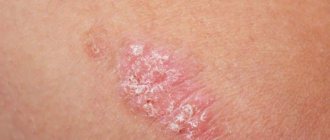
Inguinal epidermophytosis is manifested by the development of pink spots on the surface of the skin, which are slightly flaky. Their diameter is initially about 1 cm, but gradually the symptoms of the disease manifest themselves as spots grow along the periphery. At the same time, the foci of inflammation in the center of the spot gradually disappear. As a result, large ring-shaped spots with a red color appear on the human body, which in the process of merging form scalloped lesions. The boundaries of the lesions are clearly defined, which are emphasized by a hyperemic ridge, on the surface of which there may be bubbles. As the rash spreads, the center of the spot gradually becomes clear. As diagnostics shows, patients most often experience damage to the groin area, the surface of the thighs, both above and inside, the scrotum, sometimes the spots spread to the intergluteal fold. It is extremely rare that the disease affects the penis, armpit area, or area under the breasts. The patient is often bothered by itching and discomfort, which means that treatment of inguinal athlete's foot and further prevention of the disease should be carried out immediately.
How to recognize epidermophytosis?
Athlete's foot has a number of consistent symptoms:
- The damaged area of the body becomes covered with red spots.
- The spots begin to itch slightly and peel off.
- The center of the affected area becomes dull and pale.
- Individual spots merge into a common formation covered with a crust (so-called plaques).
- In place of the plaques, blisters filled with clear or cloudy liquid develop.
- The bubbles burst and the skin becomes wet.
- Unbearable itching appears.
Typically, at any stage of the disease, the affected area is surrounded by a border, which is both swelling and a border beyond which the lesion does not extend.
If a person sweats frequently and heavily, this accelerates the development of the disease and increases discomfort. Seeing a doctor is necessary.
Diagnostics
As a rule, diseases can be diagnosed based on a thorough examination of the affected area, as well as taking into account where exactly the rash is located. The doctor evaluates all the signs of the disease and studies the history of its development. To confirm the diagnosis, skin flakes from the affected areas are examined using a microscope. The final diagnosis can be made by identifying the causative agent of the disease during a microscopic examination. Culture is also sometimes used to confirm the diagnosis.
Treatment
If inguinal athlete's foot is acute, then to treat the disease, it is practiced to use lotions from a solution of silver nitrate , a solution of resorcinol , as well as the use of antifungal ointments. In addition, oral antihistamines are prescribed. Later, after the elimination of vesiculation, tincture of iodine , sulfur-tar ointment , and antimycotic agents for topical use are used. To prevent the disease, it is important to follow personal hygiene standards and prevent excessive sweating. All other preventive methods are similar to the preventive measures that are relevant for athlete's foot.
Consequences and complications
When athlete's foot complications are considered:
- Transition from one form to another.
- Onychomycosis.
- The addition of a bacterial infection is secondary pyoderma , lymphangitis , lymphadenitis and recurrent erysipelas, resistant to antibiotic treatment. Against the background of such lesions, thrombophlebitis , lymphostasis and elephantiasis occur .
- Allergization of the body: urticaria , bronchial asthma , allergic rhinitis , the occurrence of mycids . These are secondary allergic rashes due to mycoses.
For inguinal athlete's foot:
- Mycotic eczema . It occurs in a chronic form, manifested by lichenification of the skin (thickening), which occurs from scratching. The process resembles neurodermatitis.
- Allergic dermatitis.
- Impetiginization is the development of impetigo as a complication of mycosis. Impetigo is a strepto-staphylococcal skin lesion.
List of sources
- Garibova L.V., Lekomtseva S.N. Fundamentals of mycology: morphology and taxonomy of fungi and fungi-like organisms. Tutorial. – M.: Partnership of Scientific Publications KMK, 2005;
- Medical mycology. Guide for doctors / Ed. prof. V.B. Sboychakova. – M.: GEOTAR-Media, 2008;
- Rukavishnikova V.M. Mycoses of the feet. M.: Elix Com; 2003;
- Dermatovenerology. National leadership / ed. Yu.K. Skripkina, Yu.S. Butova, O.L. Ivanova. - M.: GEOTAR-Media, 2011;
- Sergeev A.Yu., Sergeev Yu.V. Fungal infections. Guide for doctors. M.: BINOM-press; 2008.
Sources
- Adam O Goldstein, et al. Dermatophyte (dermatophyte) infections. 2022 UpToDate.
- El-Gohary M, et al. Topical antifungals for herpes zoster and body rash. Cochrane Database Syst Rev 2014.
- Greenberg H.L. et al. Clotrimazole/betamethasone dipropionate: a review of costs and complications in the treatment of common cutaneous fungal infections. Pediatric Dermatol 2002.
- Rosen T., Elewski B.E. Ineffectiveness of clotrimazole-betamethasone dipropionate cream in the treatment of Microsporum canis infections. J Am Acad Dermatol 1995.
- Goldstein A.O. etc. Fungal infections. Effective treatment of diseases of the skin, hair and nails. Geriatrics. 2000.
- Crawford F, Hollis S. Topical treatments for fungal infections of the skin and toenails. Cochrane Database Syst Rev 2007.
- Gupta A. K., Cooper E. A. Update in antifungal therapy for dermatophytosis. Mycopathology 2008.
ONLINE REGISTRATION at the DIANA clinic
You can sign up by calling the toll-free phone number 8-800-707-15-60 or filling out the contact form. In this case, we will contact you ourselves.