But there are cases when a mole rubs against clothes, gets touched when shaving and is constantly injured:
- areas of skin with large folds,
- scalp,
- beard zones for men and others.
In some cases, moles are removed for aesthetic reasons at the request of the patient and to prevent injury. In addition, there is a risk of the mole degenerating into a malignant tumor. If a malignant skin tumor is suspected, the cosmetologist will refer it to a specialized oncology institution for removal.
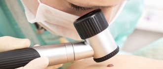
Only a dermatologist, cosmetologist or oncologist can determine the type of nevus (mole) using a simple device - a dermatoscope. The essence of the method is that, using a special magnifying glass (or other magnifying device), the doctor examines skin tumors under high magnification directly on the patient’s body.
This allows you to evaluate the structure of the tumor and identify small changes that are invisible to the naked eye, which can be important in making a diagnosis and prescribing treatment. Thus, dermatoscopy allows a specialist to determine whether a skin tumor is benign or not and choose a method of removal.
Benign moles can be removed by a cosmetologist, but always with a pathomorphological examination.
What to do with moles?
If there are no clear signs of a malignant process, then you can go in 2 ways:
The first way is observation. In this case, we take a picture, a photo of the mole, warn the person about the signs in case the size, shape, symmetry changes, and schedule a return visit in 3-6 months, depending on how much we fear for this formation.
The second way is removal of the formation with histological examination. We even resort to this path more often. There is a common belief that a mole should not be touched. In fact, there is no danger in removal. Moreover, this is even a preferable option, since in this case a suspicious formation can be sent for histological examination. So, we excise the formation, sometimes if it is a suspicious mole, then not with a laser, but with a scalpel, with minimal grip (1-2 mm is enough) and send the material for histological examination.
If we receive the result that it is a benign formation, then we have removed it as benign and no further action is required.
Is histology necessary when removing a mole, fibroma or other neoplasm? Doctor's opinion
Most neoplasms are not life-threatening. They are removed for aesthetic reasons and to prevent injury (for example, convex moles that constantly rub against clothing or get touched when shaving). But we cannot exclude the risk of the neoplasm degenerating into a malignant tumor.
Patients who want to remove a mole, papilloma or other skin tumor often ask: Is it necessary to send the removed tumor for histology?
Natalia Genrievna Yezhova, Deputy Director for the Medical Department of CLINIC21, answers:
“I often come across the desire of patients to simply remove any tumor on the skin without conducting a pathological examination.
Many people think that they just have a harmless growth and there is no danger.
People do not have developed cancer alertness. I will give an example from my experience. Patient X underwent excision of a 'soft fibroma' in the area of the shoulder joint with pathomorphological examination. The study showed that it was nodular basalioma, that is, an oncological formation. The patient was taken under observation, the progress in treatment is positive, because the disease was detected on time.”
Histological analysis allows us to determine the presence of cancer cells in the tumor. This is an important diagnostic procedure that can save lives. Thanks to this study, it is possible to timely determine the malignant nature of the tumor and undergo treatment to prevent the progression of oncology.
Melanoma and dysplasia
If, according to histology, we receive a result - severe dysplasia or early melanoma, then this can also be regarded as a positive result, since the disease was detected in the initial stage. In this case, we refer the patient to the oncology clinic, where he undergoes further treatment.
Early (thin) melanoma is very treatable, with a good survival prognosis. In contrast to melanomas, which are left untouched, which grow and patients treat them at stages 3-4, when effective treatment is no longer possible.
Signs of malignancy
The clinical signs of a malignant mole are very diverse. This is manifested in size, shape, outline, surface character, consistency, color, and dynamics of change.
Common to all forms is a set of characteristics, which is expressed by the initial letters by the abbreviation “AKORD”, developed by dermatologists and cosmetologists:
- Asymmetry (A) - lack of symmetry in the shape and contours of a spot, with the exception of birthmarks present on the child’s body at birth.
- Edges (K) are most often uneven and indistinct (blurry).
- Coloring (O) - uneven; The presence of dots and stripes of various tones of dark brown and black is noted.
- Size (P) - in diameter from 7 mm or more.
- Dynamics (D) of development - an increase in the previous birthmark or a rapid increase in the size of a new pigmented formation.
Removal methods
There are several of them. The doctor prescribes it individually for each patient.
- Excision, "Traditional method". The mole is removed with a scalpel. Possibly histological examination. A small scar remains.
- Laser vaporization. A special beam evaporates the changed cells. This method is undesirable if melanoma is suspected, since histological examination of the mole is impossible. Not a trace remains of her.
- Laser cutting. With this method, histological examination is possible, but the skin receives a thermal burn. Healing proceeds quite quickly.
- Radio wave removal. Exposure to radio waves is effective for small moles. Histological examination is possible. The wound is healing successfully.
Prices for all types of removal can be found in the price list.
“Can I not take away the answer? Will they call me if something is wrong?”
Of course, you don’t have to take the answer, and then write messages like this on the forums:
Please don't think I'm laughing at tragedy. This is just a way to get your attention.
As far as I know, there is no law according to which the morphology laboratory must notify the patient that cancer has been detected. At the end of the conclusion after the examination, moles may be written: “Re-examination with the results of histological examination.” But no one reads these conclusions.
And then everything happens according to the classic scenario, even if in an institution calls of this kind are in the order of things, the human factor comes into play. For example, the person responsible for this quit, or someone replaced him, or he got sick, didn’t make an appointment, it was a birthday, etc. - in the end, no one called, and the patient didn’t even know that they could have done this. Remember that your health is yours and no one owes you anything.
Please always take the result of the histological examination and always show it to the oncologist who performed the removal.
“I took the histological answer, what should I do with it? Will I figure it out myself?”
No. If you received the result of the study in paper or electronic form, you must show it to the oncologist who performed the removal. Let me give you an example.
Here are two diagnoses:
- Dysplastic nevus with severe dysplasia. There are tumor cells at the resection margin.
- Squamous cell papilloma with inflammatory component. Removed within unaffected tissues.
In the first case, repeated excision of the mole removal site is required, but in the second - not. Therefore, do not be lazy, show the conclusion to the doctor.
Stages of histological examination: timing and reasons for delays
There are several stages of histological examination. I won’t go into detail about their details, I’ll just list them and briefly explain the essence:
1. Biopsy, i.e. removal of a mole. The main thing I want to note here is that there is never “too little” material for research. They often write to me: “The doctor said that the formation was too small, there would be nothing to send for examination, so I did not have a histology.” In my practice, there have been cases when a normal conclusion was received for a mole up to 1 mm in size. Therefore, no matter how huge a “soldering iron” a mole is removed, it is important to send a fragment of the largest possible size for histology. This is certainly worse than examining the entire nevus, but much better than nothing.
2. Fixation of the material. To prevent the mole from decomposing while it is being transported to the laboratory, it is necessary to destroy microorganisms and deactivate the enzymes of the mole cells. This is done using special solutions. Immediately after removal, the mole is placed in a formaldehyde solution. You can replace it with alcohol, but this option is less common
3. Washing the material. To remove traces of the fixing liquid, the material is washed, as a rule, for several hours in running water.
4. Dehydration . Preliminary preparation for paraffin filling.
5. Compaction (filling). The washed and dehydrated mole is filled with liquid paraffin. This is necessary so that the thinnest sections can be made, suitable for examination under a microscope. The result is paraffin blocks like these.
6. Preparation of sections. Using a special device - a microtome - sections are made with a thickness of about 0.05 mm.
7. Coloring the material. This is necessary to distinguish different cells and tissues under a microscope. In an unpainted material, all structures refract light equally and nothing can be seen.
8. Conclusion of sections. This is the name for the arrangement of colored sections between two glasses.
9. The final stage - the pathologist places the glass under the microscope lens and makes his conclusion on the material being studied.
All stages of histological examination take no more than 10 days. The fastest stage, in my opinion, is the examination of the preparations by a pathologist. In some advanced laboratories, due to the high automation of all of the above processes, histological examination is completed within 48 hours.
And sometimes I am surprised by patients who tell me that the histological examination took two months or more. If this happens to you, you should call the laboratory and find out what the difficulties are.
Is a histological examination of a wart necessary?
To be honest, I’m already tired of answering this question. I will simply give 2 histological conclusions, which are united by a clinical (on examination) diagnosis - a wart.
First case:
I was unable to trace the fate of the patient and the result of the immunohistochemical study. However, in a conversation with a morphologist, I realized that the outcome here can be very different and not necessarily successful.
And the second case:
Intraepithelial carcinoma is not the worst thing that can happen. Bowen's disease (and that's exactly what it is) is just stage zero squamous cell carcinoma, which can be successfully cured by removal within healthy tissue. However, another year or two, and the consequences for the man could be dire.
Conclusion: warts should be sent for histology. Always. No matter how close they may seem to you.
Are there errors in the histological examination of nevi?
Yes, unfortunately, they are possible. I wrote about this. Histological examination is carried out by people, and they can make mistakes. If you have the slightest doubt about the results of the conclusion, take your histological preparations and send them for review to another laboratory. The preparations include both paraffin blocks with mole tissue and glasses with sections.
You ask: “How can I go and pick something up? They’ll send me…” If they don’t give you histological preparations, then you need to go to the head doctor and demand it. In my experience, after that everyone gives away.
Methodology
Histological analysis takes place in several stages. This includes the preparation of sections, the use of microscopy methods, and quantitative and qualitative analysis of the results obtained.
The object of research is living and dead tissues, images obtained using electron and light microscopes. Material for analysis – smear, thin section of tissue, film.
Finished samples can be studied after the material has been collected, but the best results are obtained from pre-processed materials. Fabrics are treated with fixatives to prevent their decomposition. They can be treated thermally and dehydrated in carbon dioxide.
To prepare sections, specialized equipment is used - ultramicrotomes for examination in electron microscopes or microtomes for studying the sample in light equipment.
Additionally, the fabrics are dyed to improve image contrast. When using an electron microscope, ultrasections are fixed in specialized grids. To improve image contrast, fabrics are treated with compounds of uranium and other metals.
Methods of histological analysis:
- Light – classical microscopes with varying degrees of magnification.
- Ultraviolet - using rays from the ultraviolet part of the spectrum. Cell and cut defects invisible under normal lighting become visible.
- Fluorescent - the study is carried out using mercury, xenon lamps.
- Phase contrast – used when other methods of studying samples are ineffective. Fabrics are pre-dyed.
- Electronic - the sample is irradiated with a stream of electrons. The advantage of this method is multiple magnification and high resolution of the equipment.
How is mole histology performed?
Thanks to histological examination, cancer cells can be detected even at the stage of their inception.
Indications for mole histology:
- redness of the skin around the tumor;
- small ulcers on the surface of the nevus;
- burning or pain in the mole area;
- change in color, size, shape of the tumor.
Histology is performed with the procedure of surgical excision of the mole. A small tissue sample is removed and sent for further examination. In the laboratory, biological material is placed in a special suspension and examined under a microscope. Diagnosis is based on the analysis of pigment-forming cells - melanocytes. Laboratory testing may take up to 14 days.
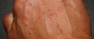
Which moles are dangerous?
Every person has moles. These are small spots on the body of various colors: from flesh-colored to almost black. These are nodules or papules consisting of pigment cells. All of them are similar to melanoma, and therefore need to be treated with special attention.
Moles at risk:
- multiple;
- atypical (irregular shape, heterogeneity of coloring).
Who needs to pay special attention to the condition of nevi:
- blondes and red-haired people, people with very fair skin and light eyes;
- people whose relatives have had cases of melanoma or precancerous skin diseases;
- people aged 40–45 years (melanoma is most often diagnosed).
Moles located in places of constant mechanical influence - friction, pressure - are most prone to malignancy (malignant degeneration). These pigment formations are localized on the neck, lower back, folds and folds of the body, in the places where clothing fits most tightly. The most common location in women is the lower leg, in men it is the back. In older people, melanoma most often develops on the skin of the neck and scalp.
In light of the craze for dark tanning and tanning salons, the risk zone includes all moles that are exposed to excessive ultraviolet radiation. People who suffer from sunburn more often are at risk, so they need to use light filter creams.
You should pay attention to the condition of your moles. If you see that they begin to enlarge, change color, bleed, or itch, you should immediately consult an oncologist. The study will rule out melanoma or atypical nevus.
The histology of the mole most accurately differentiates:
- atypical moles;
- intradermal nevi;
- non-pigmented neoplasms.
It helps to prescribe optimal therapy based on the most accurate diagnosis.
What are the risks of melanoma?
Melanoma and mole are very similar in appearance. They consist of identical melanocyte cells. These cells produce melanin. A pigment that protects the skin from high doses of ultraviolet rays. However, when a mole becomes injured or burned, it becomes dangerous. Melanocytes begin to multiply rapidly. They crowd out healthy cells.
A tumor forms. From the surface of the skin it quickly makes its way deeper. At the same time, it affects all layers of the skin. It is important to stop the tumor in time! Otherwise, metastases may appear - cancer cells that separate and affect internal organs (brain, liver, lungs, etc.).
How long does it take to perform histology?
The duration of processing of tissue samples should be checked with your doctor. There are 2 types of analysis:
- Emergency - the cut is examined during the operation. This allows, if necessary, to expand the scope of surgical measures and remove more tissue. And immediately after removal of the tumor, begin drug treatment. Used in controversial cases, when new circumstances are identified during surgery.
- Planned - used when the diagnosis is beyond doubt, and an emergency study confirmed the doctor’s assumption. The processing time for results is up to 7 days.
The timing of the study is influenced by the following factors:
- legal status of the laboratory or clinic - private or public;
- analysis methods and equipment used in a particular medical center;
- laboratory workload;
- force majeure circumstances.
A long sample processing time is not synonymous with bad news!
Information for foreign patients
Foreign patients wishing to receive qualified oncological care in Israel can count on correspondence consultation with a specialist at the pre-hospital stage. In addition, if necessary, a histological examination of the material can be carried out.
In Israeli clinics, a sentinel lymph node biopsy procedure is used, according to which lymph nodes with possible metastatic lesions are eliminated.
In Russia and the CIS countries, the procedure of sentinel lymph node biopsy has not yet become widespread.
The use of this technique is most relevant if histological examination reveals an increased mitotic index and ulceration in the area of primary melanoma.
The type of melanoma, its classification and determination of the stage of tumor growth are the main characteristics of histological examination.
In specialized cancer centers such as the Melanoma Unit, histological testing of a tissue sample is carried out for more than 20 parameters. In addition to structural features, the pathologist takes into account many additional characteristics, including the properties of cell nuclei and enzyme activity.
Patient Andrey M., 31 years old, diagnosed with melanoma of the right shoulder
The conclusion of Russian pathologists speaks of increased mitotic activity (from 2 to 5 mitoses in the field of view), melanoma is described as a tumor of the 3rd level of invasive growth according to Clark with a thickness of 2.5 mm. Israeli oncologists established level 4 melanoma according to Clark with a tumor thickness of 4.25 mm. The mitotic activity rate was 20/mm2.
Examples of differences in the histological reports of Russian and Israeli pathologists.
The study of tumor material involves determining its following properties:
- Tumor thickness
- Ulceration
- Clark level
- Histological type
- Cell type
- Primary localization
- Signs of regression
- Number of mitoses
- Lymphocytic infiltration
- Vertical growth stage
- Invasion of blood vessels
- Invasion into the lymphocytic zone
- Ploidy
- S phase of the cell cycle
- DR1 gene expression
- DNA Index
- Heat shock protein expression
- Positive staining for HLD-DR
- P53 protein mutation
- Cell adhesion factor expression
- Protease expression
- Migration marker molecule
- Angiogenesis factor
- Oncogene expression
- Presence of an estrogen receptor
- Cytokine, growth factor
Our clinic offers correspondence consultations and remote examination of histological samples. This greatly facilitates diagnosis and increases the efficiency of medical care.