Dermatoscopy is an instrumental non-invasive study in which the doctor examines moles and other formations on the skin using a special device that creates multiple magnification. In oncology, dermatoscopy helps to distinguish harmless formations from skin cancer and identify melanoma in the early stages.
- Indications for dermatoscopy
- Why are moles dangerous?
- How is dermatoscopy performed?
- Equipment for dermatoscopy at Euroonco
- How to assess the malignancy of a mole
- Advantages of dermatoscopy using the FotoFinder system
- Prices for dermatoscopy at Euroonko
Indications for dermatoscopy
- the appearance of any red, brown or black formation on the skin;
- injury to a mole; uneven change in its color;
- an increase in the total number of moles and age spots;
- itching and tingling in the area of education;
- increase in size of the mole.
For preventive purposes, dermatoscopy is recommended:
- fair-skinned people with moles and age spots;
- with a family history of melanoma (if it was diagnosed in close relatives);
- people who have many moles and freckles on their body;
- when taking oral contraceptives for more than 1 year;
- when working in hazardous industries;
- with regular trips to hot countries;
- For dysplastic nevi, it is recommended to undergo dermatoscopy every six months to a year.
No special preparation is required for dermatoscopy. It is not recommended to use cosmetics or topical medications on the day of the examination. To obtain a clearer image, a small amount of gel is sometimes applied to the test site to reduce the reflection of light from the surface of the skin.
Modern digital dermatoscopes have many advantages over conventional ones. The device tube is connected to a computer monitor, on which the image is displayed. Digital dermatoscopy serves as a monitoring method for patients at high risk for developing melanoma. It also allows you to map moles over the entire surface of the body and observe their changes in dynamics, comparing the results with previous ones. The technique of digital dermatoscopy is simple, safe and fully automated. Within 3 minutes you can get a detailed analysis of all neoplasms.
It should be remembered that dermatoscopy does not allow a definitive diagnosis of melanoma; this requires a biopsy and histological examination of a tissue sample. [1,3]
Briefly about the main thing:
I think that scraping or puncture before removing a mole is not necessary, because... The accuracy of diagnosis is only 95%, in contrast to histology (close to 100%).
Instead of scraping or puncture, in my practice I always remove all moles with an indentation of 1 MM and always send them for histological examination (excisional biopsy). If histology shows melanoma, the biopsy site is excised again with an indentation of 1 to 3 CM, which fully complies with safety standards in oncology.
Why are moles dangerous?
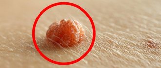
Moles (pigmented nevi) are benign neoplasms, but some of them can transform into an aggressive, dangerous malignant tumor - melanoma. Risks are increased in the following conditions:
- A large number of moles on the body.
- Dysplastic nevi are special moles that are large in size, uneven edges, and uneven in color.
- Congenital melanocytic nevi are moles that are present on a child’s body from birth. Usually children are born without moles - they appear throughout life. The most dangerous are giant congenital nevi with a diameter of more than 10 cm. They degenerate into melanoma with a probability of 30%.
Important to know: Scientific research shows that only 30% of melanomas develop from pre-existing pigmented nevi. In 70% of cases, a malignant tumor occurs on unchanged skin, where there were no moles. [2]
Why examine a mole if it has already been removed? Will it be too late?
A logical question arises - what to do if the histology shows melanoma? Will we do any harm by removing it? Will this lead to tumor progression?
No.
A large number of clinical studies have found that performing an excisional biopsy
does not worsen the course of melanoma.
In other words, the chances of recovery in patients who first underwent such a biopsy, and then wide excision, are the same as those whose melanoma was immediately excised widely.
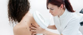
How is dermatoscopy performed?
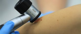
Classic dermatoscopy is carried out using a special instrument resembling a magnifying glass - a dermatoscope. Using it, the doctor examines the skin, assesses the size and appearance of the detected tumors.
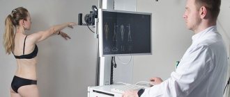
A more modern technique, which is used at Euroonco, is videodermatoscopy using the German PhotoFinder. The device takes pictures of the entire surface of the patient's skin, creates a “mole map” and stores the images in a computer. The procedure is absolutely painless. [3]
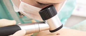
Hardware diagnostics of melanoma
The doctor examines suspicious moles using a special device - a dermatoscope. This device provides a tenfold magnification, which allows you to examine the skin area in detail, identifying pigment inclusions and heterogeneity of the structure.
With confocal laser scanning microscopy (CLSM), the doctor takes images of “slices” of the skin without damaging it. It is a painless and quick alternative to a biopsy. The result is confirmed in 97% of cases.
A biopsy for suspected melanoma is rarely used, as it can provoke the degeneration and growth of malignant cells. The method is more often used on distant tumors. When the diagnosis is confirmed, the nearest lymph nodes are punctured.
Equipment for dermatoscopy at Euroonco
At Euroonko, dermatoscopy is performed using a modern PhotoFinder installation from the German company FotoFinder Systems GmbH. This manufacturer has been producing high-tech turnkey solutions for visualizing skin tumors and image analysis for more than 20 years. The device takes pictures of the entire surface of the body and loads them into a computer, where they are saved and processed by a special program.
Why is a dermatoscope better than an oncologist's eye?
This device has 2 serious advantages:
- Tenfold (or more) magnification
- The deep structures of the mole are examined, not the superficial ones. When examining a mole, the human eye perceives only those rays of light that are reflected from its surface layers. In a dermatoscope, thanks to immersion oil, special conditions are created. With them, the rays of light from the built-in illuminator are reflected not from external, but from internal structures. Thanks to this, the doctor looks into the depths of the mole through a dermatoscope and can evaluate its structure.
I think comments are unnecessary here. The capabilities of human vision are limited.
How to assess the malignancy of a mole
During dermatoscopy, a number of characteristics are assessed, such as size, its elevation above the skin, shape, symmetry, state of the borders, color, nature of the surface, and the presence of ulcerations. However, the assessment of these indicators is quite subjective. In this regard, various methods for determining the malignancy of a neoplasm have been developed. The most commonly used are the following:
- Scale of 3 signs. Asymmetry, the typical pigment network and the presence of blue-white structures are assessed.
- Scale of 7 signs (G. Argenziano). The main criteria (atypical pigment network and vascular pattern, blue-white veil) and additional ones (atypical branches, pigmentation, spots, as well as areas of regression) are assessed.
- 11 signs scale (S.Menzies). Within this system, positive criteria are identified (black dots along the edge of the formation, a blue-white veil, many brown dots, the presence of more than 5 colors in the formation, many blue and gray dots, etc.), as well as negative ones (symmetry of shape, the presence of one color ).
- ABCD system (W. Stolz). This algorithm takes into account asymmetry, unevenness and clarity of boundaries and color, as well as the presence of dermoscopic structures.
Each of the described systems has demonstrated high practical significance and has its own counting algorithm. For the presence of one or another attribute, a certain number of points are added. If their summation exceeds the threshold value, the neoplasm is considered suspicious, it is biopsied or immediately removed as a malignant neoplasm. [4]
In the PhotoFinder system, a computer program automatically analyzes images, thereby achieving greater diagnostic accuracy.
Melanoma symptoms
There are five types of melanoma, differing in appearance, growth rate and metastasis:
- Superficial. 70% of cases of the disease belong to this form. The tumor grows in the upper layer of the skin and does not metastasize for a long time. The cancer appears as a flat spot with uneven borders and heterogeneous coloring, most often occurring on the lower half of the body, legs and back.
- Lentigo maligna is similar to superficial melanoma. Developing for a long time in the upper layers of the skin, the tumor does not metastasize. Forms on the ears, face and upper half of the body. When it grows deep into the skin it becomes lentigo-melanoma.
- Acral lentigenic melanoma appears as black or dark brown spots on the palms, soles, and under the nails. The disease progresses, growing deep into the skin layers.
- The nodular form is extremely malignant, grows upward, metastasizes early and leads to death after 6–18 months. This dangerous type accounts for 10–15% of cases of the disease.
- Amelanotic melanoma is light pink or flesh-colored. This is a type of nodular form that, due to its unusual color and rarity (less than 5% of cases of the disease), is difficult to diagnose. Due to aggressive growth and metastasis, most cases of the disease have an unfavorable outcome.
Advantages of dermatoscopy using the FotoFinder system
- The diagnostic accuracy is 99%.
- While a dermatologist using a conventional dermatoscope does not examine the entire surface of the skin and may miss pathological neoplasms in hard-to-reach places, the FotoFinder system provides the most complete scanning of the body surface.
- The ability to detect melanoma and other small skin tumors in the early stages.
- If videodermatoscopy is performed regularly, the doctor is able to assess the condition of the skin over time, and this further increases the accuracy of diagnosis.
- The system can automatically analyze images.
- The study is completely safe, painless, and has virtually no contraindications. [5,6]
Self-examination
It is useful for people of any age to regularly examine their skin for new growths. Of course, it is not necessary to spend a lot of time and run to doctors with every mole that you find. But there are some symptoms that will help you pay attention to changes occurring in the tissues when examining moles. A mole that is not yet going to turn into melanoma:
- symmetrical;
- has clear outlines;
- evenly colored;
- retains its size, does not increase or decrease;
- It does not itch and does not cause any discomfort.
Accordingly, if the nevus begins to change somehow (be it a blurring of the border or a change in color), itches, hurts or causes any other unpleasant sensations, then it’s time to consult a doctor - this is a mole that needs to be observed professionally. Remember that a healthy organ does not remind you of itself.
Prices for dermatoscopy at Euroonko
- Initial consultation with a clinical oncodermatologist—RUB 5,100.
- Consultation with professor, doctor of medical sciences — 10,500 rub.
- Examination of the skin under magnification (dermatoscopy) - 4,600 rubles.
- Screening using FotoFinder - 13,400 rub.
Book a consultation 24 hours a day
+7+7+78
Bibliography:
- Bakulev A. L., Konopatskova O. M., Stanchina Yu. V. Dermatoscopy in the diagnosis of pigmented skin nevi. Bulletin of dermatology and venereology. 2019;95(4):48–56. https://doi.org/10.25208/0042-4609-2019-95-4-48-56
- V.G. Polyakov, R.V. Shishkov. Clinical guidelines for the diagnosis and treatment of melanoma in children and adolescents. All-Russian Union of Public Associations. Association of Oncologists of Russia.
- Recommendations for performing dermatoscopy of skin tumors, protocol for dermoscopic examination: a textbook for doctors. - Ekaterinburg: SV - 96, 2022. - 23 p.
- Korovin S.I., Litus A.I., Litvinenko B.V., Kukushkina M.N., Palivets A.Yu. Dermatoscopy of skin melanoma, applied significance and prospects. Clinical Oncology, No. 8 (4), 2012
- Julia K. Winkler, Katharina Sies, Christine Fink. Melanoma recognition by a deep learning convolutional neural network – Performance in different melanoma subtypes and localisations. European Journal of Cancer 127 (2020) 21-29 https://doi.org/10.1016/j.ejca.2019.11.020
- D.V. Sokolov, N.N. Potakaev, L.V. Demidov. Experience in automatic recognition of skin melanoma based on digital epiluminescence dermatoscopy. Clinical dermatology and venereology 3, 2010.
"Pearls" on the Internet.
We will not analyze in detail the “pearls” that can be found on the Internet on the topic of dermatoscopy.
Just follow the first link for this request:
The author suggests that we urgently do dermatoscopy immediately after traumatization of a mole. In order not to waste precious time, suddenly the mole has already become malignant...
The article has a signature, but it is impossible to understand from it whether the doctor wrote it or not.
I am sure that any oncologist will agree with me - a person who gives such recommendations, at best, is far from diagnosing skin cancer. At worst, it has nothing to do with medicine at all.
Very often I have to deal with manifestations of depression in people who have read such publications.
But we only examined one of them...
When should you schedule a visit to an oncologist?
- The mole has changed color or surface
- You notice bleeding from a mole (even minor)
- The hair growing on the mole began to fall out
- The mole began to grow quickly and asymmetrically
- An unusual-looking neoplasm has appeared on the skin
- The mole is often injured
- You have discovered long-term non-healing wounds on your skin
A tandem of oncologist specialists - a surgeon and a dermatologist - will help eliminate the problem in a short time and prevent the development of skin cancer.
Injury to nevi
Another factor that can trigger the onset of malignancy is trauma to the mole. Moreover, the formation can be injured either over a long period of time, for example, due to constant friction or squeezing by clothing, or as a result of a more serious external influence, for example, when a mole is cut.
If a mole is accidentally damaged, it must be thoroughly treated with a disinfectant solution. If bleeding occurs, it should be stopped using a sterile bandage or gauze. The injured formation should be examined by a doctor as soon as possible in order to determine a further course of action. The specialist may recommend removing the remaining part of the mole or undergoing routine examinations for malignancy over a period of time.
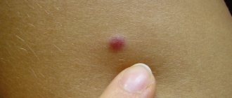
What changes in education are alarming?
The following symptoms should not be ignored:
- The mole has increased to 6 or more millimeters in diameter.
- The color of education has changed. Light or dark spots appeared on its surface.
- The texture, texture, and density of the nevus have changed.
- The edges of the mole have become uneven.
- Both during palpation and at rest, the mole hurts and itches. The skin around the formation is red and swollen.
These changes are unfavorable signs. It is recommended that you seek professional advice immediately.
How is the diagnosis done?
The most accurate method for diagnosing moles is dermatoscopy. The digital device dermatoscope allows you to “enlighten” the formation and enlarge its image. It becomes possible to study the texture and nature of skin pathology. Diagnosis is painless, safe and does not take much time.
Interesting: After dermatoscopy, all examined formations are recorded. At a subsequent visit to the doctor, you can monitor changes in nevi, their size, and shape.
During the examination, the dermatologist decides on the need for histological analysis. Most often, there are no contraindications for mole removal. It remains for the doctor to choose the optimal method.