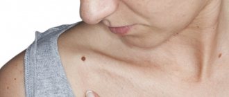
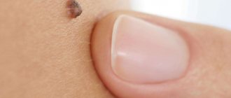
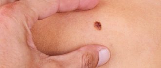
Moles (nevi) are benign pigmented formations on the skin. They do not pose a health hazard, but under certain conditions they can transform into malignant neoplasms (melanoma). For this reason, many people worry about moles and want to have them removed.
- When should you visit a specialist?
- Causes of moles
- Symptoms of degeneration
- Injury to nevi
- Which doctor should I contact about a mole?
- Diagnostics
Causes of moles
Some moles are congenital. They are formed in connection with local disturbances in the division of skin cells during the period of intrauterine development of the fetus, and appear as the child grows. The location and size of some nevi can be inherited.
The main factor that provokes the appearance of new moles is prolonged and intense exposure to ultraviolet rays. The formation of nevi is also often observed during hormonal changes (puberty, pregnancy).
Prevention
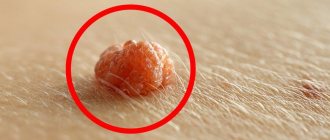
To prevent a mole from being torn off, it is important to follow the advice of doctors. The first thing doctors recommend is to monitor the hygiene of your hands and nails. This is one of the principles, since nevi are often injured by nails. You can accidentally comb the formation, scratch it or tear it off. Therefore, hand hygiene should always be carried out thoroughly. There are many bacteria under the nails, due to which microorganisms that provoke inflammatory processes enter the wound formed from tearing off a mole. If you damage a mole with your nails, skin cancer can even develop.
In addition, you need to watch out for clothing, which can tear the nevus. If moles are located in places of increased contact with it, then choose spacious wardrobe items. Clothing should not put pressure on nevi and constantly rub them. In addition, doctors recommend giving preference to those things that are made from natural materials and abandoning synthetic ones.
It is not recommended to use rough washcloths. Instead, it is better to opt for soft and delicate hygiene items. Often moles are injured during the process of bathing, when people subject the skin to excessive massaging with washcloths. It would also be useful to carefully use abrasive products, for example, scrubs. It is recommended not to apply the products to areas of the body where hanging moles are located or where there is an increased accumulation of flat ones. Substances that exfoliate rough skin sometimes injure nevi, causing a number of complications. It is also important to be careful after taking a bath, when wiping the body with a towel, especially when there is a mole that is torn. You should not make too fast or aggressive movements; you need to blot the skin carefully.
Symptoms of degeneration
Doctors have identified the main signs that may indicate malignant degeneration of moles. These include:
- Rapid increase in size. One of the alarming symptoms is asymmetrical growth, which leads to a change in the shape of the nevus.
- Change in color, namely rapid and uneven darkening of the mole.
- The appearance of pain, burning at the site of localization of the formation.
- The appearance of ulcerations and cracks on the surface of the mole.
- Changes in surrounding tissues in the form of a swollen reddish corolla.
If you detect at least one of these signs, you should immediately consult a doctor. Within a short time, an ordinary mole can degenerate into a malignant skin tumor - melanoma. This tumor metastasizes quickly and has high mortality rates.
The location of a mole on areas of the skin that are constantly exposed to ultraviolet radiation (face, hands) is also a reason to consult a doctor. Intense exposure to sunlight is one of the main triggers for malignant degeneration.
How to detect melanoma at an early stage
Since melanoma is dangerous, a person needs to take strange growths very seriously, be it a mole, wart, or pimple. Under no circumstances should you delay visiting a doctor, since in just a couple of months radical treatment may turn out to be useless, and the specialist will only offer maintenance therapy.
Diagnosis of melanoma includes:
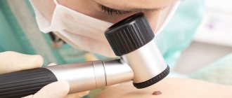
- Dermatoscopy. Suspicious moles and areas of skin are examined through a dermatoscope - a special microscope that allows you to study the skin in incident light (electroluminescence effect).
- Smear-imprint. Used if there are cracks or ulcers on the surface of the formation. The smear looks for tumor cells.
- Biopsy. Allows you to accurately determine the type of tumor: malignant or benign. To do this, the tumor area is examined under a microscope.
- Computer, magnetic resonance or positron emission tomography. Any of the tests to check other organs for the presence of metastases.
- Sentinel lymph node biopsy. Helps clarify the diagnosis and determine whether there are metastases. Typically, tumor cells are retained in the nearest lymph node, which is why it is checked for the presence of metastases.
Special biopsy method
Carrying out a biopsy is a controversial issue in the case of melanoma, since injuring the tumor can cause its rapid growth. In some cases, an excisional biopsy is resorted to. The surgeon excises the tumor, capturing 2-10 mm of healthy skin around it. While the patient is being sutured, the lesion is urgently sent for histology, often right in the operating room. If the diagnosis is confirmed, the surgeon immediately begins a repeat operation. The task is to remove all the tumor that could grow deep into the skin. Here everything depends on the results of histology, which determines the stage of the disease.
Injury to nevi
Another factor that can trigger the onset of malignancy is trauma to the mole. Moreover, the formation can be injured either over a long period of time, for example, due to constant friction or squeezing by clothing, or as a result of a more serious external influence, for example, when a mole is cut.
If a mole is accidentally damaged, it must be thoroughly treated with a disinfectant solution. If bleeding occurs, it should be stopped using a sterile bandage or gauze. The injured formation should be examined by a doctor as soon as possible in order to determine a further course of action. The specialist may recommend removing the remaining part of the mole or undergoing routine examinations for malignancy over a period of time.
Benefits of dermatoscopy
The advantages of the examination are undeniable:
- High accuracy (up to 97%);
- Simplicity;
- Ability to study the smallest moles;
- Delicacy towards the skin and the object of study;
- Painless;
- Safety;
- No contraindications;
- Quick results.
Another advantage is the fact that the use of the latest generation devices makes it possible to save an image of an object for further observation.
Histological examination
The Onco.Rehab Integrative Medicine Clinic will help you navigate and complete the study efficiently.
Which doctor should I contact about a mole?
There are several medical specialties that treat nevi: dermatologist, oncologist, surgeon. There is also a separate doctor for skin tumors - an oncodermatologist.
Visit to the dermatologist
A dermatologist is the first doctor you should contact about a mole. It is this specialist who, in most cases, is able to exclude a malignant process based on the results of examination and dermatoscopy.
Why go to a surgeon?
If the mole turns out to be benign, but is constantly subjected to friction or pressure, then it can be removed by a surgeon. You can contact a plastic surgeon if you have an aesthetic problem such as a mole on the face or other visible areas of the skin. For such nevi, if they do not raise questions regarding their benignity, laser removal is recommended.
When should you contact an oncologist?
If you have a mole with signs of malignant degeneration, you can immediately contact an oncologist. A doctor in this specialty has the necessary knowledge and skills for the surgical treatment of melanoma and other malignant skin tumors. The earlier cancer is detected, the more favorable the future prognosis will be.
Can the problem be solved by a cosmetologist?
Removal of moles in beauty salons and offices is possible only by a cosmetologist who has a higher medical education. There are several ways to remove a benign formation on the skin: laser method, cryodestruction, electrocoagulation. The patient needs to consult an oncologist before having the nevus removed by a cosmetologist, since, for example, after laser removal the possibility of histological examination of the tissue is lost.
Oncologist-mammologist
Women who have moles on the skin of the breast with signs of a possible malignant process should be examined by a breast oncologist. A doctor in this specialty selects a treatment plan taking into account the characteristics of the structure and functioning of the mammary glands.
When should you schedule a visit to an oncologist?
- The mole has changed color or surface
- You notice bleeding from a mole (even minor)
- The hair growing on the mole began to fall out
- The mole began to grow quickly and asymmetrically
- An unusual-looking neoplasm has appeared on the skin
- The mole is often injured
- You have discovered long-term non-healing wounds on your skin
A tandem of oncologist specialists - a surgeon and a dermatologist - will help eliminate the problem in a short time and prevent the development of skin cancer.
Diagnostics
In the process of diagnosing nevi, the doctor uses both standard methods (history collection, visual examination) and instrumental diagnostic methods. In this case, studies such as dermatoscopy, biopsy and histological analysis of the mole are highly informative.
Dermatoscopy
The essence of the dermatoscopy method is to examine the skin under magnification. During the examination, the doctor evaluates the color and structure of the epidermis, identifies the main characteristics of skin rashes, examines the epidermal-dermal junction and the papillary layer of the dermis.
To perform the study, a special device is used - a dermatoscope. This device may resemble a small magnifying glass (manual dermatoscopy) or be a special device that can be equipped with a digital photography function with the possibility of subsequent detailed, even computerized, analysis of various skin structures.
The dermatoscopy method is simple and accessible, but at the same time very informative. Examination of moles by a doctor using a dermatoscope significantly increases the chance of detecting malignant skin tumors, primarily melanoma.
Conducting research
During the procedure, the doctor has the opportunity to examine in detail the color and microstructure of different layers of the skin, conduct a differentiated diagnosis of any skin tumors, and determine their danger.
Thanks to modern instruments, it is possible to examine structural changes as small as 0.2 microns.
There are criteria by which a doctor can immediately evaluate tumors. The following are analyzed:
- Pigmentation;
- Color uniformity;
- Structure;
- Increase in size;
- Outlines and drawing;
- The presence of peeling and inflammatory areola;
- Availability of seal;
- Presence of cracks and areas of ulceration.
What does a dermato-oncologist do?
The specialist studies nevi and prescribes tests to determine the nature of the neoplasm.
Conducts differential diagnostics, assesses the likelihood of transforming a benign process into a malignant one.
First, a visual inspection is carried out.
Then specialized equipment is used for a more detailed study - a dermatoscope.
If there are risks of malignancy or the process of malignancy has already started, surgical intervention is prescribed.
The operation is performed by a surgeon using general or local anesthesia.
Healing after mole removal: can it hurt and for how long?
What to do if a mole hurts after removal, as well as the area around it?
Sometimes after surgery you may experience discomfort, pain and even a burning sensation.
More often this is observed after surgical removal of the nevus and adjacent tissues.
Minor soreness and discomfort may be present for up to one week, and an itchy sensation may also occur.
After removing the nevus, the wound and surrounding areas are treated with antibacterial ointments or a weakly concentrated solution of potassium permanganate twice a day for 5-7 days.
After surgery, a scab (crust) is formed at the site of mole removal, which protects the damaged area from infection.
In order not to injure the healing surface in the first 7-10 days, the use of a washcloth, taking a bath, sauna, or applying cosmetics is strictly prohibited.
After 8-9 days, the scab disappears, and healthy pink skin appears underneath.
Very often, patients are interested in whether it can hurt and how long it will take for the skin to heal after removal of a mole?
Pain may be present, but not always, and as a rule, if all medical recommendations are followed, it disappears within 2-3 days.
The main condition for rapid recovery is the complete exclusion of direct sunlight.
If the operation is planned to be performed in the summer, it is necessary to avoid going to the beach for 3-4 weeks.
Also reduce the frequency of being outside in the open sun.
FAQ
Who should I contact to remove a large birthmark?
Not all moles or birthmarks degenerate into malignant melanomas.
But large birthmarks can cause discomfort.
Many consider them a cosmetic defect and want to remove or significantly reduce them.
The removal procedure itself is performed by a surgeon.
But before the operation, you need to pass the prescribed tests, visit an oncologist and a dermatologist, who will assess all the risks and conduct the necessary studies.
The removal method depends on this.

